
Solid-State Preparation of Metal and Metal Oxides Nanostructures and Their Application in Environmental Remediation


Abstract
:1. Introduction
2. A General Survey of Methods for Preparation of Metal Oxide Nanoparticles
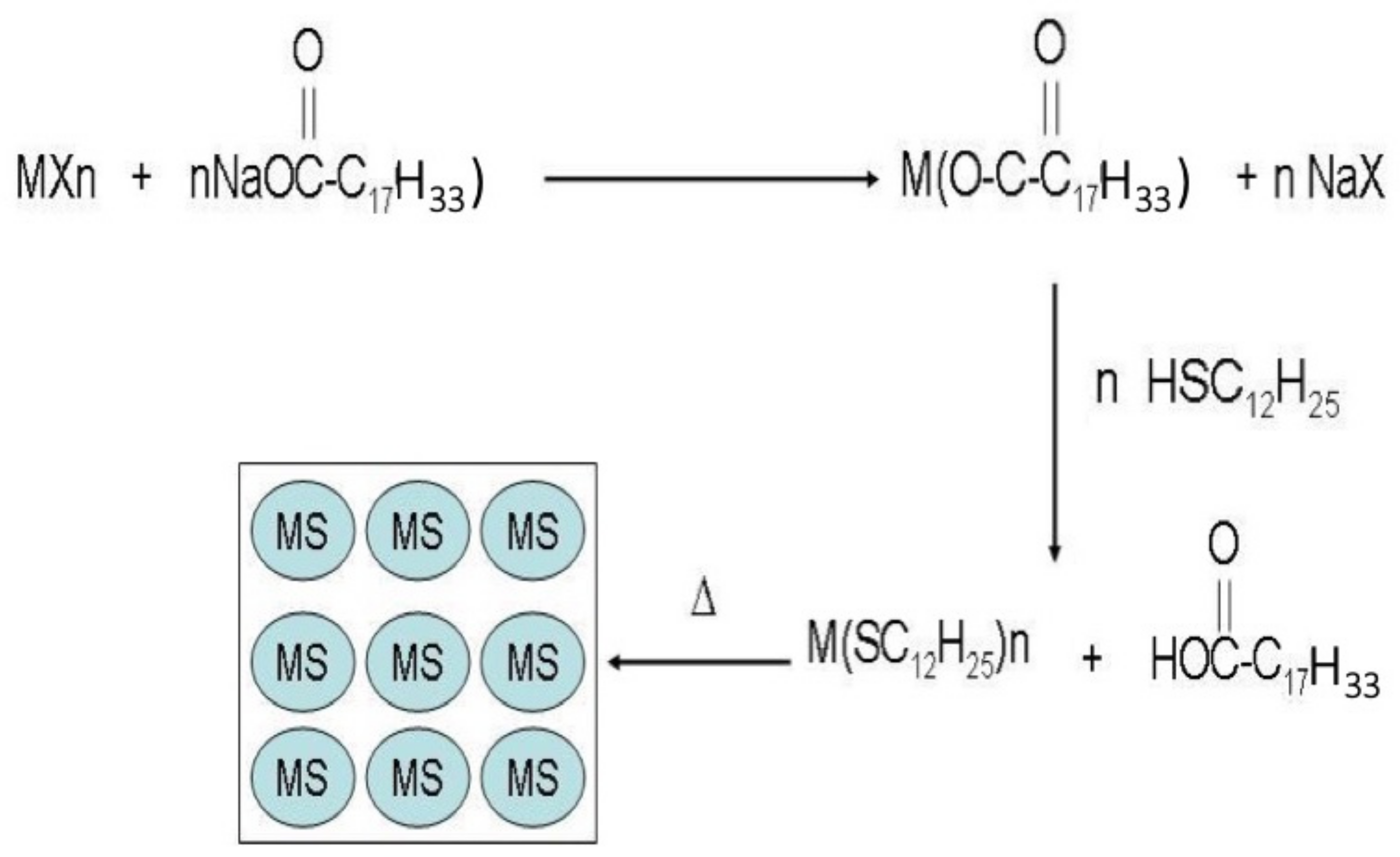
2.1. The Korgel’s Method
2.2. The Molecular and Macromolecular Complex Decomposition Method
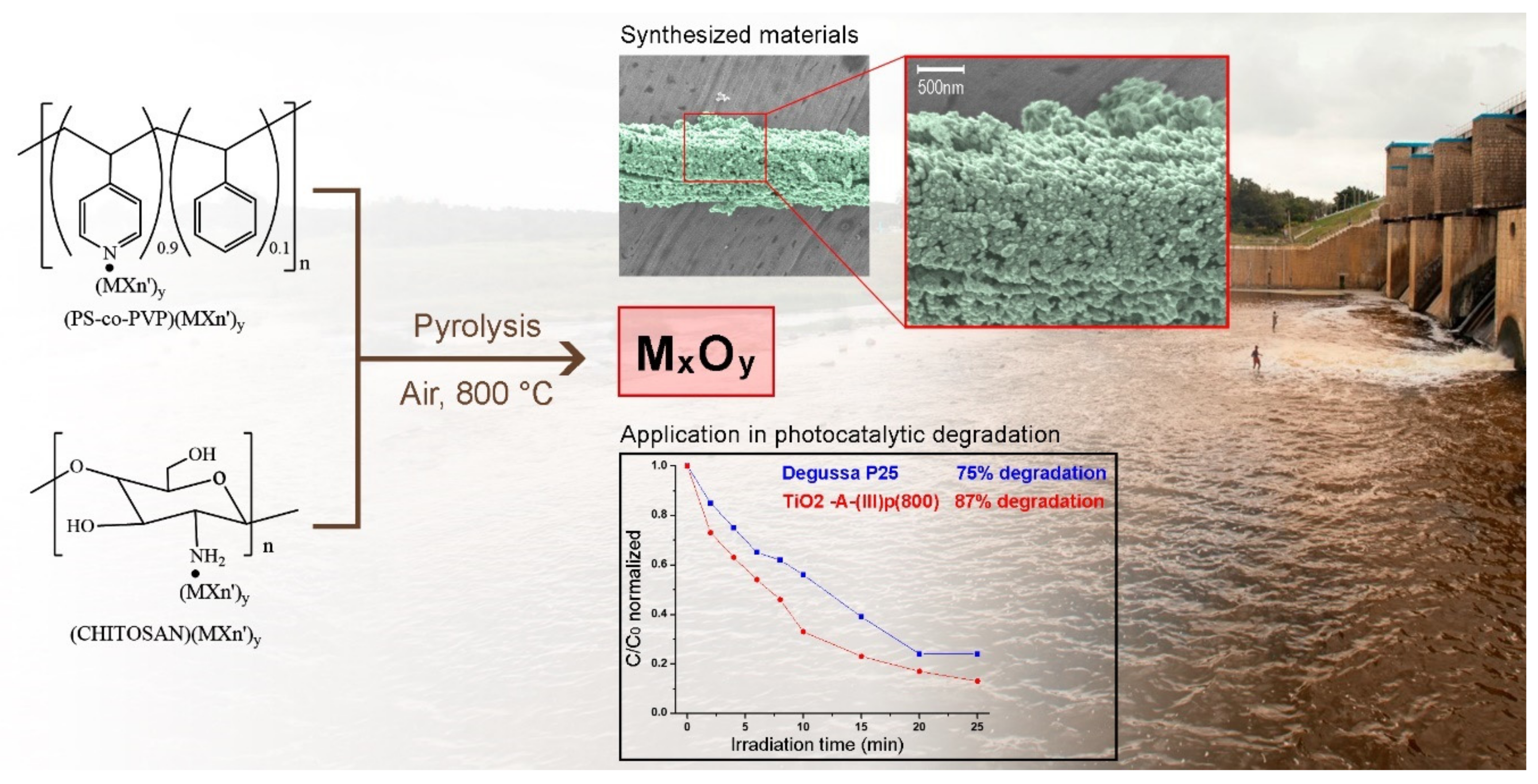
2.3. A Novel Solid-State Approximation
- The preparation of macromolecular precursors of general formula chitosan●MXn and PS-co-4-PVP·MXn;
- Pyrolysis of the chitosan·MXn and PS-co-4-PVP●MXn.
2.3.1. Transition Metal (First Row)
Titanium
Vanadium
Chromium, Molybdenum, and Tungsten
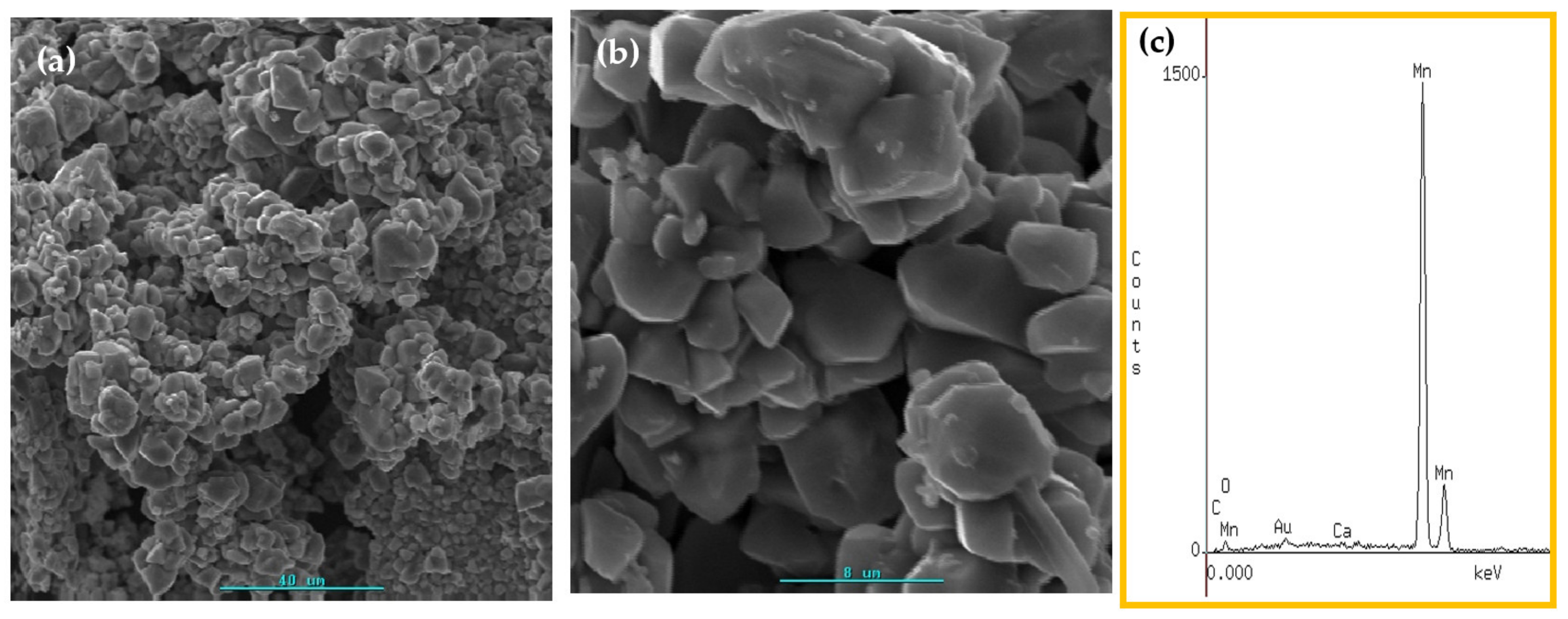
Manganese
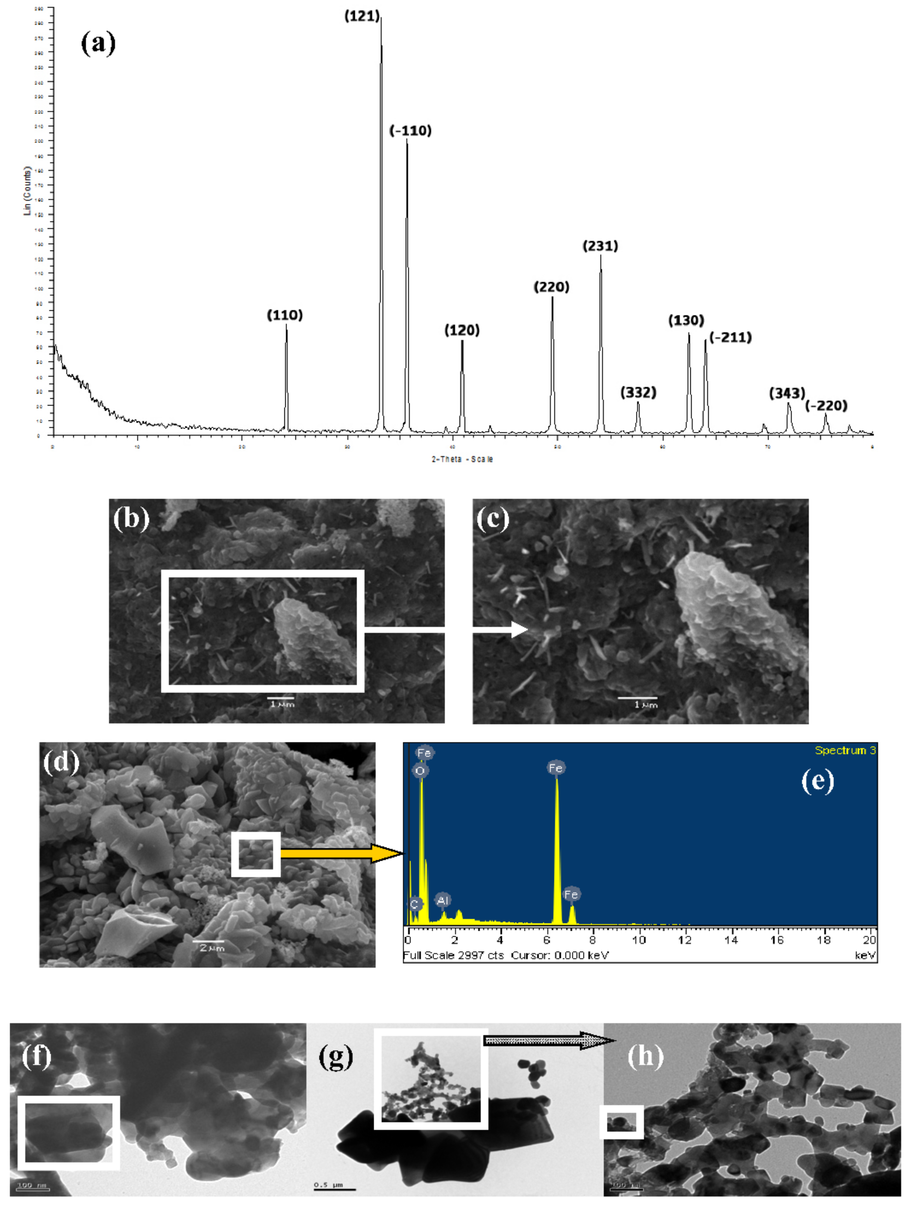
Iron
Cobalt
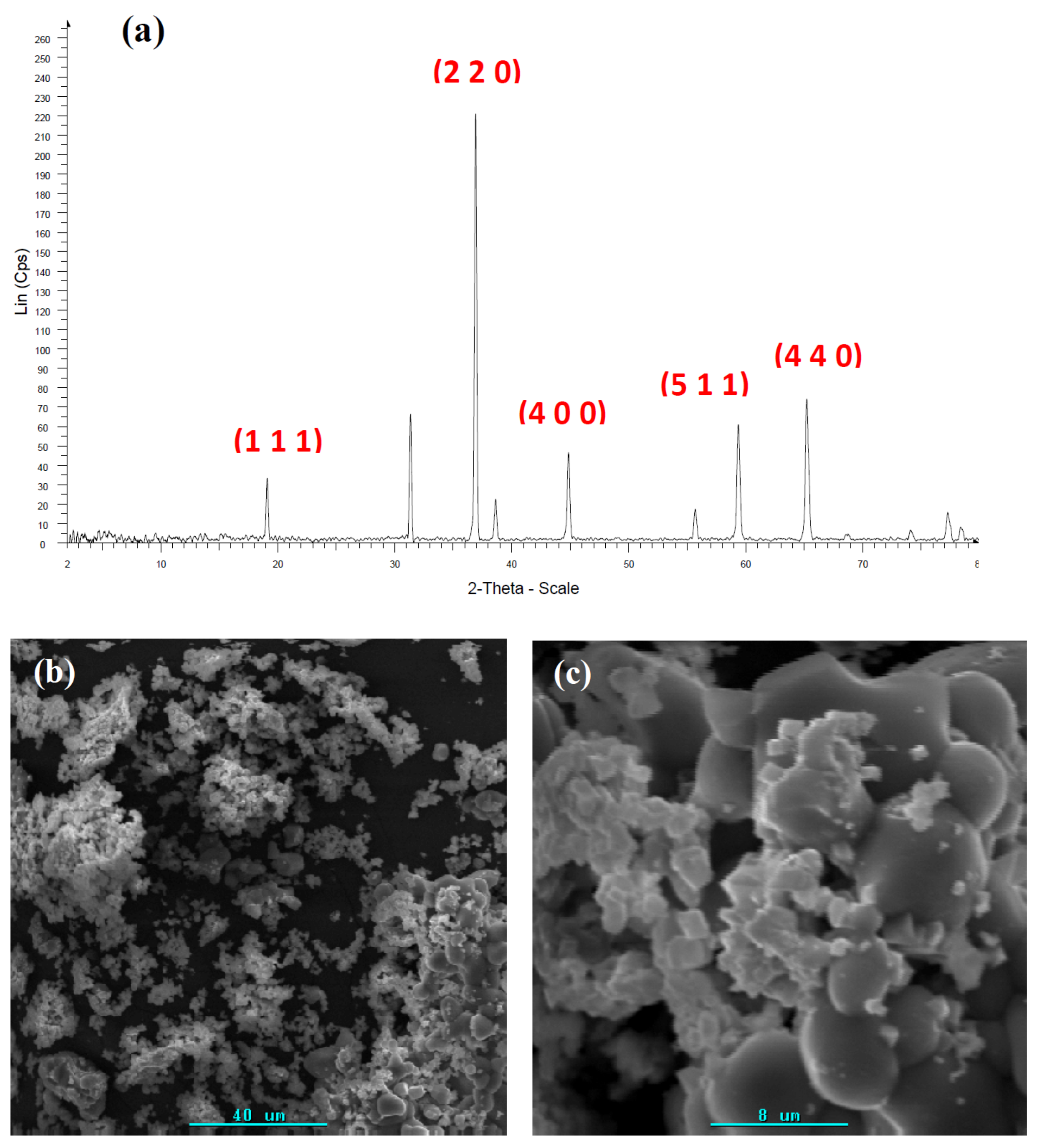
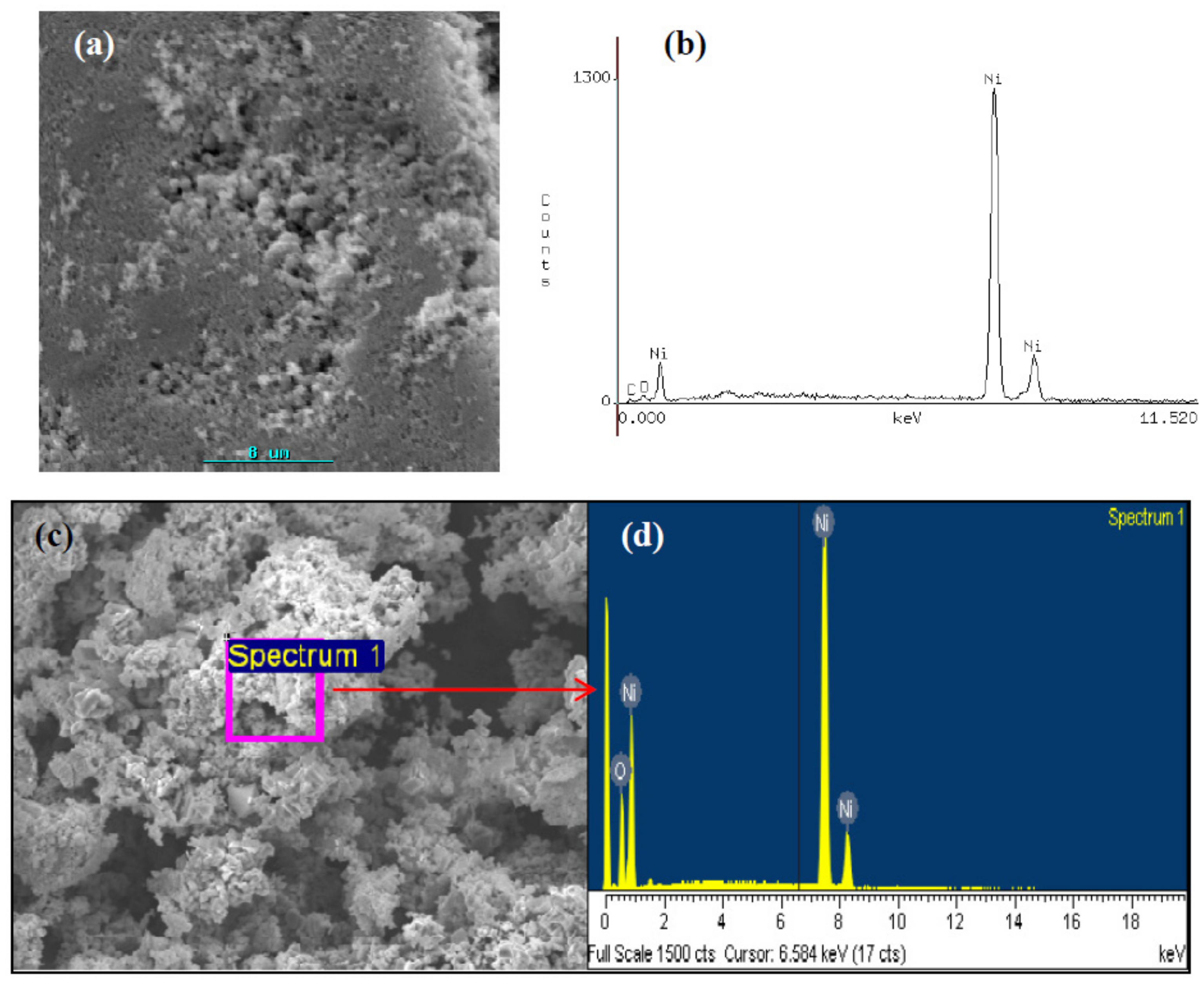
Nickel
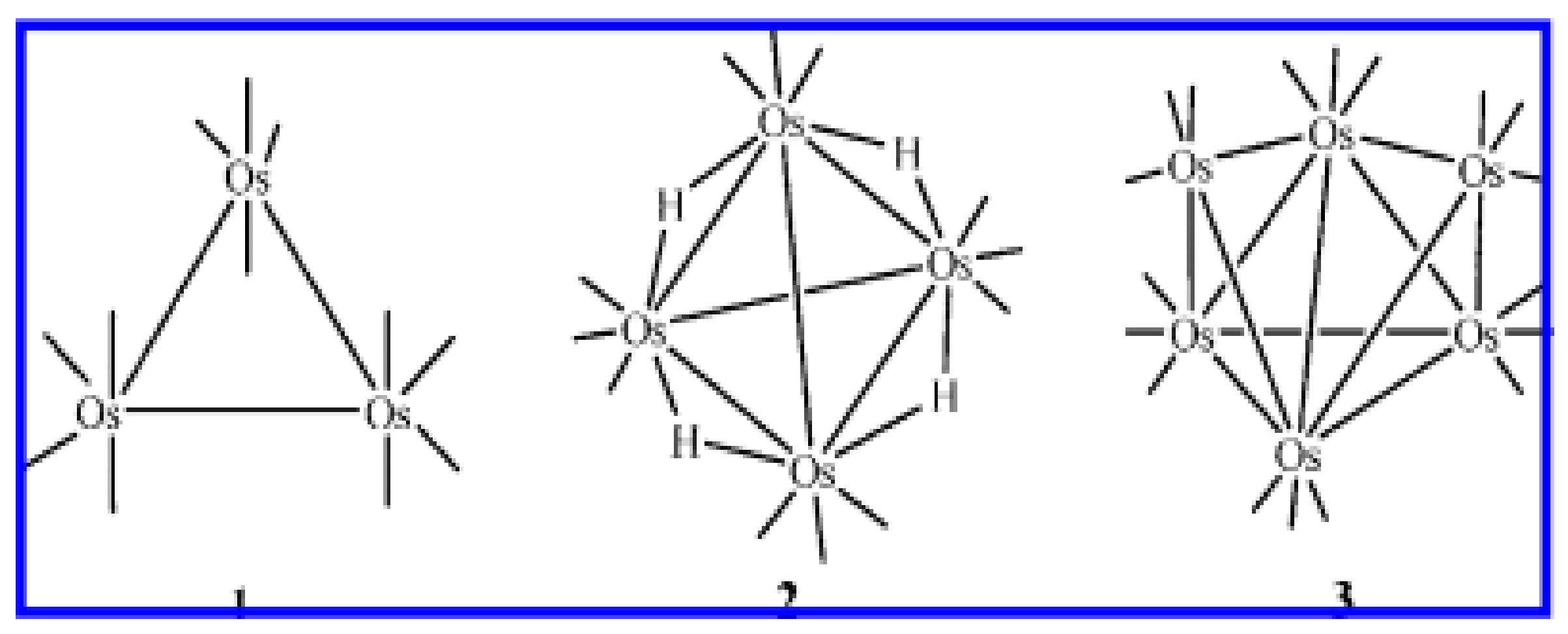
2.3.2. Noble and Precious Metals
Gold
Silver
Platinum
Iridium
Rhodium
2.3.3. Representative Metals
2.3.4. Rare Metals
2.3.5. Actinides
3. Incorporation of Metallic and Metal Oxides into Solid Matrix
4. Photocatalytic Applications
- (i)
- Ambient operating temperatures and pressure;
- (ii)
- Complete mineralization of contaminants and their intermediates compounds without leaving secondary pollutants;
- (iii)
- Low operating costs [146].
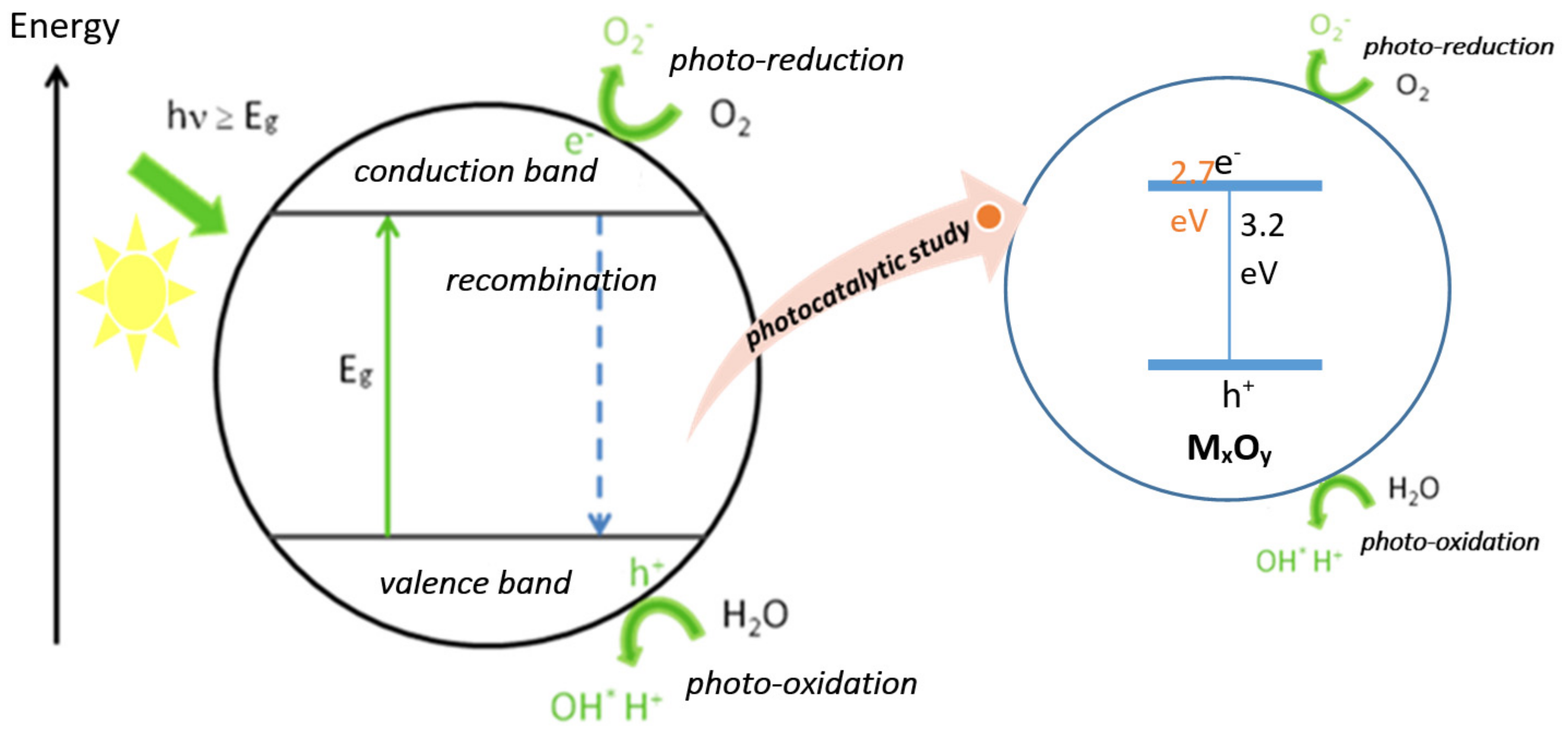
- An adequate band-gap;
- Suitable morphology;
- High surface area;
- Stability and reusability.
- Generation of •OH radicals by oxidation of OH− anions;
- Generation of O2− radicals by reduction in O2.
5. Probable Formation Mechanism of Nanostructures Metallic and Metal Oxides
6. Concluding Remarks
Author Contributions
Funding
Conflicts of Interest
References
- Hornyak, G.; Tibbals, H.F.; Dutta, J.; Moore, J. Introduction to Nanoscience and Nanotechnology; CRC Press Taylor and Francis: Boca Raton, NY, USA, 2009. [Google Scholar]
- Altavilla, C.; Ciliberto, E. Inorganic Nanoparticles; CRC Press Taylor and Francis: Boca Raton, NY, USA, 2011. [Google Scholar]
- Rao, C.N.; Muller, A.; Cheetham, A.K. The Chemistry of Nanomaterials; Wiley–VCH: Weinheim, Germany, 2004. [Google Scholar]
- Edelstein, A.S.; Cammarata, R.C. Nanomaterials, Synthesis and Applications; Institute of Physics Publishing: Bristol, UK, 2002. [Google Scholar]
- Cao, G. Nanostructures and Nanomaterial, Synthesis, Properties and Applications; Imperial College Press: London, UK, 2011. [Google Scholar]
- Díaz, C.; Valenzuela, M.L. Metallic Nanostructures Using Oligo and Polyphosphazenes as Template or Stabilizer in Solid State. In Encyclopedia of Nanoscience and Nanotechnology; Nalwa, H.S., Ed.; American Scientific Publishers: Valencia, CA, USA, 2010; Volume 16, pp. 239–256. [Google Scholar]
- Walter, E.C.; Ng, K.; Zach, M.P.; Penner, R.M.; Favier, F. Electronic Devices from electrodeposited metal nanowires. Microelectron. Eng. 2002, 61–62, 555–561. [Google Scholar] [CrossRef]
- Walkers, G.; Parkin, I.P. The Incorporation of noble nanoparticles into hast matrix thin films, synthesis, characterization and applications. J. Mater. Chem. 2009, 19, 574–590. [Google Scholar] [CrossRef]
- Scott, C.A. Epitaxial growth and properties of doped transition metal and complex oxide films. Adv. Mater. 2010, 21, 219–248. [Google Scholar]
- Teo, B.; Sun, X. Silicon-Based Low-Dimensional Nanomaterials and Nanodevices. Chem. Rev. 2010, 107, 1454–1532. [Google Scholar] [CrossRef] [PubMed]
- Khomutov, G.B.; Kislov, V.V.; Antipina, M.N.; Gainutdinov, R.V.; Gubin, S.P.; Obydenov, A.Y.; Pavlov, S.A.; Rakhnyanskaya, A.A.; Sergeev-Cherenkov, A.N.; Soldatov, E.S.; et al. Interfacial nanofabrication strategies in development of new functional nanomaterials and planar supramolecular nanostructures for nanoelectronic and nanotechnology. Microelectron. Eng. 2003, 69, 373–383. [Google Scholar] [CrossRef]
- Diaz, C.; Valenzuela, M.L.; Bobadilla, D. Bimetallic Au/Ag metal superstructures from macromolecular metal complexes in solid-state. J. Chil. Chem. Soc. 2013, 58, 1194–1997. [Google Scholar] [CrossRef] [Green Version]
- Díaz, C.; Valenzuela, M.L. Small-Molecule and High-Polymeric Phosphazenes containing oxypyridine side groups and their organometallic derivatives, Useful precursors for metal nanostructured materials. Macromolecules 2006, 39, 103–111. [Google Scholar] [CrossRef]
- Díaz, C.; Valenzuela, M.L. Organometallic Derivatives of Polyphosphazenes as Precursors for Metallic Nanostructured Materials. J. Inorg. Organomet. Polym. 2006, 16, 419–435. [Google Scholar] [CrossRef]
- Díaz, C.; Valenzuela, M.L.; Zuñiga, L.; O’Dwyer, C. Organometallic derivatives of cyclotriphosphazene as precursors of Nanostructured metallic materials, A new solid state Method. J. Inorg. Organomet. Polym. Mater. 2009, 19, 507–520. [Google Scholar] [CrossRef] [Green Version]
- Díaz, C.; Valenzuela, M.L.; Lavayen, V.; O’Dwyer, C. Layered Graphitic Carbon Host Formation during Liquid-free Solid State Growth of Metal Pyrophosphates. Inorg. Chem. 2012, 51, 6228–6236. [Google Scholar] [CrossRef] [Green Version]
- Díaz, C.; Valenzuela, M.L.; Carriedo, G.A.; Zuñiga, L.; O’Dwyer, C. Polymer/Trimer/Metal Complex Mixtures as Precursors of Gold Nanoparticles, Tuning the Morphology in the Solid-State. J. Inorg. Organomet. Polym. 2012, 22, 447–454. [Google Scholar]
- Díaz, C.; Valenzuela, M.L.; Cáceres, S.; O’Dwyer, C. Solution and surfactant-free growth of supported high index facet SERS active anoparticles of rhenium by phase demixing. J. Mater. Chem. A 2013, 1, 1566–1572. [Google Scholar]
- Díaz, C.; Valenzuela, M.L.; Cáceres, S.; O’Dwyer, C.; Diaz, R. Solvent and stabilizer free growth of Ag and Pd nanoparticles using Metallic salts/cyclotriphosphazenes mixtures. Mater. Chem. Phys. 2013, 143, 124–132. [Google Scholar] [CrossRef]
- Díaz, C.; Valenzuela, M.L.; Zuñiga, L.; O’Dwyer, C. Solid State Pathways to Complex Shape Evolution and Tunable Porosity During Metallic Crystal Growth. Sci. Rep. 2013, 3, 2642. [Google Scholar]
- Díaz, C.; Valenzuela, M.L. Organometallic-Metallic-Cyclotriphosphazene Mixtures, Solid-State Method for Metallic Nanoparticle Growth. In Nanostructures, Properties, Production Methods and Application; Nova Science Publishers: New York, NY, USA, 2013; Chapter 5. [Google Scholar]
- Díaz, C.; Valenzuela, M.L. A General Solid-State approach to metallic, metal oxides and Phosphates Nanoparticles. In Advances in Chemical Research; Nova Science Publishers: New York, NY, USA, 2011. [Google Scholar]
- Díaz, C.; Valenzuela, M.L. A general Solid-State approach to Metallic, Metal oxides and Phosphate nanoparticles. Gold Nanoparticles, Properties Synthesis and Fabrication. In Solution and Solid State Methods to Prepare Au Nanoparticles: A Comparison; Chow, P.E., Ed.; Nova Science Publishers: New York, NY, USA, 2010; Chapter 14. [Google Scholar]
- Larsen, T.H.; Sigman, M.; Ghezelbash, A.; Christopher-Doty, R.; Korgel, B.A. Solventless Synthesis of Copper Sulfide Nanorods by Thermolysis of a Single Source Thiolate-Derived Precursor. J. Am. Chem. Soc. 2003, 125, 5638–5639. [Google Scholar] [CrossRef] [PubMed]
- Sigman, M.; Ghezelbash, A.; Hanrath, T.; Saunders, A.E.; Lee, F.; Korgel, B.A. Solventless Synthesis of Monodisperse Cu2S Nanorods, Nanodisks, and Nanoplatelets. J. Am. Chem. Soc. 2003, 125, 16050–16057. [Google Scholar] [CrossRef] [PubMed]
- Ghezelbash, A.; Sigman, M.; Korgel, B.A. Solventless Synthesis of Nickel Sulfide Nanorods and Triangular Nanoprisms. Nano Lett. 2004, 4, 537–542. [Google Scholar] [CrossRef]
- Sigman, M.; Korgel, B.A. Solventless Synthesis of Bi2S3 (Bismuthinite) Nanorods, Nanowires, and Nanofabric. Chem. Mater. 2005, 17, 1655–1660. [Google Scholar] [CrossRef]
- Han, Y.C.; Cha, H.G.; Kim, C.h.W.; Kim, Y.H.; Kang, Y.S. Synthesis of Highly Magnetized Iron Nanoparticles by a Solventless Thermal Decomposition Method. J. Phys. Chem. C 2007, 111, 6275–6280. [Google Scholar] [CrossRef]
- Farhadi, S.; Roostaei-Zaniyani, Z. Preparation and characterization of NiO nanoparticles from thermal decomposition of the Ni(en)3(NO3)2 complex, A facile and low-temperature route. Polyhedron 2011, 30, 971–975. [Google Scholar] [CrossRef]
- Li, X.; Zhang, X.; Li, Z.; Qian, Y. Synthesis and characteristics of NiO nanoparticles by thermal decomposition of nickel dimethylglyoximate rods. Solid State Commun. 2006, 137, 581–584. [Google Scholar] [CrossRef]
- Davar, F.; Salavati-Niasari, M.; Mir, N.; Saberyan, K.; Monemzadeh, M. Thermal decomposition route for synthesis of Mn3O4 nanoparticles in presence of a novel precursor. Polyedron 2010, 29, 1747–1753. [Google Scholar] [CrossRef]
- Yanh, Z.; Zhang, Y.; Zhang, W.; Wang, X.; Qian, Y.; Weng, X.; Yang, S. Nanorods of manganese oxides, Synthesis, characterization and catalytic application. J. Solid State Chem. 2006, 179, 679–684. [Google Scholar]
- Soofivand, F.; Salavati-Niasari, M.; Mohandes, F. Novel precursor-assisted synthesis and characterization of zinc oxide nanoparticles/nanofibers. Mater. Lett. 2013, 98, 55–58. [Google Scholar] [CrossRef]
- Farhadi, S.; Pouzare, K.; Sadedhinejad, S. Simple preparation of ferromagnetic CO3O4 nanoparticles by thermal dissociation of the CoII(NH3)6(NO3)2 complex at low termperature. J. NanoStruct. Chem. 2013, 3, 16. [Google Scholar] [CrossRef] [Green Version]
- Randhawa, B.S.; Gandotra, K. A comparative study on the thermal decomposition of some transition metal carboxylates. J. Therm. Anal. Calorim. 2006, 85, 417–424. [Google Scholar] [CrossRef]
- Chunxiang Li Weng, K.L. Thermolysis of Polymeric Ru(CO)4.n to Metallic Ruthenium, Molecular Shape of the Precursor Affects the Nanoparticle Shape. Langmuir 2008, 24, 12040–12041. [Google Scholar]
- Nelson, J.M.; Nguyen, P.; Petersen, R.; Rengel, H.; Macdonald, P.M.; Lough, I.J.; Manners, I.; Raju, N.P.; Greedan, J.E.; Barlow, S.; et al. Thermal Ring-Opening Polymerization of Hydrocarbon-Bridged 2.Ferrocenophanes, Synthesis and Properties of Poly(ferrocenylethy1ene)s and Their Charge-Transfer Polymer Salts with Tetracyanoethylene. Chem. Eur. J. 1997, 3, 573–584. [Google Scholar] [CrossRef]
- Tang, B.Z.; Petersen, R.; Foucher, D.A.; Lough, A.; Coombs, N.; Sodhi, R. Novel Ceramic and Organometallic Depolymerization Products from PoIy(ferrocenyIsiIanes) via PyroIysis. J. Chem. Soc. Chem. Commun. 1993, 523–525. [Google Scholar] [CrossRef] [Green Version]
- Chang, B.S.; Brijith, T.; Chen, J.; Tevis, I.D.; Karanja, P.; Çınar, S.; Venkatesh, A.; Rossini, A.J.; Thuo, M.M. Ambient synthesis of nanomaterials by in situ heterogeneous metal/ligand reactions. Nanoscale 2019, 11, 14060–14069. [Google Scholar] [CrossRef] [PubMed] [Green Version]
- Li, C.; Zhong, Z.; Leong, W.K. Organometallic Clusters As Precursors for Metallic Nanoparticles, Effect of Cluster Size, Ligand Set, and Decomposition Method. Langmuir 2008, 24, 10427–10431. [Google Scholar] [CrossRef] [PubMed]
- Cai, B.; Akkiraju, K.; Mounfield, W.P.; Wang, Z.; Li, X.; Huang, B.; Yuan, S.; Su, D.; Roman-Leshkov, Y.; Shao-Horn, Y. Solid-State Gelation for Nanostructured Perovskite Oxide Aerogel. Chem. Mater. 2019, 31, 9422–9429. [Google Scholar] [CrossRef]
- Wang, B.; Zhang, C.; Zheng, W.; Zhang, Q.; Bao, Z.; Kong, L.; Li, L. Large-Scale Synthesis of Highly Luminescent Perovskite Nanocrystals by Template-Assisted Solid-State Reaction at 800. Chem. Mater. 2020, 32, 308–314. [Google Scholar] [CrossRef]
- Mao, Y.; Banerjee, S.; Wong, S.S. Large-Scale Synthesis of Single-Crystalline Perovskite Nanostructures. J. Am. Chem. Soc. 2003, 125, 15718–15719. [Google Scholar] [CrossRef]
- Schmitt, W.; Hill, J.P.; Malik, S.; Volkert, C.A.; Ichinose, I.; Anson, C.; Powell, A.K. Thermolysis of a Hybrid Organic–Inorganic Supramolecular Coordination Assembly, Templating the Formation of Nanostructured Fibrous Materials and Carbon-Based Microcapsules. Angew. Chem. Int. Ed. 2005, 44, 7048–7053. [Google Scholar] [CrossRef] [PubMed]
- Reda, G.M.; Fan, H.; Tian, H. Room-temperature solid state synthesis of Co3O4/ZnO p–n heterostructure and its photocatalytic activity. Adv. Powder Technol. 2017, 28, 953–963. [Google Scholar] [CrossRef]
- Nalluri, S.R.; Nagarjuna, R.; Patra, D.; Ganesan, R.; Balaj, G. Large Scale Solid-state Synthesis of Catalytically Active Fe3O4@M (M =Au, Ag and Au-Ag alloy) Core-shell Nanostructures. Sci. Rep. 2019, 9, 6603. [Google Scholar] [CrossRef]
- Dey, A.; Zubko, M.; Kusz, J.; Reddy, V.R.; Banerjee, A.; Bhattacharjee, A. Thermal synthesis of Hematite nanoparticles, Structural, magnetic and morphological characterizations. Int. J. Nano Dimens. 2020, 11, 188–198. [Google Scholar]
- Das, R.; Pachfule, P.; Banerjee, R.; Poddar, P. Metal and Metal oxide nanoparticle synthesis from metal organic frameworks (MOFs), finding the border of metal and metal oxides. Nanoscale 2012, 4, 591–599. [Google Scholar] [CrossRef] [PubMed]
- Rong, K.; Wei, J.; Huang, L.; Fang, Y.; Dong, S. Synthesis of low dimensional hierarchical transition metal oxides via a direct deep eutectic solvent calcining method for enhanced oxygen evolution catalysis. Nanoscale 2020, 12, 20719–20725. [Google Scholar] [CrossRef]
- Zhu, J.; Jiang, Y.; Lu, Z.; Zhao Ch Xie, L.; Chen, L.; Duan, J. Single-crystal Cr2O3 nanoplates with differing crystalinities, derived from trinuclear complexes and embedded in a carbon matrix, as an electrode material for supercapacitors. J. Colloid Interface Sci. 2017, 498, 351–363. [Google Scholar] [CrossRef] [PubMed]
- Yuan, Y.; Chen, L.; Yang, R.; Lu, X.; Peng, H.; Luo, Z. Solid-state synthesis and characterization of core–shell CoFe2O4–carbon composite nanoparticles from a heterometallic trinuclear complex. Mater. Lett. 2012, 71, 123–126. [Google Scholar] [CrossRef]
- Díaz, C.; Valenzuela, M.L.; Lavayen, V.; Mendoza, K.; Peña, O.; O’Dwyer, C. Nanostructured copper oxides and phosphates from a new solid-state route. Inorg. Chim. Acta 2011, 377, 5–11. [Google Scholar] [CrossRef]
- Díaz, C.; Platoni, S.; Molina, A.; Valenzuela, M.L.; Geaney, H.; O’Dwyer, C. Novel Solid-State Route to Nanostructured Tin, Zinc and Cerium Oxides as Potential Materials for Sensors. J. Nanosci. Nanotechnol. 2014, 14, 7648–7653. [Google Scholar] [CrossRef]
- Diaz, C.; Barrera, G.; Segovia, M.; Valenzuela, M.L.; Osiak, M.; O’Dwyer, C. Solvent-less method for efficient photocatalytic α-Fe2O3 nanoparticles for using macromolecular polymeric precursors. New J. Chem. 2016, 40, 6768–6776. [Google Scholar] [CrossRef]
- Diaz, C.; Valenzuela, M.L.; Laguna, M.A.; Orera, A.; Bobadilla, D.; Abarca, S.; Peña, O. Synthesis and Magnetic Properties of Nanostructured metallic Co, Mn and Ni oxide materials obtained from solid-state macromolecular complex precursors. RSC Adv. 2017, 7, 27729–27736. [Google Scholar] [CrossRef] [Green Version]
- Diaz, C.; Valenzuela, M.L.; Bobadilla, D.; Laguna-Bercero, M.A. Bimetallic Au//Ag Alloys Inside SiO2 using a solid-state method. J. Clust. Chem. 2017, 28, 2809–2815. [Google Scholar] [CrossRef] [Green Version]
- Diaz, C.; Valenzuela, M.L.; Garcia, C.; De la Campa, R.; Soto, A.-P. Solid-state synthesis of pure and doped lanthanides oxide nanomaterials by using polymer templates. Study of their luminescent properties. Mater. Lett. 2017, 209, 111–114. [Google Scholar] [CrossRef]
- Diaz, C.; Valenzuela, M.L.; Segovia, M.; De la Campa, R.; Soto, A.-P. Solution, Solid-State Two Step Synthesis and Optical Properties of ZnO and SnO Nanoparticles and Their Nanocomposites with SiO2. J. Clust. Sci. 2018, 29, 251–266. [Google Scholar] [CrossRef] [Green Version]
- Allende, P.; Laguna, M.A.; Barrientos, L.; Valenzuela, M.L.; Diaz, C. Solid State tuning Morphology, Crystal Phase and Size through Metal Macromolecular Complexes and Its Significance in the Photocatalytic Response. ACS Appl. Energy Mater. 2018, 1, 3159–3170. [Google Scholar] [CrossRef]
- Diaz, C.; Carrillo, D.; De la Campa, R.; Soto, A.-P.; Valenzuela, M.L. Solid-State synthesis of LnOCl/Ln2O3 (Ln = Eu, Nd) by using chitosan and PS-co-P4VP as polymeric supports. J. Rare Earth 2018, 36, 1326–1332. [Google Scholar] [CrossRef]
- Allende, P.; Barrientos, L.; Orera, A.; Laguna-Bercero, M.A.; Salazar, N.; Valenzuela, M.L.; Diaz, C. TiO2/SiO2 Composite for Efficient Protection of UVA and UVB Rays Through of a Solvent-Less Synthesis. J. Clust. Sci. 2019, 30, 1511–1517. [Google Scholar] [CrossRef]
- Diaz, C.; Valenzuela, M.L.; Cifuentes-Vaca, O.; Segovia, M.; Laguna-Bercero, M.A. Incorporation of Nanostructured ReO3 in Silica Matrix and Their Activity Toward Photodegradation of Blue Methylene. J. Inorg. Organomet. Polym. Mater. 2020, 30, 1726–1734. [Google Scholar] [CrossRef]
- Diaz, C.; Valenzuela, M.L.; Cifuentes-Vaca, O.; Segovia, M.; Laguna-Bercero, M.A. Iridium nanostructured metal oxide, its inclusion in Silica matrix and their activity toward Photodegradation of Methylene Blue. Mater. Chem. Phys. 2020, 252, 123276–123286. [Google Scholar] [CrossRef]
- Diaz, C.; Valenzuela, M.L.; Cifuentes-Vaca, O.; Segovia, M. Polymer precursors effect in the macromolecular metal-polymer on the Rh/RhO2/Rh2O3 phase using solvent-less synthesis and its photocatalytic activity. J. Inorg. Organomet. Polym. Mater. 2020, 30, 4702–4708. [Google Scholar] [CrossRef]
- Diaz, C.; Valenzuela, M.L.; Cifuentes-Vaca, O.; Segovia, M.; Laguna-Bercero, M.A. Incorporation of NiO into SiO2, TiO2, Al2O3, and Na4.2Ca2.8(Si6O18) Matrices, Medium Effect on the Optical Properties and Catalytic Degradation of Methylene Blue. Nanomaterials 2020, 10, 2470. [Google Scholar] [CrossRef]
- Diaz, C.; Valenzuela, M.L.; Laguna-Bercero, M.A.; Mendoza, K.; Cartes, P. Solventless preparation of thoria, their inclusion inside SiO2 and TiO2, their luminiscent properties and their photocataltytic behavior. ACS Omega 2021, 6, 9391–9400. [Google Scholar] [CrossRef] [PubMed]
- Chen, X.; Mao, S.S. Titanium Dioxide Nanomaterials, Synthesis, Properties, Modifications, and Applications. Chem. Rev. 2007, 107, 2891–2959. [Google Scholar] [CrossRef] [PubMed]
- Ismail, A.A.; Bahnemannb, D.W. Mesoporous titania photocatalysts, preparation, characterization and reaction mechanisms. J. Mater. Chem. 2011, 21, 11686–11707. [Google Scholar] [CrossRef] [Green Version]
- Wang, Y.; Cao, G. Synthesis and Enhanced Intercalation Properties of Nanostructured Vanadium Oxides. Chem. Mater. 2006, 18, 2787–2804. [Google Scholar] [CrossRef]
- Mao, C.J.; Pan, H.C.; Wu, X.C.; Zhu, J.J.; Chen, H.Y. Sonochemical Route for Self-Assembled V2O5 Bundles with Spindle-like Mosphology and their Novel Application in Serum Albumin Sensing. J. Phys. Chem. 2006, 110, 14709–14713. [Google Scholar]
- Avansi, W.; Ribeiro, C.; Leite, E.; Mastelaro, V. Vanadium Pentoxide Nanostructured, An Effective Control of Morphology and Crystal Strucutred in Hydrothermal Conditions. Cryst. Growth Des. 2009, 9, 3626–3631. [Google Scholar] [CrossRef]
- Diaz, C.; Barrera, G.; Segovia, M.; Valenzuela, M.L.; Osiak, M.; O’Dwyer, C. Crystallizing Vanadium Pentoxide Nanostructures in the Solid State using Modified Block co-Polymer and Chitosan Complexes. J. Nanomater. 2015, 2015, 105157. [Google Scholar] [CrossRef] [Green Version]
- Fei, H.L.; Liu, M.; Zhou, H.J.; Sun, P.C.; Ding, D.T.; Chen, T.H. Synthesis of V2O5 micro-architectures via in situ generation of single-crystalline nanoparticles. Solid State Sci. 2009, 11, 102–107. [Google Scholar] [CrossRef]
- Díaz, C.; Valenzuela, M.L.; Yutronic, N.; Villalobos, V.; Barrera, G. Nanostructured VOx/VO(PO4)n Using Solid-State Vanadium Containing Phosphazene Precursors, A Uselful Potential Bi-Catalyst System. J. Clust Sci. 2011, 22, 693–704. [Google Scholar] [CrossRef]
- Zhou, Y.; Qiu, Z.; Lu, M.; Zhang, A.; Ma, Q. Preparation and characterization of V2O5 macro-plates. Mater. Lett. 2007, 61, 4073–4075. [Google Scholar] [CrossRef]
- Diaz, C.; Valenzuela, M.L.; Zepeda, L.; Herrera, P.; Valenzuela, C. General group VI transition nanostructured metal oxides and their inclusion into solid matrices by a solution-solid approach. J. Chil. Chem. Soc. 2021, 66. in press. [Google Scholar]
- Han, Y.; Chen, F.; Zhong, Z.; Ramesh, K.; Chen, L.; Widjaja, E. Controlled Synthesis, Characterization, and Catalytic Properties of Mn2O3 and Mn3O4 Nanoparticles Supported on Mesoporous Silica SBA-15. J. Phys. Chem. B 2006, 110, 24450–24456. [Google Scholar] [CrossRef]
- Laurent, S.; Forge, D.; Port, M.; Roch, A.; Robic, C.; Vander Elst, L.; Muller, R. Magnetic Iron Oxide Nanoparticles, Synthesis, Stabilization, Vectorization, Physicochemical Characterizations and Biological Applications. Chem. Rev. 2008, 108, 2064–2110. [Google Scholar] [CrossRef]
- Teja, A.; Koh, P.Y. Synthesis propieties and applications of magnetic iron oxide nanoparticles. Prog. Cryst. Growth Charact. Mater. 2009, 55, 22–45. [Google Scholar] [CrossRef]
- Patra, A.K.; Kundu, S.K.; Bhaumik, A.; Kim, D. Morphology evolution of single-crystalline hematite nanocrystals: Magnetically recoverable nanocatalysts for enhanced facet-driven photoredox activity. Nanoscale 2016, 8, 365. [Google Scholar] [CrossRef] [PubMed]
- Zheng, Y.; Cheng, Y.; Wang, Y.; Bao, F.; Zhou, L.; Wei, X.; Zhang, Y.; Zheng, Q. Quasicubic α-Fe2O3 Nanoparticles with Excellent Catalytic Performance. J. Phys. Chem. 2006, 110, 3093–3097. [Google Scholar] [CrossRef]
- Hua, J.; Gengsheng, J. Hydrothermal synthesis abd characterization of monodispere α-Fe2O3 Nanoparticles. Mater. Lett. 2009, 63, 2725–2727. [Google Scholar] [CrossRef]
- Liu, Y.; Yu, C.; Dai, W.; Gao, X.; Qian, H.; Hu, Y.; Hu, X. One-post solvothermal synthesis of multi-shelled α-Fe2O3 hollow spheres with enhaced visible-light photocatalytic activity. J. Alloy. Compd. 2013, 551, 440–443. [Google Scholar] [CrossRef]
- Jiu, J.; Ge, Y.; Li, X.; Nie, L. Preparation of Co3O4 nanoparticles by a polymer conbution route. Mater. Lett. 2002, 54, 260–263. [Google Scholar] [CrossRef]
- Li, Y.; Tan, B.; Wu, Y. Mesoporous Co3O4 Nanowires Arrays for Lithium Ion Batteries with High Capacity and rate Capability. Nano Lett. 2008, 8, 265–270. [Google Scholar] [CrossRef]
- Deng, J.; Kang, L.; Bai, G.; Li, Y.; Li, P.; Liu, X.; Yang, Y.; Gao, F.; Liang, W. Solution combustion synthesis of cobalt oxides (Co3O4 and Co3O4/CoO) nanoparticles as supercapacitor electrode materials. Electrochim. Acta 2014, 132, 127–135. [Google Scholar] [CrossRef]
- Salvati-Niasari, M.; Khansari, A.; Davar, F. Synthesis and characterization of cobalt oxides nanoparticles by thermal process. Inorg. Chem. Acta 2009, 362, 4937–4942. [Google Scholar] [CrossRef]
- Nassar, M.Y.; Ahmed, I.S. Hydrothermal synthesis of cobalt carbonates using different counter ions, An efficient precursor to nano-sized cobalt oxide (Co3O4). Polyhedron 2011, 30, 2431–2437. [Google Scholar] [CrossRef]
- Palacios-Hernandez, P.; Hirata-Flores, G.; Contreras-Lopez, O.; Mendoza-Sanchez, M.; Valeriano-Arreola, I.; Gonzalez-Vergara, E.; Mendez-Rojas, M. Synthesis of Cu and Co metal oxide nanoparticles from thermal decomposition of tastrate complexes. Inorg. Chem. Acta 2012, 392, 277–282. [Google Scholar] [CrossRef]
- Kalam, A.; Al-Sehemi, A.; Al-Shihri, A.; Du, G.; Tokeer, A. Synthesis and Characterization if NiO nanoparticles by thermal decomposition of nickel linoleate and their optical properties. Mater. Charact. 2012, 68, 77–81. [Google Scholar] [CrossRef]
- Wang, C.; Li, J.; Liang, X.; Zhang, Y.; Guo, G. Photocatalytic organic pollutants degradation in metal–organic frameworks. Energy Environ. Sci. 2014, 7, 2831–2867. [Google Scholar] [CrossRef]
- Ukoba, K.O.; Eloka-Eboca, A.C.; Inambao, F.L. Review of nanostructured NiO thin film deposition using the spray pyrolysis technique. Renew. Sustain. Energy Rev. 2018, 82, 2900–2915. [Google Scholar] [CrossRef]
- Rowashden-Omary, M.; Lopez-Luzuriaga, J.M.; Rashdan, M.; Elbjeirami, O.; Monge, M.; Rodriguez-Castillo, M.; Laguna, A. Golden Metallopolymers with an Active T State via Coordination of Poly(4-vinyl)pyridine to Pentahalophenyl-Gold (I) Precursors. J. Am. Chem. Soc. 2009, 131, 3824–3825. [Google Scholar] [CrossRef]
- Diaz, C.; Valenzuela, M.L.; Soto, K.; Laguna-Bercero, M.A. Incorporation of Au and Ag Nanostructures inside SiO2. J. Chilean Chem. Soc. 2019, 64, 4502–4506. [Google Scholar] [CrossRef] [Green Version]
- Tappan, B.; Steiner, S.; Luther, E. Nonporous Metal Foams. Angew. Chem. Int. Ed. 2010, 49, 4544–4565. [Google Scholar] [CrossRef]
- Yaqoob, A.A.; Umar, K.; Nasir, M.; Ibrahim, M. Silver nanoparticles, various methods of synthesis, size afecting factors and their potential applications—A review. Appl. Nanosci. 2020, 10, 1369–1378. [Google Scholar] [CrossRef]
- Syafiuddin, A.; Salmiati Salim, M.R.; Hong Kueh, A.B.; Hadibarata, T.; Nure, H.A. Review of Silver Nanoparticles, Research Trends, Global Consumption, Synthesis, Properties, and Future Challenges. J. Chin. Chem. Soc. 2017, 64, 732–756. [Google Scholar] [CrossRef]
- Chen, A.; Holt-Hindle, P. Platinum-Based Nanostructured Materials, Synthesis, Properties and Applications. Chem. Rev. 2010, 110, 3767–3804. [Google Scholar] [CrossRef]
- Diaz, C.; Valenzuela, M.L.; Baez, R.; Segovia, M. Solid State Morphology and Size Tuning of Nanostructured Platinum Using Macromolecular Complexes. J. Chil. Chem. Soc. 2015, 60, 2986–2990. [Google Scholar] [CrossRef] [Green Version]
- Wu, J.; Qi, L.; You, H.; Gross, A.; Li, J.; Yang, H. Icosahedral Platinum Alloy Nancrystals with Enhanced Electrocatalytic activities. J. Am. Chem. Soc. 2012, 134, 11880–11883. [Google Scholar] [CrossRef]
- Leong, G.J.; Schulze, M.C.; Strand, M.B.; Maloney, D.; Frisco, S.L.; Dinh, H.N.; Pivovar, B.; Richards, R.M. Shape-directed platinum nanoparticle synthesis, nanoscale design of novel catalysts. Appl. Organomet. Chem. 2014, 28, 1–17. [Google Scholar] [CrossRef]
- Peng, H.; Yang, H. Designer platinum nanoparticles, Control of shape, composition in alloy, nanostrucutred and electrocatalytic property. Nano Today 2009, 4, 143–164. [Google Scholar] [CrossRef]
- Chen, J.; Lim, B.; Lee, E.P.; Xia, Y. Shape-controlled synthesis of platinum nanocrystals for catalytic and electrocatalytic applications. Nano Today 2009, 4, 81–95. [Google Scholar] [CrossRef]
- Cotton, F.A.; Wilkinson, G. Chapter 22 and 30. In Advanced Inorganic Chemistry; John Wiley and Sons: New York, NY, USA, 1980. [Google Scholar]
- Jin, R. The impacts of nanotechnology on catalysis by precious metal nanoparticles. Nanotechnol. Rev. 2012, 1, 31–56. [Google Scholar] [CrossRef]
- Liu, L.; Corma, A. Metal Catalysts for Heterogeneous Catalysis: From Single Atoms to Nanoclusters and Nanoparticles. Chem. Rev. 2018, 118, 4981–5079. [Google Scholar] [CrossRef] [Green Version]
- Chen, R.S.; Korotcov, A.; Huiang, A.S.; Tsai, D. One-dimensional conductive IrO2 nanocrystals. Nanotechnology 2006, 17, 67–87. [Google Scholar] [CrossRef] [Green Version]
- Woo, H.; Shim, H.S.; Myung, J.H.; Lee, C. Annealing effect on the structural properties of IrO2. Vacuum 2008, 82, 1400–1403. [Google Scholar]
- Ortel, E.; Reier, T.; Strasser, P.; Kraehnert, R. Mesoporous film template by PEO-PB-PEO block-copolymers, Self-Assembly, Cristalización behaviour and electrocatalytic performance. Chem. Mater. 2011, 23, 3201–3209. [Google Scholar] [CrossRef]
- Zhao, Y.; Hernandez, E.A.; Vargas-Barbosa, N.M.; Dysart, J.L.; Mallouk, T.E. A high yield Synthesis of ligand-free iridium oxide nanoparticles with high electrocatalytic activity. J. Phys. Chem. Lett. 2011, 2, 402–406. [Google Scholar] [CrossRef]
- Brewer, D.; Wicajksana, J.; Maria, A.; Kingon, S. Franzen, Investigation of the electrical and optical properties of iridium oxide by reflectance FTIR spectroscopy and density functional theory calculations. Chem. Phys. 2005, 313, 25–31. [Google Scholar] [CrossRef]
- Xu, D.; Diao, P.; Jin, T.; Wu, Q.; Liu, X.; Guo, X.; Gong, H.; Li, F.; Xiang, M.; Ronghai, Y. Iridium oxide nanoparticles and Iridium/Iridium Oxide Nanocompósites, Photochemical Fabrication and Application in Catalytic Rediction of Notrophenol. ACS Appl. Interfaces 2015, 7, 16738–16749. [Google Scholar] [CrossRef] [PubMed]
- Biswas, K.; Rao, C.N. Synthesis and characterization of Nano crystals of oxide metals RuO2, IrO2, and ReO3. J. Nanosci. Nanotechnol. 2007, 7, 1969–1974. [Google Scholar] [CrossRef] [PubMed]
- Lee, Y.M.; Suntivich, J.; May, K.J.; Perry, E.E.; Shao, H. Synthesis and activities of Rutile IrO2 and RuO2 nanoparticles for oxygen evolution in acid alkaline solutions. J. Phys. Chem. Lett. 2011, 2, 402–406. [Google Scholar] [CrossRef]
- Quinson, J. Surfactant-Free Precious Metal Colloidal Nanoparticles for Catalysis. Front. Nanotechnol. 2021, 3, 770281. [Google Scholar] [CrossRef]
- Fernandez-Garcia, M.; Martinez-Arias, A.; Hanson, J.; Rodriguez, C. Nanostructured Oxides in Chemistry, Characterization and Properties. J. Am. Chem. Rev. 2004, 104, 4063–4104. [Google Scholar] [CrossRef]
- Kim, Y.L.; Ha, Y.; Lee, N.S.; Kim, J.G.; Baik, J.M.; Le, C.; Yoon, K.; Lee, Y.; Kim, M.H. Hybrid architecture of rhodium oxide nanofibers and ruthenium oxide nanowires for electrocatalysts. J. Alloy. Compd. 2016, 663, 574–580. [Google Scholar] [CrossRef]
- Bai, J.; Han, S.-H.; Peng, R.; Zheng, J.-H.; Jiang, J.-X.; Chen, Y. Ultrathin Rhodium Oxide Nanosheet Nanoassemblies, Synthesis, Morphological Stability, and Electrocatalytic Application. ACS. Appl. Mater Interfaces 2017, 9, 17195–17200. [Google Scholar] [CrossRef]
- Shimura, K.; Kawai, H.; Yoshida, T.; Yoshida, H. Simultaneously photodeposited rhodium metal and oxide nanoparticles promoting photocatalytic hydrogen production. Chem. Commun. 2011, 47, 8958–8960. [Google Scholar] [CrossRef] [PubMed]
- Saric, A.; Popovic, S.; Music, S. Formation of crystalline phases by thermal treatment of amorphous rhodium hydrous oxide. Mater. Lett. 2002, 55, 145–151. [Google Scholar] [CrossRef]
- Saric, A.; Popovic, S.; Trojko, R.; Music, S. The thermal behavior of amorphous rhodium hydrous oxide. J. Alloys Compd. 2011, 320, 140–148. [Google Scholar] [CrossRef]
- Kibis, L.S.; Stadnichenko, A.I.; Koscheev, S.V.; Zaikovskii, V. Boron in A.I. XPS Study of Nanostructured Rhodium Oxide Film Comprising Rh4+ Species. J. Phys. Chem. 2016, 120, 1942–19150. [Google Scholar]
- Tricoli, A.; Righettoni, M.; Dupont, L.; Teleki, A. Semiconductor Gas Sensors, Dry Synthesis and Application. Angew. Chem. Int. Ed. 2010, 49, 7632–7659. [Google Scholar] [CrossRef]
- Wang, H.; Rogach, A.L. Hierarchical SnO2 Nanostructures, Recent Advances in Desing, Synthesis and Applications. Chem. Mater. 2004, 26, 123–133. [Google Scholar] [CrossRef]
- Ahmad, M.; Zhu, J. ZnO based advanced functional nanostructures, synthesis, properties and applications. J. Mater. Chem. 2011, 21, 599–614. [Google Scholar] [CrossRef]
- Li, H.; Wang, X.; Huang, D.; Chen, G. Recent advances of lanthanide-doped upconversion nanoparticles for biological applications. Nanotechnology 2020, 31, 072001. [Google Scholar] [CrossRef]
- Qin, X.; Xu, J.; Wu, Y.; Liu, X. Energy-Transfer Editing in Lanthanide-Activated Upconversion Nanocrystals: A Toolbox for Emerging Applications. ACS Cent. Sci. 2019, 5, 29–42. [Google Scholar] [CrossRef]
- Binnemans, K. Lanthanide-Based Luminescent Hybrid Materials. Chem. Rev. 2009, 109, 4283–4374. [Google Scholar] [CrossRef] [Green Version]
- Bunzli, J.C. Benefiting from the Unique Properties of Lanthanide Ions. Acc Chem Res. 2006, 39, 53–61. [Google Scholar] [CrossRef] [PubMed]
- Dong, H.; Sun, L.D.; Yan, C.H. Basic understanding of the lanthanide related up conversion emissions. Nanoscale 2013, 5, 5703–5714. [Google Scholar] [CrossRef] [PubMed]
- Si, R.; Zhang, Y.W.; You, L.P.; Yan, C. Rare-Earth Nanopolyhedra, Nanoplates and Nanodisks. Angew. Chem. Int. Ed. 2005, 44, 3256–3260. [Google Scholar] [CrossRef] [PubMed]
- Kort, K.R.; Banerjee, S. Shape-Controlled Synthesis of Well-Defined Matlockite LnOVl (Ln, La, Ce, Gd, Dy) Nanocrystals by a Novel Non-Hydrolytic Approach. Inorg. Chem. 2011, 50, 5539–5544. [Google Scholar] [CrossRef]
- Du, Y.P.; Zhang, Y.W.; Sun, L.D.; Yan, C.H. Atomically Efficient Synthesis of Self Assembled Monodisperse and Ultrathin Lanthanide Oxychloride Nanoplates. J. Am. Chem. Soc. 2009, 131, 3162–3163. [Google Scholar] [CrossRef] [PubMed]
- Cotton, A.; Wilkinson, G.; Murillo, C.A.; Bochmann, M. Chapter 20. In Advanced Inorganic Chemistry; John Wiley and Sons: New York, NY, USA, 1999. [Google Scholar]
- Lin, Z.W.; Kuang, Q.; Lian, W.; Jiang, Z.Y.; Xie, Z.X.; Huang, R.B.; Zheng, L.S. Preparation and Optical Properties of ThO2 and Eu-Doped ThO2 Nanotubes by the Sol-Gel Method Combined with Porous Anodic Aluminum Oxide Template. J. Phys. Chem. B 2006, 110, 23007–23011. [Google Scholar] [CrossRef]
- Hudry, D.; Apostolidis, C.; Walter, O.; Gouder, T.; Courtois, E.; Kubel, C.; Meyer, D. Non-aqueous Synthesis of Isotropic and Anisotropic Actinide Oxide Nanocrystals. Chem. Eur. J. 2012, 18, 8283–8287. [Google Scholar] [CrossRef]
- Hudry, D.; Apostolidis, C.; Walter, O.; Gouder, H.; Courtois, E.; Kubel, C.; Meyer, D. Controlled Synthesis of Thorium and Uranium Oxide Nanocrystals. Chem. Eur. J. 2013, 19, 5297–5305. [Google Scholar] [CrossRef]
- Tripathi, V.K.; Narajan, R. Sol–Gel Synthesis of High-Purity Actinide Oxide ThO2 and Its Solid Solutions with Technologically Important Tin and Zinc Ions. Inorg. Chem. 2016, 55, 12798–12806. [Google Scholar] [CrossRef]
- Armelao, L.; Barreca, D.; Bottaro, G.; Gasparotto, A.; Gross, S.; Maragnob, C.; Tondello, E. Recent trends on nanocomposites based on Cu, Ag and Au clusters: A closer look. Coord. Chem. Rev. 2006, 250, 1294–1314. [Google Scholar] [CrossRef]
- Liu, S.; Han, M.Y. Silica-Coated Metal Nanoparticles. Chem. Asian J. 2010, 5, 36–45. [Google Scholar] [CrossRef] [PubMed]
- Zhang, Y.; Mei, J.; Yan, C.; Liao, T.; Bell, J.; Sun, Z. Bioinspired 2D Nanomaterials for Sustainable Applications. Adv. Mater. 2019, 32, 1902806. [Google Scholar] [CrossRef]
- Adeyemo, A.A.; Adeoye, I.O.; Bello, O.S. Metal organic frameworks as adsorbents for dye adsorption, overview, prospects and future challenges Toxicol. Environ. Chem. 2012, 94, 1845–1863. [Google Scholar]
- Ali, I.; Asim, M.; Khan, T.A. Low cost adsorbents for the removal of organic pollutants from wastewater. J. Environ. Manage. 2012, 113, 267–276. [Google Scholar] [CrossRef] [PubMed]
- Padmanabhan, O.P.; Sreekumar, K.; Sengupta, P.; Dey, G.; Werrier, K. Nano-crystalline titanium dioxide formed by reactive plasma synthesis. Vacuum 2006, 80, 1252–1255. [Google Scholar] [CrossRef]
- Gaya, U.I.; Abdullah, A.H. Heterogeneous photocatalytic degradation of organic contaminants over titanium dioxide, A review of fundamentals, progress and problems. J. Photochem. Photobiol. C 2008, 9, 1–12. [Google Scholar] [CrossRef]
- Chong, M.N.; Jin, B.; Chow, C.W.; Saint, C. Recent developments in photocatalytic water treatment technology: A review. Water Res. 2010, 44, 2997–3027. [Google Scholar] [CrossRef]
- Ray, C.; Pai, T. Recent advances of metal–metal oxide nanocomposites and their tailored nanostructures in numerous catalytic applications. J. Mater. Chem. 2017, 5, 9465–9478. [Google Scholar] [CrossRef]
- Preeti, S.; Abdullah, M.M.; Saiga, I. Role of Nanomaterials and Their Applications as Photo-catalst and Sensor: A review. Nano Res. Appl. 2016, 2, 1–10. [Google Scholar]
- Nikam, A.V.; Prasad, B.L.V.; Kulkarnia, A.A. Wet Chemical Synthesis of Metal Oxide Nanoparticles, A Review. Cryst. Eng. Comm. 2018, 20, 5091–5107. [Google Scholar] [CrossRef]
- Oska, G. Metal oxide nanoparticles, synthesis, characterization and application. J. Sol.-Gel. Sci. Techn. 2006, 37, 161–164. [Google Scholar] [CrossRef]
- Guo, H.; Li, H.; Fernandez, D.; Willis, S.; Jarvis, K.; Henkelman, G.R.; Humphrey, S.M. Stabilizer-Free CuIr Alloy Nanoparticle Catalysts. Chem. Mater. 2019, 31, 10225–10235. [Google Scholar] [CrossRef]
- Mozaffari, S.; Li, W.; Thompson, C.; Ivanov, S.; Seifert, S.; Lee, B.; Kovarik, L.; Karim, A. Colloidal nanoparticle size control, experimental and kinetic modeling investigation of the ligand–metal binding role in controlling the nucleation and growth kinetics. Nanoscale 2017, 9, 13772–13785. [Google Scholar] [CrossRef] [PubMed]
- Nguyen, T.; Thah, T.K.; Maclean, N.; Mahiddine, S. Mechanisms of Nucleation and Growth of Nanoparticles in Solution. Chem. Rev. 2014, 114, 7610–7630. [Google Scholar]
- Finney, E.; Finke, R. Nanocluster nucleation and growth kinetic and mechanistic studies, A review emphasizing transition-metal nanoclusters. J. Colloid Interface Sci. 2008, 317, 351–374. [Google Scholar] [CrossRef] [PubMed]


| a (Cp2TiCl2)·(Chiotosan) | ||||
| Temperature (°C) | Phase | Size (nm) | Dispersion (nm) | Morphology |
| 500 | Anatase | 11 | +/−1 | Lamellar |
| 600 | Mixture | 13 | +/−1 | Lamellar |
| 700 | Mixture | 11 | +/−1 | Porous mixture with sheets |
| 800 | Rutile | 24 | +/−2 | Highly porous |
| (Cp2TiCl2)·(PS-co-4-PVP) | ||||
| Temperature (°C) | Phase | Size (nm) | Dispersion | Morphology |
| 500 | Anatase | 13 | +/−1 | Porous and sheets |
| 600 | Anatase | 25 | +/−9 | Granular |
| 700 | Anatase | 32 | +/−3 | Porous and granular |
| 800 | Mixture | 33 | +/−2 | Porous and sheet |
| (TiOSO4)·(Chitosan) | ||||
| Temperature (°C) | Phase | Size (nm) | Dispersion | Morphology |
| 500 | Anatase | 27 | +/−1 | Nanoparticulated microfibers |
| 600 | Anatase | 17 | +/−5 | Nanoparticulated microfibers |
| 700 | Anatase | 32 | +/−2 | Nanoparticulated microfibers |
| 800 | Anatase | 7 and 32 | +/−1 and +/−2 | Nanoparticulated microfibers |
| (TiOSO4)·(PS-co-4-PVP) | ||||
| Temperature (°C) | Phase | Size (nm) | Dispersion | Morphology |
| 500 | Anatase | 11 | +/−1 | Irregular porous and microfibers |
| 600 | Anatase | 20 | +/−1 | Irregular porous and microfibers |
| 700 | Mixture | 35 | +/−1 | Nanoparticulated microfibers |
| 800 | Mixture | 30 | +/−1 | Nanoparticulated microfibers |
| (TiO(acac)2)·(Chitosan) | ||||
| Temperature (°C) | Phase | Size (nm) | Dispersion | Morphology |
| 500 | Anatase | 12 | +/−1 | Mainly porous |
| 600 | Anatase | 14 | +/−1 | Irregular porous and sheets |
| 700 | Mixture | 25 | +/−1 | Irregular porous and sheets |
| 800 | Mixture | 18 | +/−1 | Mainly smooth |
| (TiO(acac)2)·PS-co-4-PVP) | ||||
| Temperature (°C) | Phase | Size (nm) | Dispersion | Morphology |
| 500 | Anatase | 8 | +/−1 | Irregular porous |
| 600 | Mixture | 11 | +/−1 | Irregular porous |
| 700 | Mixture | 39 | +/−3 | Highly porous |
| 800 | Rutile | 62 | +/−2 | Irregular porous and sheets |
| Composite | Precursor Formula | Eg(eV) |
|---|---|---|
| NiO | Chitosan·NiCl2 | 5.2 |
| NiO | PSP-4-PVP·NiCl2 | 5.2 |
| NiO/SiO2 | Chitosan·NiCl2 | 5.0 |
| NiO/SiO2 | PSP-4-PVP·NiCl2 | 5.5 |
| NiO/TiO2 | Chitosan·NiCl2 | 5.2 |
| NiO/TiO2 | PSP-4-VP·NiCl2 | 5.2 |
| NiO/Al2O3 | Chitosan·NiCl2 | 5.4 |
| NiO/Na4.2Ca2.8(Si6O18) | Chitosan·NiCl2 | 5.6 |
| Photocatalyst | Discoloration Rate (%) |
|---|---|
| NiO-CHITOSAN | 71% |
| NiO-PS-4-PVP | 68% |
| NiO/SiO2-CHITOSAN | 69% |
| NiO/SiO2-PS-4-PVP | 48% |
| NiO/TiO2-CHITOSAN | 91% |
| NiO/TiO2-PS-4-PVP | 81% |
| NiO/Al2O3-CHITOSAN | 45% |
| NiO/Na4.2Ca2.8(Si6O18) | 75% |
| Photocatalyst | Photodegradation Rate Constant k (10−3 M·min−1) | Discoloration Rate (%) |
|---|---|---|
| ReO3-PS-4-PVP | 2.8 | 64% |
| ReO3-Chitosan | 2.8 | 53% |
| ReO3/SiO2-PS-4-PVP | 3.7 | 67% |
| ReO3/SiO2-Chitosan | 1.9 | 57% |
| Photocatalyst | Discoloration Rate (%) |
|---|---|
| ThO2-chitosan | 67 |
| ThO2-PS-4-PVP | 66 |
| ThO2/SiO2-chitosan | 24 |
| ThO2/SiO2-PS-4-PVP | 25 |
| ThO2/TiO2-chitosan | 39 |
| ThO2/TiO2-PS-4-PVP | 27 |
Publisher’s Note: MDPI stays neutral with regard to jurisdictional claims in published maps and institutional affiliations. |
© 2022 by the authors. Licensee MDPI, Basel, Switzerland. This article is an open access article distributed under the terms and conditions of the Creative Commons Attribution (CC BY) license (https://creativecommons.org/licenses/by/4.0/).
Share and Cite
Diaz, C.; Valenzuela, M.L.; Laguna-Bercero, M.Á. Solid-State Preparation of Metal and Metal Oxides Nanostructures and Their Application in Environmental Remediation. Int. J. Mol. Sci. 2022, 23, 1093. https://doi.org/10.3390/ijms23031093
Diaz C, Valenzuela ML, Laguna-Bercero MÁ. Solid-State Preparation of Metal and Metal Oxides Nanostructures and Their Application in Environmental Remediation. International Journal of Molecular Sciences. 2022; 23(3):1093. https://doi.org/10.3390/ijms23031093
Chicago/Turabian StyleDiaz, Carlos, Maria Luisa Valenzuela, and Miguel Á. Laguna-Bercero. 2022. "Solid-State Preparation of Metal and Metal Oxides Nanostructures and Their Application in Environmental Remediation" International Journal of Molecular Sciences 23, no. 3: 1093. https://doi.org/10.3390/ijms23031093