Article

Dependence of Structural, Morphological and Magnetic Properties of Manganese Ferrite on Ni-Mn Substitution

1
Faculty of Science, Technical University of Cluj-Napoca, 76 Victoriei Street, 430122 Baia Mare, Romania
2
INCDO-INOE 2000, Research Institute for Analytical Instrumentation, 67 Donath Street, 400293 Cluj-Napoca, Romania
3
Faculty of Physics, Babes-Bolyai University, 1 Kogalniceanu Street, 400084 Cluj-Napoca, Romania
4
Faculty of Chemistry and Chemical Engineering, Babes-Bolyai University, 11 Arany Janos Street, 400028 Cluj-Napoca, Romania
*
Author to whom correspondence should be addressed.
Int. J. Mol. Sci. 2022, 23(6), 3097; https://doi.org/10.3390/ijms23063097
Submission received: 6 February 2022 / Revised: 1 March 2022 / Accepted: 11 March 2022 / Published: 13 March 2022

Abstract

:
This paper presents the influence of Mn2+ substitution by Ni2+ on the structural, morphological and magnetic properties of Mn1−xNixFe2O4@SiO2 (x = 0, 0.25, 0.50, 0.75, 1.00) nanocomposites (NCs) obtained by a modified sol-gel method. The Fourier transform infrared spectra confirm the formation of a SiO2 matrix and ferrite, while the X-ray diffraction patterns show the presence of poorly crystalline ferrite at low annealing temperatures and highly crystalline mixed cubic spinel ferrite accompanied by secondary phases at high annealing temperatures. The lattice parameters gradually decrease, while the crystallite size, volume, and X-ray density of Mn1−xNixFe2O4@SiO2 NCs increase with increasing Ni content and follow Vegard’s law. The saturation magnetization, remanent magnetization, squareness, magnetic moment per formula unit, and anisotropy constant increase, while the coercivity decreases with increasing Ni content. These parameters are larger for the samples with the same chemical formula, annealed at higher temperatures. The NCs with high Ni content show superparamagnetic-like behavior, while the NCs with high Mn content display paramagnetic behavior.

1. Introduction

Nanoscale materials have remarkable optical, magnetic, electrical, and catalytic properties [1,2,3,4,5,6]. The structure and composition of spinel nano-ferrites control the functional properties of magnetic nanosized materials [4,5]. Nanocomposites (NCs) are mixtures of different components at the nanometer scale, with properties that depend on the contribution of each component in the mixture [5].
Magnetic spinel ferrite (MFe2O4, where M = Zn, Co, Mn, Ni, etc.) nanoparticles are of high interest for materials science and nanotechnology, due to their high reactivity, chemical stability, and reusability [7,8,9,10]. The structure, magnetic, and electrical properties of nanosized ferrites depend upon the synthesis method, annealing temperature, as well as on the concentration, nature, and distribution of the cations between the tetrahedral (A)- and octahedral (B) sites [2]. Thus, by selecting the suitable synthesis parameters it is possible to design ferrites with the expected properties [7,11]. Particle size, shape, and enhanced surface-to-volume ratio also influence the magnetic characteristics of the nanomaterials [12]. Accordingly, the magnetization parameters are enhanced by the surface spins and spin canting [11]. Below the critical single domain, the nanomaterials have a single domain blocked state and exhibit optimum magnetic properties. In such single-domain systems, the magnetic anisotropy determines the spin alignment along the easy axis of magnetization, while the thermal fluctuations cause these spins to undergo Brownian motion along their axes [12,13,14]. As the magnetic field allows the control of the shape-memory effect, new types of microstructures may be produced by applying an external magnetic field [6]. The coercivity (HC), remanent magnetization (MR), saturation magnetization (MS), and anisotropy constant (K) are the main magnetic properties that determine the spinel ferrites applications [14].
The nickel ferrite, NiFe2O4, has remarkable magnetic and electrical characteristics such as high MS, permeability, resistivity, Curie temperature, and low eddy current loss [1,2,7,8,9]. NiFe2O4 has an inverse spinel structure with the Fe3+ ions placed equally in tetrahedral (A) and octahedral (B) sites and the Ni2+ ions placed in octahedral (B) sites [2,8]. Moreover, due to the magnetic moments of antiparallel spins between Fe3+ ions at the tetrahedral (A) sites and Ni2+ ions at the octahedral (B) sites of the spinel structure, NiFe2O4 displays ferromagnetic behavior [8]. The partial substitution of NiFe2O4 with magnetic divalent transition metal ions (Mn2+, Cu2+, Zn2+, Cd2+, Mn2+, etc.) results in exceptional properties.
The manganese ferrite, MnFe2O4, is a soft ferrite characterized by high magnetic permeability and low hysteresis losses [2]. MnFe2O4 has a spinel crystal structure with the Mn2+ ions occupying only the tetrahedral (A) sites, while the Fe3+ ions populate the octahedral (B) sites. The substitution of Ni2+ ions in MnFe2O4 changes its structure, magnetic, electrical, and dielectric properties [8]. When the Mn2+ ions are substituted by Ni2+ ions, Ni2+ ions are expected to occupy the octahedral (B) sites, while Mn2+ ions are randomly distributed between tetrahedral (A) and octahedral (B) sites [12,14]. Moreover, when substituting one Mn2+ ion with one Ni2+ ion, the atomic magnetic moment increases from 2 μB to 5 μB [9]. Mixed Ni-Mn ferrites present attractive magnetic properties with applications as soft and hard magnets due to their high electrical resistivity, MS and permeability, and low dielectric losses [1,9,12].
For the development of new applications, it is important to tailor the magneto-optic properties of spinel ferrites. The main routes that allow the properties tailoring for a specific application are the optimization of the synthesis parameters and selection of the optimum spinel ferrite composition [13]. Thus, the development of new ways to control the properties, especially the particle size and shape of spinel ferrites by the preparation route become of great interest [13]. The large-scale applications of nanosized spinel ferrites promoted the development of various chemical preparation methods as alternatives to solid-state reactions which produce large agglomerated particles with limited homogeneity and low sinterability [2]. Generally, the chemical methods produce fine-grained particles, but the poor crystallinity and wide particle size distribution can alter the expected properties. Moreover, the use of long reaction time and post-synthesis thermal treatment is needed [7,15,16,17,18]. Spinel ferrites are usually prepared by a standard ceramic technique that uses high temperatures and produces particles with a low specific surface [2]. Therefore, in order to obtain nanosized ferrites with high specific surface and homogeneity, alternative methods such as co-precipitation, polymeric gel, hydrothermal, micro-emulsion, heterogeneous precipitation, sono-chemistry, combustion, and sol-gel methods are used. These methods require expensive equipment, energy overriding, and high processing temperature as well as long reaction time [2,7]. The sol-gel route is the most popular way to prepare nanosized ferrites due to its simplicity, low cost, and good control over the structure and properties [10]. The microwave-assisted sol-gel method combines the advantages of microwave and sol-gel methods, being a faster, energy-saving procedure for obtaining single-phase nanopowders of high purity with accurate control of stoichiometry and capability of industrial scale-up [15,16,17,18,19,20]. The homogenous dispersion of the ferrite particles into an organic matrix also allows the production of composite materials with highly dispersed fine magnetic particles [10]. Embedding ferrites into silica (SiO2) matrix allows the control of the particle growth, minimizes the particle agglomeration, and enhances the magnetic guidability and biocompatibility [19].
The objective of the study was to investigate the effect of Ni content and annealing temperature on the structure, morphology, and magnetic behavior of Mn1−xNixFe2O4@SiO2 (x = 0, 0.25, 0.50, 0.75, 1.00) NCs. The formation of ferrite and SiO2 matrix was investigated by Fourier transform infrared (FT-IR) spectroscopy, the formation of crystalline phases was studied by X-ray diffraction (XRD), while the shape, morphology, size, and rugosity of nanoparticles were investigated by atomic force microscopy (AFM). The variation of magnetization saturation (MS) vs. the coercive field (HC) of Mn1−xNixFe2O4@SiO2 NCs was studied by magnetic measurements.

2. Results and Discussion

2.1. X-ray Diffraction

The XRD patterns of MnxNi1−xFe2O4@SiO2 NCs (x = 0, 0.25, 0.50, 0.75, 1.00) annealed at 400, 800, and 1200 °C are presented in Figure 1. At 400 °C, the baseline noise and the amorphous halo between 10 and 30° (2θ) indicate the formation of poorly crystalized ferrite, while at higher annealing temperatures, the formation of highly crystalline mixed spinel ferrites is confirmed by the sharp diffraction peaks. At higher annealing temperatures, the presence of other crystalline secondary phases is also remarked. The variation of the relative intensities and signal-to-noise ratio indicates distinct crystallinity degrees or different crystallite sizes [7].
In the case of MnxNi1−xFe2O4@SiO2 (x = 0.00), the poorly crystallized MnFe2O4 (JCPDS card no 74-2403 [21]) is accompanied by α-Fe2O3 (JCPDS card no. 87-1164 [21]), cristobalite (JCPDS card no. 89-3434 [21], quartz (JCPDS card 85-0457 [21]) and Fe2SiO4 (JCPDS card no.87-0315 [21]) at 800 °C, and α-Fe2O3, cristobalite and quartz at 1200 °C. The diffraction peaks matching with the MnFe2O4 reflection planes (2 2 0), (3 1 1), (2 2 2), (4 0 0), (4 2 2), (5 1 1), and (4 4 0) confirm the cubic spinel structure corresponding to the space group Fd-3m [22]. The formation of α-Fe2O3 might be explained by the partial embedding of ferrite in the SiO2 matrix and the unsatisfactory annealing temperature or time needed to produce pure crystalline MnFe2O4 phase [7,8,9,19]. The formation of Fe2SiO4 could be a consequence of the reducing conditions produced by the decomposition of carboxylate precursors in the matrix pores that partially reduce the Fe3+ ions into Fe2+ ions, which react with SiO2 leading to the formation of Fe2SiO4 [7,8,9,10].
In the case of MnxNi1−xFe2O4@SiO2 (x = 1.00), NiFe2O4 (JCPDS card no. 10-0325 [21]) is conveyed by α-Fe2O3, cristobalite, quartz and Fe2SiO4 at 800 °C, and cristobalite, quartz, and Fe2SiO4 at 1200 °C. The distinct formation of secondary phases of α-Fe2O3 and SiO2 could be attributed to the instability of Mn2+ ions [23,24,25]. The SiO2 matrix avoids the aggregation of nanoparticles through steric repulsion [24,25]. The possible oxidation-reduction reactions are also determined by the oxygen partial pressure and the presence of air during the annealing process [23,25].
In the case of MnxNi1−xFe2O4@SiO2 (x = 0.25–0.75), at 800 °C, the ferrite is accompanied by cristobalite and quartz (x = 0.25) and cristobalite, quartz, α-Fe2O3, and Fe2SiO4 (x = 0.50 and x = 0.75). At 1200 °C, the crystalline phase of mixed Mn-Ni ferrite is accompanied by cristobalite and quartz (x = 0.25 and x = 0.50), and cristobalite, quartz, and Fe2SiO4 (x = 0.75). Some possible explanations for the formation of secondary phases could be the higher mobility of cations and the strain variation induced by the annealing process which also causes a small shift in 2θ positions and peak broadening, concomitantly with the increase of crystallite sizes [3,26].
The XRD parameters are presented in Table 1. The average crystallite size was estimated using the full width at half-maximum (whkl) of the most intense (311) peak via Scherrer’s equation [11]. For the cubic structure, the lattice parameter (a) can be calculated from Miller indices (h, k, l) and inter-planar spacing (d) using the equation a = d(h2 + k2 + l2)1/2 and Bragg’s law [11]. Larger crystallite sizes were obtained at high annealing temperatures since the small nanoparticles join and form larger nanoparticles during the annealing process [12].
In NCs with a low Ni content (x = 0.25–0.50), the expansion of crystallite size is delayed, while at high Ni content (x = 0.75–1.00), the growth of crystallite size at the nucleation centers is preferred [7]. The metal ions are distributed between the tetrahedral (A) and octahedral (B) sites with oxygen as the nearest neighbor [12]. The increase of the lattice parameters at a low Ni content can be ascribed to the replacement of the smaller ionic radii Ni2+ (tetrahedral: 0.55Å; octahedral 0.69 Å) by the larger ionic radii Mn2+ (tetrahedral: 0.655 Å; octahedral: 0.80 Å). The replacement of Ni2+ ions by Mn2+ ions causes an increase of the interatomic space and, consequently, the lattice constant increase in accordance with Vegard’s law [11,22,26]. The variation of the lattice constant generates internal stress and suppresses additional grain growth during the annealing process. The difference between the theoretical and experimental values can be accredited to the approximation which considers the ions as spheres distributed in a rigid manner [1]. The obtained results are in good agreement with previous studies [8]. The crystallites are more compact in the case of NC (x = 1.00), as a Ni2+ ion is smaller and dissolves more easily in the spinel lattice. The decrease of unit cell volume is also observed with the introduction of smaller-sized Ni2+ ions in the crystal lattice [1]. There is no significant difference between the molecular weight of the obtained NCs, thus, the decrease of the unit cell volume with the increase of Ni content leads to the increase of X-ray density [1]. The X-ray density also increases with the increase of Ni content and annealing temperature. The variation of X-ray density as a consequence of small fluctuations of the lattice constant is attributed to the variation of the distribution of cations within tetrahedral (A) and octahedral (B) sites [1]. The substitution of Mn2+ ion generates an increase in the porosity of grains due to its greater ionic radius, the grains becoming less compact and causing an increase in particle size [26]. The hopping length (LA and LB) between the magnetic ions in the tetrahedral (A)- and octahedral (B)- sites increases with the increase of annealing temperature and decreases with the Ni content, probably due to the higher ionic radius of Mn2+ in comparison to that of Ni2+ [1]. Furthermore, the Mn2+ and Ni2+ ions have a very low tendency for tetrahedral (A) site occupancy, while Fe3+ ions are unevenly divided between tetrahedral (A) and octahedral (B) sites, depending on the Ni content in the sample [1].

2.2. Fourier-Transform Infrared Spectroscopy

At all annealing temperatures, the FT-IR spectra show the characteristic peaks for ferrite and SiO2 matrix in the range of 1500–400 cm−1, while outside this range only the specific bands of adsorbed water are remarked (Figure 1). The specific bands of the SiO2 matrix appear at 1068–1098 cm−1 with a shoulder around 954–960 cm−1 attributed to the stretching and bending vibration of Si-O-Si chains, 793–807 cm−1 attributed to the symmetric and asymmetric vibrations of SiO4 tetrahedron, and 449–469 cm−1 attributed to the vibration of the Si-O bond that overlaps the vibration band of the Fe-O bond [19,20,27]. The high intensity of these bands suggests a low polycondensation degree of the SiO2 network. Additionally, to the specific bands of SiO2, the vibration of tetrahedral Zn-O and Ni-O bonds (555–590 cm−1) and the octahedral Fe-O bonds (449–469 cm−1) are observed [2,19,27]. These bands confirm the formation of cubic spinel structure and are in good agreement with XRD analysis [12]. The vibration band at 555–590 cm−1 was not observed at 400 °C, but appears at 800 and 1200 °C and increases with the increasing of annealing temperature, most probably due to the increase of the ferrite crystallization degree [19,27]. The shift of the vibration bands (555–590 cm−1) towards lower wavenumbers observed in samples with high Mn content is a consequence of the displacement of Fe, Mn, and Ni ions in the octahedral (B) and tetrahedral (A) sites that further leads to changes of the Fe3+–O2− (M3+–O2−) and M2+–O2− distances, respectively. This shift indicates a lower degree of occupancy of tetrahedral sites with Fe3+ ions [2].

2.3. Atomic Force Microscopy

As the powder samples are slightly agglomerated, the aqueous dispersion facilitates the release of free nanoparticles that are transferred onto a solid substrate as thin films prior to AFM scanning [26,28,29]. AFM images of tailored nanostructures obtained via liquid dispersion of ferrite nanoparticles were previously reported [30,31,32]. Liquid dispersion of ferrite nanoparticles allows the production of tailored nanostructures with possible application in magnetic resonance imaging [33] and 3D inkjet printing to produce ferrite nanomaterial thin films for magneto-optical devices [34].
AFM images reveal that the annealing process has a high influence on the particle size, smaller size particles being obtained at 400 °C. The particle size increases considerably with the annealing temperature, as follows: 18 nm at 400 °C, 24 nm at 800 °C, and 30 nm at 1200 °C. The smallest nanoparticles were obtained at 400 °C and the largest at 1200 °C. Mn ferrite nanoparticles are smaller than Ni ferrite nanoparticles, while the particle size of mixed Ni-Mn composition increases with the increasing of Ni content. The obtained particle sizes are slightly higher than those reported for Mn ferrite (10 nm) obtained by thermal decomposition [35] and lower than those reported for Mn ferrite annealed at high temperatures [34,36].
The obtained NiFe2O4 particle size (Figure 2m–o) of 22 nm at 400 °C, 30 nm at 800 °C, and 58 nm at 1200 °C are in good agreement with the data reported by Tong et al. [37] and Ashiq et al. [38] for nanoparticles obtained by reverse micelle technique. The progressive replacement of the Mn2+ ions by Ni2+ ions has direct consequences on the particle size (Figure 2d–l), which progressively increases with the increase of the Ni content and the annealing temperature. In this regard, the finest nanoparticles were obtained for Mn0.75Ni0.25 Fe2O4 annealed at 400 °C, while the bigger particles for Mn0.25Ni0.75 Fe2O4 were annealed at 1200 °C. The obtained results confirmed that the modified sol-gel method resulted in very fine, highly dense, homogenous, and single-phase ferrite nanoparticles.
The nanoparticles size is slightly higher than the ferrite crystallite size estimated by the Scherrer equation, most probably due to the presence of secondary phases at high annealing temperatures. In all cases, round shape particles with a marked tendency to be adsorbed in uniform layers onto a solid substrate are remarked (Figure 3). The short deposition time allows the optimal arrangement of the particles onto the substrate and prevents their overlapping and agglomeration. Thus, the deposed thin film roughness (Table 2), depends mainly on the nanoparticle’s size.
The smoothest thin film was obtained for the powders annealed at 400 °C, while the rougher films result from the powders annealed at 1200 °C. The tridimensional aspect is almost clogged for the ferrites with high Ni content annealed at 1200 °C due to their relatively higher size and agglomeration tendency.

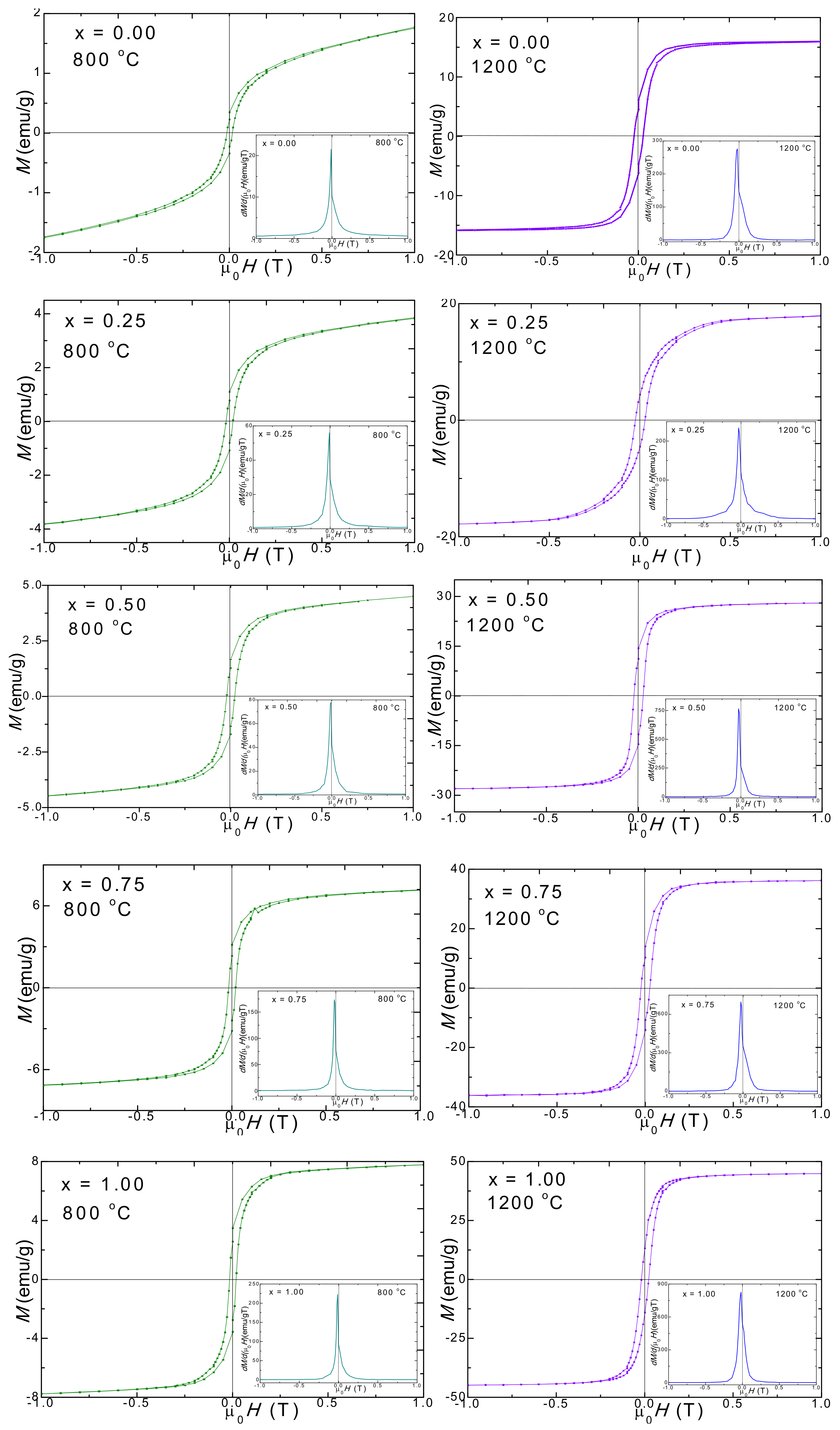
2.4. Magnetic Properties

Generally, the magnetic properties of the ferrites are affected by the chemical formula, by the cation distribution between the tetrahedral (A) and octahedral (B) sites of the lattice, as well as by the particle sizes and their distribution [22]. The main magnetic properties of Mn1−xNixFe2O4@SiO2 NCs annealed at 800 and 1200 °C are displayed in Figure 4. In all cases, the hysteresis loops have a typical shape for ferrimagnetic materials. The main magnetic parameters, namely saturation magnetization (MS), remanent magnetization (MR), squareness (S), coercivity (HC), the magnetic moment per formula unit (nB) expressed in numbers of Bohr magnetons and anisotropy constant (K) extracted from the hysteresis loops are presented in Table 3.
The magnetic parameters MS, MR, nB, and K increase, while HC decreases with increasing Ni content. All the magnetic parameters are larger for the NCs with the same Ni content, annealed at higher temperatures. This behavior is different from that reported for bulk ferrites with the same chemical formula, for which an increase of the saturation magnetization was found with increasing Mn content [39]. The difference between the two systems consists in the presence of SiO2 coating in our samples. The largest Ms value was recorded for the samples with x = 1.00 (NiFe2O4@SiO2) with the largest particle sizes, for both annealing temperatures. MS increases almost linearly with increasing Ni content for both annealing temperatures. The MS is strongly affected by the so-called “surface spin effect” which is a result of the defects and broken chemical bonds which disrupt the parallel alignment of the magnetic moments and give rise to spin canting and spin disorder in the layer from the surface of the particles. The smaller the size of the particle, the larger the surface-to-volume ratio. Increasing the fraction of this layer will make dominant the magnetic behavior of the shell over that from the interior, and the magnetization of the smaller size particles will be reduced. In addition to this size effects, the XRD analysis also showed an increase of the hematite (which is known to have low magnetic properties) and quartz content with increasing Mn content in the samples and this can contribute additionally to the decrease of the MS value The MS value is also affected by the cation’s distribution between the tetrahedral (A) and octahedral (B) sites [22].
The increase of the MS with increasing Ni content can also suggest that in the octahedral (B) sites Fe3+ ions (5 µB) were replaced by Ni2+ (2 µB) ions with a smaller magnetic moment which force the Fe3+ ions to migrate in the tetrahedral (A) sites. This results in an inverse spinel structure since the Fe3+ ions are rearranged in both tetrahedral (A) and octahedral (B) sites and the antiferromagnetic interaction becomes weaker, while the ferromagnetic super-exchange interaction increases. Therefore, the normal spinel Mn ferrite is converted to a dominant inverse spinel ferrite as a result of the Mn2+ ions substitution by Ni2+ ions. This may be a consequence of the coating of Mn1−xNixFe2O4 nanoparticles by the SiO2 matrix. Comparative Ms values were reported by Airimioaei et al. [2] for Ni-Mn ferrites obtained by combustion reaction, while Jessudoss et al. [22] reported higher MS and MR values for Ni1−xMnxFe2O4 obtained by a microwave combustion reaction route. Köseoğlu reported MS between 31 and 56 emu/g at room temperature and 41–70 emu/g at 10 K [8]. Opposite to our results, the higher MS value of undoped MnFe2O4 of 66.93 emu/g steadily decreased from 64.68 emu/g (x = 0.2) to 35.43 emu/g (x= 1.0) with increasing Ni content, with NiFe2O4 showing the lower value; the linear decrease of Ms values of the samples could be mainly due to the difference in the magnetic moments of Mn2+ and Ni2+ ions [7].
The coercive field (HC) slightly decreases with increasing Ni content for both the samples annealed at 800 and 1200 °C (Table 3). This behavior can be attributed to the well-known dependence of the HC on the sizes of the nanoparticles in the magnetic multidomain range. In this region, the size of the nanoparticles causes them to be composed of many magnetic domains which allow an easy domain wall motion and magnetization reversal, reducing the value of the coercive field by lowering the value of the domain wall energy [1,19,28]. The HC is a measure of the magneto-crystalline anisotropy of a sample. By increasing the Ni content, the nanoparticle and the crystallite sizes also increase, leading to a decrease of the magnetocrystalline anisotropy. The HC is strongly affected by the particle’s sizes and their shape as well as by their distribution, crystallinity and magnetic domain sizes, and micro-strains induced by the SiO2 matrix [1]. Similarly, Airimioaei et al. reported that the HC slightly increases with increasing the amount of Mn from 37.4 Oe to 53.7 Oe for x = 0–0.5, respectively [2]. In accordance with Mathubala [7], the HC and the MR values decrease with the increase of the Ni content in MnFe2O4 lattice.
The magnetic moment per formula unit increases with the increasing of Ni content for the same reasons used to explain the behavior of the MS since the ratio of MS/nB is nearly constant. The increase of remanent magnetization (MR) with the increase of Ni content also needs to be correlated with the variation of the particle’s size and the related surface effects as the presence of defects and of secondary phases may act as a pinning center for the magnetic domain walls.
The anisotropy constant (K) reveals the energy required to rotate the magnetic moment inside the particle. The K increases with increasing Ni content with a factor of 1.8 for the NCs annealed at 800 °C and with a factor of 1.98 for the samples annealed at 1200 °C. A possible explanation could be the increase of magneto-crystalline anisotropy which originates in spin-orbital contribution since for MnFe2O3 the orbital quantum number is L = 0. Another explanation could be the presence of the spin disorder in the surface layer of the nanoparticles which needs a higher magnetic field for MS, a field that depends on the size of the particles and their distribution within the samples [8].
The squareness ratio (S = MR/MS) is a measure of how square the hysteresis loop is. A theoretical value of MR/MS lower than 0.5 indicates the presence of non-interacting uniaxial single domain particles with the easy axis being randomly oriented [1]. The S increases from 0.064 to 0.333 for the NCs annealed at 800 °C and from 0.246 to 0.357 for the NCs annealed at 1200 °C. Generally, the derivatives of the hysteresis loops exhibited small and broad single peaks indicating partially crystalline samples with a main magnetic phase in the presence of crystal defects. The presence of a high magnetic purity phase is indicated by the sharp peaks. The broad peaks correspond to large particle size distributions and wide coercive fields distributions. The horizontal shifts of peaks from the origin are rather small for all the samples, suggesting that the coercivities distributions are not large as a result of the magnetic interaction between the particles.

2.5. Potential Applications

Magnetic nanoparticles that are small enough to remain in circulation after injection and are able to pass through the capillary systems of various organs are non-toxic, well dispersed, and biocompatible and are potential candidates for biomedical applications such as cancer therapy, drug delivery, magnetic resonance imaging, or magnetic hypothermia [19,40]. The magnetic parameters of nanosized Mn1−xNixFe2O4 ferrites are related to the synthesis route. The synthesized Mn-Ni ferrite nanoparticles are prospective candidates for biomedicine due to their easy synthesis process, controllable structure and size, stoichiometry control, high magnetization value, and superparamagnetic nature. Moreover, their embedding into mesoporous SiO2 enhances their biocompatibility and reduces their agglomeration and degradation. SiO2 matrix is an excellent non-toxic coating material that can create cross-linking, giving rise to an inert outer shield, avoiding the acute toxicity by ferrite inoculation [24]. However, their biocompatibility, cytotoxicity, pharmacokinetics, and other potential side effects are still underexplored. Although there are several in vitro studies that indicate the cytotoxicity through suppression of proliferation and apoptosis induction of different nanosized magnetic ferrites against human colon cancer (HT29), breast cancer (MCF7), and liver cancer (HepG2) cells, data on the Mn-Ni ferrite cytotoxicity is limited [41]. The dose-dependent cytotoxic effects of Mn1−xNixFe2O4 nanoparticles against J774 E murine macrophages and U2OS human osteosarcoma was reported [42]. The effective light-to-heat conversion upon exposure of Mn-Ni ferrites to near-infrared irradiation could also be attractive for different bio-applications [42]. In order to increase their chemical stability in biological systems and for enhancing their magnetic properties different doping elements (Ni, Co, Mn, Zn, Mg, etc.) might be added [43]. Intravenous inoculation of ferrite nanosized particles can be useful as contrast agents for magnetic resonance imaging (MRI) [44]. Mn ferrite was found as a very effective MRI contrast agent in comparison with magnetite since it has large MS and high crystalline anisotropy resulting in a slower magnetic moment of relaxation [45].

3. Materials and Methods

3.1. Synthesis of NCs

Ferric nitrate nonahydrate (Fe(NO3)3∙9H2O) of 99.6% purity, nickel nitrate hexahydrate (Ni(NO3)2∙6H2O) of 99.8% purity, manganese nitrate trihydrate (Mn(NO3)2∙3H2O) of 100.0% purity, 1,4-butanediol (1,4BD) of 99.9% purity, tetraethyl orthosilicate (TEOS) of 100.0% purity, and ethanol of 99.9% purity were purchased from Merck (Germany) and used for synthesis without additional purifications.
The Mn1−xNixFe2O4@SiO2 NCs (60% wt. ferrite, 40% wt. SiO2) were prepared by sol-gel route using Mn:Ni:Fe molar ratios of 0:1:2 (x = 0.00), 0.25:0.75:2 (x = 0.25), 0.50:0.50:2 (x = 0.50), 0.75:0.25:2 (x = 0.75) and 1:0:2 (x = 1.00). The sols were obtained by mixing the nitrate mixture with 1,4BD, TEOS and ethanol. After 4 weeks at room temperature, the gelation takes place by the formation of an SiO2 matrix that contains the nitrates and 1,4BD. The gels were annealed at 400, 800, and 1200 °C for 4h in air using a LT9 muffle furnace (Nabertherm, Lilienthal, Germany).

3.2. Characterization of NCs

The crystallinity and structure of the ferrite were investigated by X-ray diffraction recorded at room temperature, using a D8 Advance (Bruker, Germany) diffractometer, operating at 40 kV and 40 mA with CuKα radiation (λ = 1.54060 Å).
The formation of ferrite and SiO2 matrix was monitored using a Spectrum BX II (Perkin Elmer, Waltham, MA, USA) Fourier transform infrared spectrometer on pellets containing 1% sample in KBr.
AFM was performed by a JSPM 4210 (JEOL, Tokio, Japan) scanning probe microscope using NSC15 cantilevers (diamond-coated silicon nitride tips) with a resonant frequency of 325 kHz and a force constant of 40 N/m in tapping mode. The samples were dispersed into ultrapure water and transferred on glass slides by vertical adsorption for 30 s, followed by natural drying. Areas of 2.5 µm × 2.5 µm to 1 µm × 1 µm of dried glass slides were scanned for three different macroscopic sites.
A cryogen-free VSM magnetometer (Cryogenic Ltd., London, UK) was used for the magnetic measurements. The MS was measured in a high magnetic field up to 10 T, while the magnetic hysteresis loops were performed between −2 and 2 T, at 300 K on samples incorporated in epoxy resin.

4. Conclusions

The microstructure, morphology, particle size, phase composition, and magnetic properties of Mn1−xNixFe2O4 (x = 0, 0.25, 0.50, 0.75, 1.00) NCs were investigated. The crystallite size (6–46 nm), X-ray density (5.050–5.249 g/cm3), lattice parameter (8.402–8.485 Å), and volume (593.1–606.6 Å3) of Mn1−xNixFe2O4 increase with the increase of Ni content. The XRD patterns showed poor crystalized Mn1−xNixFe2O4 at 400 °C and highly crystalline Mn1−xNixFe2O4, accompanied by secondary phases of Fe2SiO4, α-Fe2O3, cristobalite, and quartz at 800 and 1200 °C. FT-IR spectroscopy confirmed the formation of the oxidic phases and SiO2 matrix. AFM investigations revealed round-shaped nanoparticles with sizes depending on the annealing temperature and Ni content. The magnetic properties of the NCs were strongly dependent on the chemical composition, cation distribution between tetrahedral (A) and octahedral (B) sites, as well as on the surface effects derived from the synthesis methods. For the NCs annealed at 1200 °C, Ms (16.4–45.7 emu/g), MR, magnetic moment per formula unit and K (2.678–5.310 erg/dm3) increased, while HC (260–185 Oe) slightly decreased with the increase of Ni content. The small S values indicated the presence of non-interacting single domain uniaxial particles. The magnetic parameters displayed similar behavior for the NCs annealed at 800 °C, but their variations are smaller. All the magnetic parameters increased with the annealing temperature. The obtained magnetic Mn-Ni ferrite nanoparticles are potential candidates for biomedical applications such are cancer therapy, drug delivery, magnetic resonance imaging, or magnetic hydrothermia. Despite the promising results, further studies of the biocompatibility, differential toxicity, and pharmacokinetics of nanosized Mn-Ni ferrites are required.

Author Contributions

T.D., conceptualization, methodology, writing—original draft, writing—review and editing, visualization, supervision; E.A.L., I.G.D., I.P. and O.C., methodology, investigation, writing—original draft, writing—review and editing. All authors have read and agreed to the published version of the manuscript.

Funding

This research received no external funding.

Acknowledgments

The authors acknowledge the Research Centre in Physical Chemistry “CECHIF” of Babes Bolyai University for AFM assistance.

Conflicts of Interest

The authors declare no conflict of interest.

References

  1. Suresh, J.; Trinadh, B.; Babu, B.V.; Reddy, P.V.S.S.S.N.; Mohan, B.S.; Krishna, A.R.; Samatha, K. Evaluation of micro-structural and magnetic properties of nickel nano-ferrite and Mn2+ substituted nickel nano-ferrite. Phys. B Condens. Matter 2021, 620, 413264. [Google Scholar] [CrossRef]
  2. Airimioaei, M.; Ciomaga, C.E.; Apostolescu, A.; Leonite, L.; Iordan, A.R.; Mitoseriu, L.; Palamaru, M.N. Synthesis and functional properties of the Ni1−xMnxFe2O4 ferrites. J. Alloys Compd. 2011, 509, 8065–8072. [Google Scholar] [CrossRef]
  3. Bonanni, B.; Cannistraro, S. Gold nanoparticles on modified glass surface as height calibration standard for atomic force microscopy operating in contact and tapping mode. J. Nanotechnol. 2005, 1, 1–14. [Google Scholar]
  4. Mohamed, W.S.; Hadia, N.M.A.; Al Bakheet, B.; Alzaid, M.; Abu-Dief, A.M. Impact of Cu2+ cations substitution on structural, morphological, optical and magnetic properties of Co1-xCuxFe2O4 nanoparticles synthesized by a facile hydrothermal approach. Solid State Sci. 2022, 125, 106841. [Google Scholar] [CrossRef]
  5. Abu-Dief, A.M.; Essawy, A.A.; Diab, A.K.; Mohamed, W.S. Facile synthesis and characterization of novel Gd2O3–CdO binary mixed oxide nanocomposites of highly photocatalytic activity for wastewater remediation under solar illumination. J. Phys. Chem. Solids 2021, 148, 109666. [Google Scholar] [CrossRef]
  6. Salaheldeen, M.; Abu-Dief, A.M.; Martinez-Goyeneche, L.; Alzahrani, S.O.; Alkhatib, F.; Alvarez-Alonso, P.; Blanco, J.A. Dependence of the magnetization process on the thickness of Fe70Pd30 nanostructured thin film. Materials 2020, 13, 5788. [Google Scholar] [CrossRef] [PubMed]
  7. Mathubala, G.; Manikandan, A.; Arul Antony, S.; Ramar, P. Photocatalytic degradation of methylene blue dye and magnetooptical studies of magnetically recyclable spinel NixMn1-xFe2O4 (x = 0.0-1.0) nanoparticles. J. Mol. Struct. 2016, 113, 79–87. [Google Scholar] [CrossRef]
  8. Köseoğlu, Y. Structural, magnetic, electrical and dielectric properties of MnxNi1-xFe2O4 spinel nanoferrites prepared by PEG assisted hydrothermal method. Ceram. Int. 2013, 39, 4221–4230. [Google Scholar] [CrossRef]
  9. Marinca, T.F.; Chicinaș, I.; Isnard, O.; Neamțu, B.V. Nanocrystalline/nanosized manganese substituted nickel ferrites—Ni1-xMnxFe2O4 obtained by ceramic-mechanical milling route. Ceram. Int. 2016, 42, 4754–4763. [Google Scholar] [CrossRef]
  10. Shobana, M.K.; Sankar, S. Structural, thermal and magnetic properties of Ni1-xMnxFe2O4 nanoferrites. J. Magn. Magn. Mater. 2009, 321, 2125–2128. [Google Scholar] [CrossRef]
  11. Abdallah, H.M.I.; Moyo, T. Superparamagnetic behavior of MnxNi1-xFe2O4 spinel nanoferrites. J. Magn. Magn. Mater. 2014, 361, 170–174. [Google Scholar] [CrossRef]
  12. Maaz, K.; Duan, J.L.; Karim, S.; Chen, Y.H.; Zhai, P.F.; Xu, L.J.; Yao, H.J.; Liu, J. Fabrication and size dependent magnetic studies of NixMn1-xFe2O4 (x = 02) cubic nanoplates. J. Alloys Compd. 2016, 684, 656–662. [Google Scholar] [CrossRef]
  13. Mohamed, W.S.; Abu-Dief, A.M. Impact of rare earth europium (RE-Eu3+) ions substitution on microstructural, optical and magnetic properties of CoFe2−xEuxO4 nanosystems. Ceram. Int. 2020, 46, 16196–16209. [Google Scholar] [CrossRef]
  14. Mohamed, W.S.; Alzaid, M.; Abdelbaky, S.M.; Amghouz, Z.; Garcia-Granda, S.; Abu-Dief, A.M. Impact of Co2+ substitution on microstructure and magnetic properties of CoxZn1-xFe2O4 nanoparticles. Nanomaterials 2019, 9, 1602. [Google Scholar] [CrossRef] [Green Version]
  15. Sudakshina, B.; Suneesh, M.V.; Arun, B.; Chandrasekhar, K.; Vasundhara, M. Effects of Cr, Co, Ni substitution at Mn-site on structural, magnetic properties and critical behaviour in Nd0.67Ba0.33MnO3 mixed-valent manganite. J. Magn. Magn. Mater. 2022, 548, 168980. [Google Scholar] [CrossRef]
  16. Siakavelas, G.I.; Charisiou, N.D.; AlKhoori, A.; Sebastian, V.; Hinder, S.J.; Baker, M.A.; Yentekakis, I.V.; Polychronopoulou, K.; Goula, M.A. Cerium oxide catalysts for oxidative coupling of methane reaction: Effect of lithium, samarium and lanthanum dopants. J. Environ. Chem. Eng. 2022, 10, 107259. [Google Scholar] [CrossRef]
  17. Yang, Y.; Li, J.; Zhang, H.; Li, J.; Xu, F.; Wang, G.; Gao, F.; Su, H. Nb5+ ion substitution assisted the magnetic and gyromagnetic properties of NiCuZn ferrite for high frequency LTCC devices. Ceram. Int. 2022, in press. [Google Scholar] [CrossRef]
  18. Junaid, M.; Oazafi, I.A.; Khan, M.A.; Gulbadan, S.; Ilyas, S.Z.; Somaily, H.H.; Attia, M.S.; Amin, M.A.; Noor, H.M.; Asghar, H.M.N.H.K. The influence of Zr and Ni co-substitution on structural, dielectric and magnetic traits of lithium spinel ferrites. Ceram. Int. 2022, in press. [Google Scholar] [CrossRef]
  19. Dippong, T.; Levei, E.A.; Cadar, O. Recent advances in synthesis and applications of MFe2O4 (M= Co, Cu, Mn, Ni, Zn) nanoparticles. Nanomaterials 2021, 11, 1560. [Google Scholar] [CrossRef] [PubMed]
  20. Barvinschi, P.; Stefanescu, O.; Dippong, T.; Sorescu, S.; Stefanescu, M. CoFe2O4/SiO2 nanocomposites by thermal decomposition of some complex combinations embedded in hybrid silica gels. J. Therm. Anal. Calorim. 2013, 112, 447–453. [Google Scholar] [CrossRef]
  21. Joint Committee on Powder Diffraction Standards. Powder Diffraction File; International Center for Diffraction Data: Swarthmore, PA, USA, 1999. [Google Scholar]
  22. Jesudoss, S.K.; Judith Vijaya, J.; John Kennedy, L.; Iyyappa Rajana, P.; Al-Lohedan, A.H.; Jothi Ramalingam, R.; Kaviyarasu, K.; Bououdina, M. Studies on the efficient dual performance of Mn1–xNixFe2O4 spinel nanoparticles in photodegradation and antibacterial activity. J. Photochem. Photobiol. B Biol. 2016, 165, 121–132. [Google Scholar] [CrossRef]
  23. Nizam, M.M.N.; Khan, S. Structural, electrical and optical properties of sol-gel synthesized cobalt substituted MnFe2O4 nanoparticles. Phys. B 2017, 520, 21–27. [Google Scholar]
  24. Aparna, M.L.; Nirmala Grace, A.; Sathyanarayanan, P.; Sahu, N.K. A comparative study on the supercapacitive behaviour of solvothermally prepared metal ferrite (MFe2O4, M=Fe, Co, Ni, Mn, Cu, Zn) nanoassemblies. J. Alloys Compd. 2018, 745, 385–395. [Google Scholar] [CrossRef]
  25. El Mendili, Y.; Bardeau, J.F.; Randrianantoandro, N.; Greneche, J.M.; Grasset, F. Structural behavior of laser-irradiated γ-Fe2O3 nanocrystals dispersed in porous silica matrix: γ-Fe2O3 to α-Fe2O3 phase transition and formation of ε-Fe2O3. Sci. Technol. Adv. Mater. 2016, 17, 597–609. [Google Scholar] [CrossRef] [PubMed] [Green Version]
  26. Hussain, A.; Abbas, T.; Niazi, S.B. Preparation of Ni1-xMnxFe2O4 ferrites by sol–gel method and study of their cation distribution. Ceram. Int. 2013, 39, 1221–1225. [Google Scholar] [CrossRef]
  27. Dippong, T.; Deac, I.G.; Cadar, O.; Levei, E.A.; Petean, I. Impact of Cu2+ substitution by Co2+ on the structural and magnetic properties of CuFe2O4 synthesized by sol-gel route. Mater. Charact. 2020, 163, 110248. [Google Scholar] [CrossRef]
  28. Fanga, P.P.; Buriez, O.; Labbé, E.; Tian, Z.Q.; Amatore, C. Electrochemistry at gold nanoparticles deposited on dendrimers assemblies adsorbed onto gold and platinum surfaces. J. Electroanal. Chem. 2011, 659, 76–82. [Google Scholar] [CrossRef]
  29. Ur. Rahman Awana, F.; Keshavarz, A.; Azhar, M.R.; Akhondzadeh, H.; Ali, M.; Al-Yaseri, A.; Abid, H.R.; Iglauer, S. Adsorption of nanoparticles on glass bead surface for enhancing proppant performance: A systematic experimental study. J. Mol. Liq. 2021, 328, 115398. [Google Scholar] [CrossRef]
  30. Fokin, N.; Grothe, T.; Mamun, A.; Trabelsi, M.; Klöcker, M.; Sabantina, L.; Döpke, C.; Blachowicz, T.; Hütten, A.; Ehrmann, A. Magnetic properties of electrospun magnetic nanofiber mats after stabilization and carbonization. Materials 2020, 13, 1552. [Google Scholar] [CrossRef] [Green Version]
  31. Anchieta, C.G.; Cancelier, A.; Mazutti, M.A.; Jahn, S.L.; Kuhn, R.C.; Gündel, A.; Chiavone-Filho, O.; Foletto, E.L. Effects of solvent diols on the synthesis of ZnFe2O4 particles and their use as heterogeneous photo-fenton catalysts. Materials 2014, 7, 6281–6290. [Google Scholar] [CrossRef] [PubMed]
  32. Januskevicius, J.; Stankeviciute, Z.; Baltrunas, D.; Mažeika, K.; Beganskiene, A.; Kareiva, A. Aqueous sol-gel synthesis of different iron ferrites: From 3D to 2D. Materials 2021, 14, 1554. [Google Scholar] [CrossRef]
  33. Van Cutsem, E.; Verheul, H.M.W.; Flamen, P.; Rougier, P.; Beets-Tan, R.; Glynne-Jones, R.; Seufferlein, T. Imaging in colorectal cancer: Progress and challenges for the clinicians. Cancers 2016, 31, 81. [Google Scholar] [CrossRef] [Green Version]
  34. Enuka, E.; Monne, M.A.; Lan, X.; Gambin, V.; Koltun, R.; Chen, M.Y. 3D inkjet printing of ferrite nanomaterial thin films for magneto-optical devices. In Quantum Sensing and Nano Electronics and Photonics XVII; International Society for Optics and Photonics: Bellingham, WA, USA, 2020. [Google Scholar] [CrossRef]
  35. Díez-Villares, S.; Ramos-Docampo, M.A.; da Silva-Candal, A.; Hervella, P.; Vázquez-Ríos, A.J.; Dávila-Ibáñez, A.B.; López-López, R.; Iglesias-Rey, V.; Salgueiriño, M.; Manganese, M. Ferrite nanoparticles encapsulated into vitamin e/sphingomyelin nanoemulsions as contrast agents for high-sensitive magnetic resonance imaging. Adv. Healthc. Mater. 2021, 10, 2101019. [Google Scholar] [CrossRef] [PubMed]
  36. Yang, L.; Ma, L.; Xin, J.; Li, A.; Sun, C.; Wei, R.; Ren, B.W.; Chen, Z.; Lin, H.; Gao, J. Composition tunable manganese ferrite nanoparticles for optimized T2 contrast ability. Chem. Mater. 2017, 29, 3038–3047. [Google Scholar] [CrossRef]
  37. Tong, S.-K.; Chi, P.-W.; Kung, S.H.; Wei, D.H. Tuning bandgap and surface wettability of NiFe2O4 driven by phase transition. Sci. Rep. 2018, 8, 1338. [Google Scholar] [CrossRef] [Green Version]
  38. Ashiq, N.M.; Ehsan, M.F.; Iqbal, M.J.; Gul, I.H. Synthesis, structural and electrical characterization of Sb3+ substituted spinel nickel ferrite (NiSbxFe2−xO4) nanoparticles by reverse micelle technique. J. Alloys Compd. 2011, 509, 5119–5126. [Google Scholar] [CrossRef]
  39. Hu, J.; Qin, H.; Wang, Y.; Wang, Z.; Zhang, S. Magnetic properties and magnetoresistance effect of Ni1−xMnxFe2O4 sintered ferrites. Solid State Commun. 2000, 115, 233–235. [Google Scholar] [CrossRef]
  40. Sánchez, J.; Cortés-Hernández, D.A.; Escobedo-Bocardo, J.C.; Jasso-Terán, R.A.; Zugasti-Cruz, A. Bioactive magnetic nanopar-ticles of Fe–Ga synthesized by sol–gel for their potential use in hyperthermia treatment. J. Mater. Sci. Mater. Med. 2014, 25, 2237–2242. [Google Scholar] [CrossRef] [PubMed]
  41. Al-Qubaisi, M.S.; Rasedee, A.; Flaifel, M.H.; Ahmad, S.H.; Hussein-Al-Ali, S.; Hussein, M.Z.; Eid, E.E.; Zainal, Z.; Saeed, M.; Ilowefah, M.; et al. Cytotoxicity of nickel zinc ferrite nanoparticles on cancer cells of epithelial origin. Int. J. Nanomed. 2013, 8, 2497–2508. [Google Scholar] [CrossRef] [PubMed] [Green Version]
  42. Pazik, R.; Zachanowicz, E.; Pozniak, B.; Małecka, M.; Ziecina, A.; Marciniak, L. Non-contact Mn1-xNixFe2O4 ferrite nano-heaters for biological applications–heat energy generated by NIR irradiation. RSC Adv. 2017, 7, 18162. [Google Scholar] [CrossRef] [Green Version]
  43. Pu, Y.; Tao, X.; Zeng, X.; Le, Y.; Chen, J.F. Synthesis of Co–Cu–Zn doped Fe3O4 nanoparticles with tunable morphology and magnetic properties. J. Magn. Magn. Mater. 2020, 322, 1985–1990. [Google Scholar] [CrossRef]
  44. Bacon, B.R.; Stark, D.D.; Park, C.H.; Saini, S.; Groman, E.V.; Hahn, P.F.; Compton, C.C.; Ferrucci, J.T., Jr. Ferrite particles: A new magnetic resonance imaging contrast agent. Lack of acute or chronic hepatotoxicity after intravenous administration. J. Lab. Clin. Med. 1987, 110, 164–171. [Google Scholar]
  45. Ravichandran, M.; Velumani, S. Manganese ferrite nanocubes as an MRI contrast agent. Mater. Res. Express 2020, 7, 016107. [Google Scholar] [CrossRef] [Green Version]
Figure 1. X-ray diffraction patterns and FT-IR spectra of Mn1−xNixFe2O4@SiO2 NCs annealed at 400, 800, and 1200 °C.
Ijms 23 03097 g001aIjms 23 03097 g001b
Figure 2. Topographical AFM images of Mn1−xNixFe2O4@SiO2 NCs annealed at 400, 800, and 1200 °C (x = 0 (ac), x = 0.25 (df), x = 0.50 (gi), x = 0.75 (jl) and x = 1.0 (mo)).
Ijms 23 03097 g002
Figure 3. AFM 3D images of Mn1−xNixFe2O4@SiO2 NCs annealed at 400, 800, and 1200 °C (x = 0 (ac), x = 0.25 (df), x = 0.50 (gi), x = 0.75 (jl) and x = 1.0 (mo)).
Ijms 23 03097 g003
Figure 4. Magnetic hysteresis loops and magnetization derivative (in insets) for Mn1−xNixFe2O4@SiO2 NCs heat-treated at 800 and 1200 °C.
Ijms 23 03097 g004
Table 1. XRD parameters of Mn1−xNixFe2O4@SiO2 annealed at 400, 800, and 1200 °C.
NCTemperature, °CCrystallite Size, nmLattice Parameter, ÅVolume, Å3X-ray Density, g·cm−3Hopping Length in A, ÅHopping Length in B, Å
x = 0.0040068.465606.65.0503.6652.993
800108.472608.15.0373.6682.995
1200238.485610.95.0143.6742.999
x = 0.2540088.441601.45.1143.6552.984
800138.448602.95.1013.6582.987
1200278.459605.35.0813.6632.991
x = 0.50400108.432599.55.1513.6512.981
800178.437600.65.1423.6532.983
1200308.443601.95.1313.6562.985
x = 0.75400128.416596.15.2023.6442.976
800218.423597.65.1893.6472.978
1200378.427598.45.1823.6492.979
x = 1.00400148.402593.15.2493.6382.971
800268.409594.65.2363.6412.973
1200468.412595.25.2303.6432.974
Table 2. AFM parameters of Mn1−xNixFe2O4@SiO2 NCs.
NC’sTemperature,
°C
Height,
nm
Rq Roughness,
nm
Average Particle Size,
nm
x = 0.00400191.1518
800161.4424
120090.8730
x = 0.25400161.1616
800181.1920
1200151.2435
x = 0.50400141.0818
800120.9325
1200394.1740
x = 0.75400121.0520
800121.0930
1200191.9760
x = 1.00400111.0822
800161.0730
120090.9258
Table 3. Saturation magnetization (MS), remanent magnetization (MR), coercivity (HC), squareness (S), magnetic moment per formula unit (nB), and anisotropy constant (K) of Mn1−xNixFe2O4@SiO2 NCs.
NCTemperature, °CMS, emu/gMR, emu/gHC, OeSnBK, erg/dm3
x = 0.008004.70.32000.0640.1940.590
120016.44.22600.2460.6772.678
x = 0.258006.81.11900.1620.2820.811
120022.45.82500.2590.9293.517
x = 0.508007.81.71830.2180.3250.896
120029.613.52400.4561.2324.461
x = 0.758009.12.81750.3080.3801.000
120037.514.62200.3891.5675.181
x = 1.0080010.23.41660.3330.4281.063
120045.716.11850.3571.9185.310
Publisher’s Note: MDPI stays neutral with regard to jurisdictional claims in published maps and institutional affiliations.

