Article

Characterisation of Trichomonas vaginalis Isolates Collected from Patients in Vienna between 2019 and 2021

by
Iwona Lesiak-Markowicz
1,*,
Julia Walochnik
1,
Angelika Stary
2 and
Ursula Fürnkranz
2,*
1
Intitute for Specific Prophylaxis and Tropical Medicine (ISPTM), Centre for Pathophysiology, Infectiology and Immunology, Medical University of Vienna, 1090 Vienna, Austria
2
Pilzambulatorium Schloesselgasse, Oupatients Centre for Diagnosis of Venero-Dermatological Diseases, 1080 Vienna, Austria
*
Authors to whom correspondence should be addressed.
Int. J. Mol. Sci. 2022, 23(20), 12422; https://doi.org/10.3390/ijms232012422
Submission received: 26 September 2022 / Revised: 13 October 2022 / Accepted: 16 October 2022 / Published: 17 October 2022
(This article belongs to the Section Molecular Biology)

Abstract

:
Trichomonas vaginalis (TV) is the causative agent of trichomoniasis, the most common nonviral sexually transmitted disease. TV can carry symbionts such as Trichomonas vaginalis virus (TVV) or Mycoplasma hominis. Four distinct strains of TV are known: TVV1, TVV2, TVV3, and TVV4. The aim of the current study was to characterise TV isolates from Austrian patients for the presence of symbionts, and to determine their effect on metronidazole susceptibility and cytotoxicity against HeLa cells. We collected 82 TV isolates and detected presence of TVV (TVV1, TVV2, or TVV3) in 29 of them (35%); no TVV4 was detected. M. hominis was detected in vaginal/urethral swabs by culture in 37% of the TV-positive patients; M. hominis DNA was found in 28% of the TV isolates by PCR. In 15% of the patients, M. hominis was detected in the clinical samples as well as within the respective TV isolates. In 22% of the patients, M. hominis was detected by culture only. In 11 patients, M. hominis was detected only within the respective cultured TV isolates (13%), while the swab samples were negative for M. hominis. Our results provide a first insight into the distribution of symbionts in TV isolates from Austrian patients. We did not observe significant effects of the symbionts on metronidazole susceptibility, cytotoxicity, or severity of symptoms.

1. Introduction

Trichomoniasis is the most common nonviral sexually transmitted infection (STI) of the human urogenital tract, with 156 million new cases estimated in 2016 [1]. The global prevalence of TV has been reported to be around 5.0% for women and 0.6% for men [1]. The majority of women and men infected with TV reportedly remain asymptomatic; however, in one-third of women a latent period of up to 6 months with subsequent appearance of symptoms has been described [2,3,4]. Trichomoniasis symptoms include vaginitis in women (vaginal erythema, itching, vaginal discharge, odour, or dysuria) and urethritis in men. Commonly known sequelae include adverse pregnancy outcomes and an increased risk of the acquisition and transmission of HIV and HPV [5,6]. Trichomonas vaginalis virus (TVV) is a nonsegmented, 4.5–5 kbp, double-stranded RNA (dsRNA) virus belonging to the Totiviridae family, members of which infect fungi [7] and a variety of parasitic protists, including Giardia duodenalis [8] and Leishmania braziliensis [9]. TVV is recognised as the first dsRNA virus observed in a protozoan parasite [10] and has been divided into four subspecies: TVV1, TVV2, TVV3, and TVV4 [11]. Different TVV subspecies can coinfect and coexist inside the same TV isolate [12]. Among the four distinct species of TVV, TVV1 is the most prevalent, followed by TVV2, TVV3, and TVV4 [11]. It has been postulated that TVVs may upregulate the expression of immunogenic surface proteins and cysteine proteinases [13]; however, the clinical significance of TVVs and their effects on the pathogenicity of TV is still not well-known. Graves et al. [11] found no association of TVV positive (TVV+) isolates with clinical symptoms, reinfections, or metronidazole resistance. Mycoplasma spp. are the smallest free-living, self-replicating, cell-wall-deficient bacteria known so far [14,15]. Mycoplasma spp. are typically extracellular and surface-attached; however, a few species can enter host cells and establish an intracellular niche [16]. TV is known to act as a niche and vector for M. hominis [17]. M. hominis is the most frequently isolated microorganism from the genital tract of both men and women, found as part of the natural flora but also considered as a putative pathogen responsible for a variety of urogenital infections, neonatal infections, and systemic infections in immunocompromised patients [18]. 5-nitroimidazoles, especially metronidazole, are the most widely used antimicrobial agents for the treatment of trichomoniasis. Metronidazole, developed in the 1950s and approved for the treatment of trichomoniasis in the early 1960s, was the first drug to have a cure rate approaching 100% when used systemically [19]. Some cases of drug resistance and treatment failure of metronidazole have been observed since 1962 [20], but resistance remained rare and could mostly be overcome with larger doses of the drug. Resistance mechanisms to metronidazole have been studied by incubating TV strains with different drug concentrations under aerobic and anaerobic conditions in vitro; however, the underlying mechanisms of resistance are still not fully understood. The current treatment guidelines recommend 500 mg metronidazole twice daily for 7 days [21]. The aim of this retrospective study was to characterise TV isolates collected from patients visiting the Outpatients Centre for the Diagnosis of Venero-Dermatological Diseases in Vienna with regard to their symbionts, drug susceptibilities to metronidazole, and cytotoxicities to human cells.

2. Results

2.1. Collection and Characteristic of TV Isolates

TV isolates were collected from 82 patients between 2019 and 2021: 10 isolates derived from men (mean age 46) and 72 from women (mean age 41.2). Thirteen patients did not report any clinical symptoms (7 women, 6 men); in four patients (only female), TV was the only pathogen detected (Table 1), and these patients reported clinical symptoms including pain, itching, discharge, and urinary tract infection symptoms. In the other 78 patients attending the Outpatients Centre, other potential pathogens such as Gardnerella vaginalis, Prevotella spp., Ureaplasma spp., Mycoplasma genitalium, and Chlamydia trachomatis, as well as Candida spp. were also detected apart from TV.

2.2. Detection of TVVs in Clinical Isolates

In 29/82 (35%) TV samples, TVVs were detected; 16 of these 29 (55%) were identified as TVV1, 2/29 (7%) as TVV2, and 5/29 (17%) as TVV3; no TVV4 was detected in our collection of TV isolates. A total of 4/29 isolates (14%) harboured both TVV1 and TVV2, and 2/29 (7%) were positive for TVV1 and TVV3. In 53/82 (65%) no TVVs were detected.

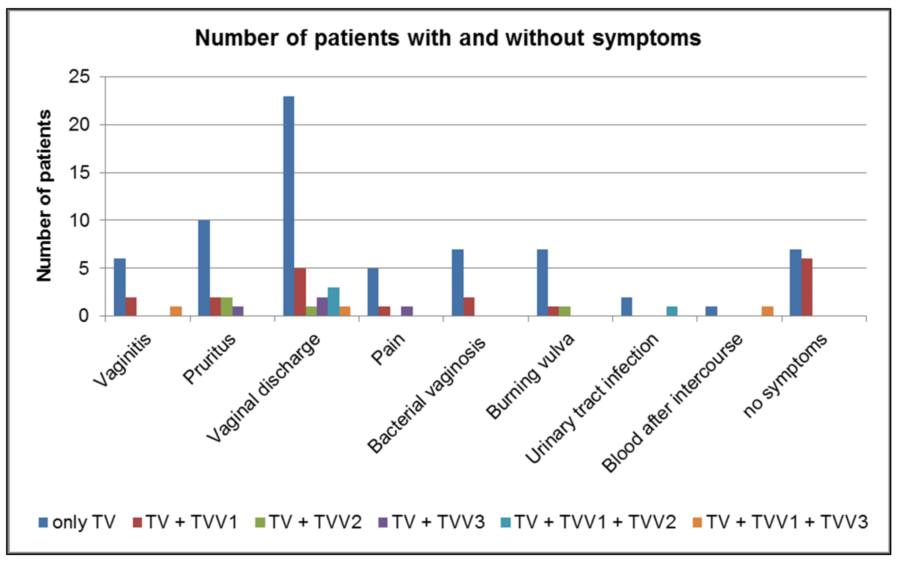
2.3. Prevalence of TVVs in Symptomatic and Asymptomatic Patients

We correlated the occurrence of symptoms as reported by the patients infected with TV with the presence of different TVVs (Figure 1). Twenty patients positive for TVV (20/29 76%) reported symptoms (e.g., vaginitis, pruritus, pain). Out of the 13 TV isolates obtained from patients without reported symptoms, 6 isolates (6/13 46%) revealed the presence of TVV1; in the other 7 isolates (7/13 54%), no TVVs were detected. Among four TV isolates from patients with TV only (no other pathogen detected), TVVs were detected in three isolates; each TV isolate harboured one TVV: TVV1, TVV2, or TVV3. The patient with TV + TVV1 had not complained about any symptoms.

2.4. Detection of M. hominis in TV and Vaginal/Urethral Swabs

M. hominis was detected in vaginal/urethral swabs (culture method: colonies on an agar plate in the Outpatients Centre) in 30 of the 82 TV-positive patients (37%) (27 female, 3 male); M. hominis DNA was proven in 23 of the 82 cultured TV isolates by PCR (28%) (Table 2). In 12 patients (12/82; 15%), with M. hominis detected by culture in the Outpatients Centre, M. hominis DNA was also detected inside TV. In 18 patients (18/82; 22%), M. hominis was detected only by culture, not inside TV by PCR. In 11 cultured TV isolates (11/82; 13%) M. hominis was only detected by PCR (Table 2).

2.5. Prevalence of M. hominis in Symptomatic and Asymptomatic Patients

Among the 30 patients with M. hominis detected in vaginal/urethral swabs by the culture method, 4 patients (2 female, 2 male) did not report any symptoms (13%). In the group of 23 patients, where M. hominis was detected inside TV by PCR, 4 patients (1 female, 3 male) were asymptomatic (17%). In the group of 12 patients where M. hominis was confirmed by culture as well as inside TV by PCR, 2 male patients (17%) were free of symptoms. In the group of 18 patients positive for M. hominis by culture but negative by PCR (not inside TV), 2 female patients (11%) were without any symptoms; and of the 11 patients who were TV-positive for M. hominis by PCR but negative by culture, 1 male and 1 female patient (18%) were asymptomatic. The remaining patients reported symptoms as mentioned above.

2.6. MICs

TV isolates were screened for metronidazole susceptibility under aerobic and anaerobic conditions in parallel. After 48 h incubation in anaerobic conditions, all tested isolates showed metronidazole sensitivity, with MICs ranging from 0.5 to 2 µg/mL. Under aerobic conditions, MICs of metronidazole ranged between 0.5 and 32 µg/mL (Table 3). We did not observe a discrepancy between MIC and MLC values, and our results did not show significant differences in the MICs in correlation to the symbionts of the respective TV isolates.

2.7. LDH Assay

Cytotoxicity of TVs was tested for four different TV groups (five isolates per group). Cytotoxicity of TVs without symbionts ranged from 0.85% to 9.17%, while TVs with TVVs showed cytotoxicities between 1.88% and 11.29%, TVs with M. hominis between 0.81% and 4.48%, and TVs with both symbionts between 0.46% and 8.02% (Figure 2). No statistically significant differences in cytotoxicity between the investigated groups of isolates were found. Microscopic examination revealed no differences in monolayer disruption between TVs with or without symbionts after two hours’ incubation of HeLa cells with the respective TVs.

3. Discussion

We detected TVVs in 35% (29/82) and M. hominis in 28% (23/82) of the collected TV isolates. To the best of our knowledge, the current study is the first reporting the prevalence of symbionts in T. vaginalis isolated from STI patients in Austria. In other European countries, TVV prevalence ranges from 33.3% to 50.4% (Czech Republic: 33–100%; Italy: 50%; the Netherlands: 50.4%) [11,22]. Masha et al. [23] investigated the prevalence of TVV subspecies in Kenya, showing that TVV1 was the most prevalent (39.1%), followed by TVV2 (26.1%), TVV3 (17.4%), and TVV4 (13%). In Brazil, TVV1 was also the most prevalent TVV species (77.5%), followed by TVV2 (30%), TVV3 (30%), and TVV4 (10%) [24]. In contrast, in Italy, the most prevalent virus was TVV2 (79.17%), followed by TVV1 and TVV3 [22]. Our data, indicating that TVV1 was the most prevalent TVV species in Austrian T. vaginalis isolates, fit into the global picture of TVV positivity in TV.
TVV prevalence and its clinical implications are still poorly understood. It has been shown that the presence of TVV can significantly enhance proinflammatory responses to TV in human epithelial culture models [25]. Double-stranded RNA viruses in other protozoan parasites (Leishmania spp., Giardia spp., Plasmodium spp., Entamoeba spp., Babesia spp.) are believed to be involved in disease outcomes. It has been demonstrated that differences in clinical manifestations between different Leishmania strains correlate to the presence of Leishmania virus (LRV), although this was not the only factor involved [26]. Moreover, L. braziliensis guyanensis infected with LRV1 induced more metastasising lesions in mice than an LRV-free strain [27]. Moreover, TVVs may upregulate the expression of immunogenic surface proteins, such as P270, and can cause phenotypic changes that may affect T. vaginalis virulence [13]. We could observe that cytotoxicity of TV isolates harbouring TVV or TVV + M. hominis was enhanced; albeit this was not statistically significant. Fraga et al. [28] reported that TV isolates infected with TVV showed increased cytoadhesion to HeLa cells. Cytotoxicity of TV is a contact-dependent mechanism. It was shown in 1984 by Alderete and Pearlman [29] that exposure of monolayer cultures of human cervical cancer (HeLa), human epithelial (HEp-2), normal baboon testicular (NBT), and monkey kidney (Vero) cells to live pathogenic T. vaginalis led to extensive disruption of the monolayers. Menezes et al. [30] observed that the LDH release from HVECs (human vaginal epithelial cells) varied between 5% and 74%, and this correlated to distinct TV strains’ cytolysis profiles. Analysis of the different groups of TV isolates (with and without symbionts) in the current study did not reveal microscopically detectable differences in the adhesion to or disruption of monolayers of HeLa cells after 2 h incubation and showed low levels of cytotoxicity. Rassmusen et al. [31] used human vaginal epithelial cells and showed that a strain of TV freshly isolated from a patient with severe symptoms caused extensive disruption of the epithelial cell monolayer, whereas a strain maintained in axenic cultures for several years did not. It has been argued that parasitic organisms maintained in culture for a long time lose their potential to infect host cells [32]. However, the observed low cytotoxicity in our study cannot be due to long culture periods, as the isolates were in culture only for several days before the experiments.
In four female patients of the current study, no other infectious agent than TV was detected; thus, we chose these patients to investigate a possible connection between symbionts and symptoms. In three of these TV isolates, TVV presence was proven; no M. hominis was detected. The patient with TV harbouring TVV1 did not complain about any symptoms, whereas the patient with TVV2 reported vaginal discharge and pruritus and the patient with TVV3 complained about pain and vaginal discharge. Among the 13 TV isolates collected from asymptomatic patients, only the presence of TVV1 was confirmed in 6 isolates; the other isolates were TVV-free. Fichorova et al. [25] reported that infections of TV with TVV1 were associated with mild clinical symptoms, whereas the presence of TVV2 inside TV was associated with severe symptoms. Thus, the assumption that the presence of TVV1 is not associated with severe symptoms was partly confirmed in our study, although the sample size was quite small. However, these data are difficult to compare, as at the time when the experiments were performed, it was not possible to clear TVVs from TV, and thus it was not possible to compare the same TV isolate with and without TVV. Recently, a cytidine nucleoside analogue has been shown to be an effective antiviral drug against TVV [33]. This surely is a major advance and will allow future studies to investigate the role of individual TVVs on pathogenicity, as well as metronidazole susceptibility.
Metronidazole has remained a highly effective drug to treat trichomoniasis until now, and drug resistance is still rare [34]. However, in some parts of the world, such as Papua New Guinea, resistance rates can exceed 15% [35]. Metronidazole resistance in TV can be either “aerobic“ (clinical) or “anaerobic“ (laboratory-induced) [36,37] with resistance being defined as >100 µg/mL and >3.1 µg/mL, respectively [38]. Our results revealed no association between TVV presence and metronidazole susceptibility, or resistance. Malla et al. [39], as well as Snipes et al. [40] showed that Trichomonas carrying TVVs showed increased susceptibility to metronidazole. However, Graves et al. [11] detected no association of TVV positivity and metronidazole susceptibility and suggested that TVV may be commensal to TV. In case of Giardia duodenalis, there was also no evidence that infection with dsRNA virus impacts resistance to metronidazole, which is also the first-line drug against Giardia [41].
The relationship between TV and Mycoplasma hominis has been investigated in different studies, indicating that the majority of women positive for TV also carry M. hominis [42]. Ioannidis et al. also showed that Ureaplasma has a tendence to be intracellular in TV [43]. A total of 37% of the patients enrolled in our study were M. hominis-positive. In a study performed in Italy, 78.6% of women with trichomoniasis were positive for M. hominis [44], and in the Netherlands, this figure was 79% [45]. Moreover, TV acts as a potential carrier for M. hominis [46], and the percentage of TV infected with M. hominis ranges from 5 to 89% depending on the geographic origin [24,28,47]. We detected M. hominis inside and/or membrane-associated to TV in 28% of the TV isolates. When M. hominis is hosted by TV, it benefits not only by evading the host immune response but can possibly also hide from antibiotics, which are unable to cross the cell membrane of TV. M. hominis is resistant to anti-Trichomonas drugs such as metronidazole and tinidazole [48,49]. Xiao et al. [50] suggested that M. hominis may confer (or contribute to) drug resistance in TV but to a varying degree, because some infected isolates were not demonstrably metronidazole-resistant when assessed in vitro. In Fürnkranz et al. [51] artificially M. hominis-infected TV strains showed 2 times higher MIC to metronidazole than the respective noninfected strains; moreover, genes involved in metronidazole resistance were influenced by the presence of M. hominis. On the other hand, Margarita et al. [52] showed that the presence of M. hominis significantly increases the sensitivity to metronidazole in TV and affects gene expression. Thus, M. hominis might have an impact on drug susceptibility of TV. It was also shown that the microbial association with M. hominis, as well as with Ca. M. girerdii, could modulate the virulence of TV [53]. Our recent results did not confirm this; however, we did not clear the TVs from M. hominis to test the same isolate with and without symbionts. Further studies would be necessary to investigate if the same TV strains with and without M. hominis show differences, and if it is possible to infect TVV-free TVs with TVV and then reveal specific impacts of the respective TVVs.
After TV treatment with metronidazole, M. hominis can be released and cause invasion not only into the foetal membranes but also into amniotic fluid, resulting in adverse pregnancy outcomes [50]. However, we must emphasise that T. vaginalis infection is not always associated with the presence of M. hominis and vice versa. In the current study, the majority of T. vaginalis-positive patients with M. hominis infection confirmed either by culture, culture and PCR (i.e., intracellular/membrane associated M. hominis), or by PCR only had symptoms such as pruritus, vaginal discharge, pain, bacterial vaginosis, etc. Plummer et al. [54] revealed that M. hominis was associated with symptoms/signs in cases of bacterial vaginosis (BV); but not associated with symptoms/signs in women without BV. On the other hand, during one year of sampling of M. hominis at the Outpatient Centre, we confirmed 56 patients (31 men and 25 women) where M. hominis was the only infectious agent found. Of these, 14 men and 20 women (45% and 80%, respectively) reported symptoms, thus leading to the assumption that M. hominis was the causative agent.
In any case, it is still controversially discussed, if M. hominis should be considered a pathogen at all. Arya et al. [55] did not find evidence that M. hominis is a vaginal pathogen in adults, and the European Academy of Dermatology and Venereology recommended in 2018 that patients should not even be screened for the presence of M. hominis [56]. On the other hand, the high frequency of M. hominis in prostate cancer patients indicates a yet hidden role of these bacteria in the development of prostate cancer during chronic, silent, and asymptomatic colonisation of the prostate [57]. Ten patients in our study would have remained undiagnosed for M. hominis because of its exclusive presence inside TV. Thus, if a screening for M. hominis should be implemented, it might also be essential to screen TV for M. hominis.
In conclusion, our results provide a first insight into the distribution of symbionts (TVVs, M. hominis) in TV isolates from Austrian patients. We detected the presence of TVV1, TVV2, and TVV3; no TVV4 was detected in our collection. M. hominis was detected in vaginal/urethral swabs by the culture method (i.e., in the patient), as well as by PCR (i.e., intracellular and, or membrane associated in TV). We did not observe significant effects of the symbionts on metronidazole susceptibility, cytotoxicity, or severity of symptoms reported by the patients.

4. Materials and Methods

4.1. Collection and Culture of Trichomonas vaginalis Isolates

Vaginal/urethral swabs were collected from symptomatic and asymptomatic patients attending the Outpatients Centre for the Diagnosis of Infectious Venero-Dermatological Diseases in Vienna between 2019 and 2021. Smears of the swabs were cultured on specific Trichomonas-culture plates [58] for 5 days and examined microscopically for the presence of mobile TV. Positive cultures were transported to the Institute of Specific Prophylaxis and Tropical Medicine (ISPTM); parasites were transferred from the plate into liquid TYM Medium (Trypticase-Pepton Medium) [59] supplemented with 10% heat-inactivated horse serum and 1 g cysteine per litre (pH 6.0), and cultured microaerobically in culture flasks at 37 °C. All patients’ data except for symptoms, coinfections, and gender were blinded. As the study was retrospective, no consent was needed from the participants.

4.2. RNA Extraction and First-Strand cDNA Synthesis

In order to detect TVV, whole-cell RNA was isolated. Briefly, TV cultures were washed twice with 1× PBS (centrifugation at 700× g for 10 min). Afterwards, nucleic acid extraction was performed using the GeneJET RNA Purification Kit (Thermo Fisher Scientific, Vilnius, Lithuania) following the manufacturer’s instructions. The isolated RNA was treated with DNase (Roche, Basel, Switzerland), in order to ensure the absence of genomic DNA. First-strand cDNA was synthesised using RevertAid First Strand cDNA Synthesis Kit (Thermo Fisher Scientific-Life Technologies); 1 µg of RNA was used as template for cDNA synthesis.

4.3. PCR for TVVs

cDNAs were used for PCR amplification, which was performed with an Eppendorf Mastercycler (Eppendorf AG, Hamburg, Germany) using primers given in Table 4 [60].
The amplification protocol was as follows: 15 min at 95 °C, followed by 35 cycles of 60 s denaturation at 95 °C, 30 s annealing at 52 °C (TVV1 and TVV3) or 54 °C (TVV2 and TVV4), and 90 s extension at 72 °C. Some 2% agarose gels stained with GelRed® Nucleic Acid Gel Stain (Biotium, Inc., Hayward, CA, USA) were used to visualise the PCR products in a Gel DocTM XR+ Imager (Bio-Rad Laboratories, Inc., Hercules, CA, USA). Additionally, bands in the expected size (see Table 4) were cut out from the gel and purified with the IllustraTM GFXTM PCR DNA and Gel Purification Kit (GE Healthcare, Buckinghamshire, UK). Amplified products were Sanger-sequenced with Thermo Fisher Scientific SeqStudio (Thermo Fisher Scientific, Waltham, MA, USA) and compared to reference sequences in GenBank to verify the specificity of the used primers.

4.4. Detection of M. hominis

The QIAGEN DNA Mini Kit (QIAGEN GmbH, Hilden, Germany) was used for extraction of total DNA from the TV cultures; the DNA was stored at −20 °C for further use. Primers listed in Table 5 were used for the amplification of a Mycoplasma genus specific 16S rRNA gene [61]; 2% agarose gel was used to visualise the PCR products. Bands of the expected size were cut out from the gel, purified, and sequenced as described above. Reference sequences were deposited in GenBank under accession numbers OP623449–OP623451.

4.5. Determination of Minimal Inhibitory Concentrations (MICs)

For metronidazole susceptibility screening, 5 × 10⁵ TV cells/mL (with and without symbionts) were incubated with increasing metronidazole (Sigma-Aldrich, M3761) concentrations (0.5–512 µg/mL) in 96-well flat-bottom plates under microaerobic and anaerobic conditions in parallel at 37 °C for 48 h. To create anaerobic conditions, the microtiter plates were incubated in plastic containers with lids and equipped with one anaerobic bag (AnaeroeGen 2.5 l bag, Thermo Scientific) per 1.5 l plastic container. Viability of the trophozoites in the culture plate wells was assessed by visual examination of motile cells with an inverted microscope at 400× magnification; the lowest drug concentration where no motile TV was observed was defined as minimal inhibitory concentration (MIC). To determine the minimal lethal concentration (MLC), TV cells from the first well with no motility observed were transferred to TYM medium and cultivated for one week to confirm death of the cells.

4.6. LDH Assay

For the determination of cytotoxicity, the LDH assay (CyQUANTTM LDH Cytotoxicity Assay Kit reaction mixture, Thermo Fisher Scientific-Life Technologies), based on the release of lactate dehydrogenase (LDH) upon cell lysis was used. Four groups of TV isolates (five isolates per group) were investigated: the first group consisted of isolates without symbionts; the second group consisted of TV with TVVs; the third group of TV with M. hominis; and the fourth of TV with TVVs and M. hominis. HeLa cells were used as target cells and cultured in IMDM Medium (Gibco, Thermo Fischer Scientific) with 10% foetal bovine serum at 5% CO2. The assays were performed in triplicate and repeated twice in independent set ups, and cytotoxicity was calculated according to the manufacturer’s protocol.

Author Contributions

Conceptualisation, U.F.; methodology, U.F. and I.L.-M.; validation, U.F. and I.L.-M.; formal analysis, I.L.-M.; investigation, I.L.-M.; resources, J.W., A.S. and U.F.; writing—original draft preparation, I.L.-M.; writing—review and editing, I.L.-M., A.S., J.W. and U.F.; visualisation, I.L.-M.; supervision, U.F.; project administration, U.F.; funding acquisition, U.F. All authors have read and agreed to the published version of the manuscript.

Funding

This research was funded by the Austrian Science Fund (FWF), project number: KLI751-B. Open Access Funding by the Austrian Science Fund (FWF).

Institutional Review Board Statement

This study was approved by the ethic commission of the Medical University of Vienna, EK Nr: 1965/2017.

Informed Consent Statement

Not applicable.

Data Availability Statement

Not applicable.

Conflicts of Interest

The authors declare no conflict of interest.

References

  1. Rowley, J.; Hoorn, S.V.; Korenromp, E.; Low, N.; Unemo, M.; Abu-Raddad, L.J.; Chico, R.M.; Smolak, A.; Newman, L.; Gottlieb, S.; et al. Chlamydia, gonorrhoea, trichomoniasis and syphilis: Global prevalence and incidence estimates. Bull. World Health Organ. 2016, 97, 548–562. [Google Scholar] [CrossRef]
  2. Sutton, M.; Sternberg, M.; Koumans, E.H.; McQuillan, G.; Berman, S.; Markowitz, L. The prevalence of Trichomonas vaginalis infection among reproductive-age women in the United States, 2001–2004. Clin. Infect. Dis. 2007, 45, 1319–1326. [Google Scholar] [CrossRef] [PubMed]
  3. Depuydt, C.E.; Leuridan, E.; Van Damme, P.; Bogers, J.; Vereecken, A.J.; Donders, G.G. Epidemiology of Trichomonas vaginalis and human papillomavirus infection detected by real-time PCR in flanders. Gynecol Obstet Investig. 2010, 70, 273–280. [Google Scholar] [CrossRef]
  4. Petrin, D.; Delgaty, K.; Bhatt, R.; Garber, G. Clinical and microbial aspects of Trichomonas vaginalis. Clin. Microbiol Rev. 1998, 11, 300–317. [Google Scholar] [CrossRef] [Green Version]
  5. Edwards, T.; Burke, P.; Smalley, H.; Hobbs, G. Trichomonas vaginalis: Clinical relevance, pathogenicity and diagnosis. Crit Rev. Microbiol. 2016, 42, 406–417. [Google Scholar] [CrossRef] [PubMed]
  6. McClelland, R.S.; Sangare, L.; Hassan, W.M.; Lavreys, I.; Mandaliya, K.; Kiarie, J.; Ndinya-Achola, J.; Jaoko, W.; Baeten, J.M. Infections with Trichomonas vaginalis increases the risk of HIV-1 acquisition. J. Infect. Dis. 2007, 195, 698–702. [Google Scholar] [CrossRef] [PubMed] [Green Version]
  7. Icho, T.; Wickner, R.B. The double-stranded RNA genome of yeast virus L-A encodes its own putative RNA polymerase by fusing two open reading frames. J. Biol. Chem. 1989, 264, 6717–6723. [Google Scholar] [CrossRef]
  8. Janssen, M.E.W.; Takagi, Y.; Parent, K.N.; Cardone, G.; Nibert, M.L.; Baker, T.S. Three-dimensional structure of a protozoal double-stranded RNA virus that infects the enteric pathogen Giardia lamblia. J. Virol. 2015, 89, 1182–1194. [Google Scholar] [CrossRef] [PubMed] [Green Version]
  9. Hartley, M.A.; Ronet, C.; Zangger, H.; Beverley, S.M.; Fasel, N. Leishmania RNA virus: When the host pays the toll. Front. Cell Infect. Microbiol. 2012, 2, 99. [Google Scholar] [CrossRef] [Green Version]
  10. Wang, A.L.; Wang, C.C. Viruses of the protozoa. Annu. Rev. Microbiol. 1991, 45, 251–263. [Google Scholar] [CrossRef] [PubMed]
  11. Graves, K.J.; Ghosh, A.P.; Schmidt, N.; Augostini, P.; Secor, W.E.; Schwebke, J.R.; Martin, D.H.; Kissinger, P.J.; Muzny, C.A. Trichomonas vaginalis virus among women with trichomoniasis and association with demographics, clinical outcomes, and metronidazole resistance. Clin. Infect. Dis. 2019, 69, 2170–2176. [Google Scholar] [CrossRef] [PubMed]
  12. Benchimol, M.; Chang, T.H.; Alderete, J.F. Trichomonas vaginalis: Observation of coexistence of multiple viruses in the same isolate. FEMS Microbiol. Lett. 2002, 215, 197–201. [Google Scholar] [CrossRef] [PubMed]
  13. Provenzano, D.; Khoshnan, A.; Alderete, J.F. Involvement of dsRNA virus in the protein composition and growth kinetics of host Trichomonas vaginalis. Arch. Virol. 1997, 142, 939–952. [Google Scholar] [CrossRef] [PubMed]
  14. Razin, S. Peculiar properties of Mycoplasmas: The smallest self-replicating prokaryotes. FEMS Microbiol. Lett. 1992, 100, 423–431. [Google Scholar] [CrossRef]
  15. Razin, S.; Yogev, D.; Naot, Y. Molecular biology and pathogenicity of Mycoplasmas. Microbiol. Mol. Biol. Rev. 1998, 62, 1094–1156. [Google Scholar] [CrossRef] [PubMed] [Green Version]
  16. Costello, E.K.; Sun, C.L.; Carlisle, E.M.; Morowitz, M.J.; Banfield, J.F.; Relman, D.A. Candidatus Mycoplasma girerdii replicates, diversifies, and co-occurs with Trichomonas vaginalis in the oral cavity of a premature infant. Sci. Rep. 2017, 7, 3764. [Google Scholar] [CrossRef] [PubMed] [Green Version]
  17. Rappelli, P.; Addis, M.F.; Carta, F.; Fiori, P.L. Mycoplasma hominis parasitism of Trichomonas vaginalis. Lancet 1998, 352, 1286. [Google Scholar] [CrossRef]
  18. Meyer, R.D.; Clough, W. Extragenital Mycoplasma hominis infections in adults: Emphasis on immunosuppression. Clin. Infect. Dis. 1993, 17 (Suppl. S1), 243–249. [Google Scholar] [CrossRef] [PubMed]
  19. Cosar, C.; Julou, L. Activity of 1-(2-hydroxyethyl)-2-methyl-5-nitroimidazole (8823 RP) against experimental Trichomonas vaginalis infection. Ann. Inst. Pasteur 1959, 96, 238–241. [Google Scholar]
  20. Robinson, S.C. Trichomonal vaginitis resistance to metronidazole. Canadian Med. Assoc. J. 1962, 86, 665. [Google Scholar]
  21. Muzny, C.; Van Gerwen, O.T.; Kissinger, P. Updates in Trichomonas treatment including persistent infection and 5-nitroimidazole hypersensitivity. Curr. Opin. Infect. Dis. 2020, 33, 73–77. [Google Scholar] [CrossRef] [PubMed]
  22. Margarita, V.; Marongiu, A.; Diaz, N.; Dessi, D.; Fiori, P.L.; Rappelli, P. Prevalence of double-stranded RNA virus in Trichomonas vaginalis isolated in Italy and association with the symbiont Mycoplasma hominis. Parasitol. Res. 2019, 118, 3565–3570. [Google Scholar] [CrossRef] [PubMed]
  23. Masha, S.C.; Cools, P.; Crucitti, T.; Sanders, E.J.; Vaneechouette, M. Molecular typing of Trichomonas vaginalis isolates by actin gene sequence analysis and carriage of T. vaginalis viruses. Parasit. Vectors 2017, 10, 537. [Google Scholar] [CrossRef] [PubMed] [Green Version]
  24. da Luz Becker, D.; dos Santos, O.; Frasson, A.P.; de Vargas Rigo, G.; Macedo, A.J.; Tasca, T. High rates of double-stranded RNA viruses and Mycoplasma hominis in Trichomonas vaginalis clinical isolates in South Brazil. Infect. Genet. Evol. 2015, 34, 181–187. [Google Scholar] [CrossRef] [PubMed] [Green Version]
  25. Fichorova, R.N.; Lee, Y.; Yamamoto, H.S.; Takagi, Y.; Hayes, G.R.; Goodman, R.P.; Chepa-Lotrea, X.; Buck, O.R.; Murray, R.; Kula, T. Endobiont viruses sensed by the human host—Beyond conventional antiparasitic therapy. PLoS ONE 2012, 7, e48418. [Google Scholar] [CrossRef] [PubMed]
  26. Nalçaci, M.; Karakuş, M.; Yılmaz, B.; Demir, S.; Özbilgin, A.; Özbel, Y.; Töz, S. Detection of Leishmania RNA virus 2 in Leishmania species from Turkey. Trop. Med. Hyg. 2019, 113, 410–417. [Google Scholar] [CrossRef]
  27. Fermin, G.; Mazumdar-Leighton, S.; Tennant, P. Viruses of Prokaryotes, Protozoa, Fungi, and Chromista. Viruses 2018, 9, 217–244. [Google Scholar]
  28. Fraga, J.; Rojas, L.; Sariego, I.; Fernández-Calienes, A.; Nuñez, F.A. Species typing of Cuban Trichomonas vaginalis virus by RT-PCR, and association of TVV-2 with high parasite adhesion levels and high pathogenicity in patients. Arch. Virol. 2012, 157, 1789–1795. [Google Scholar] [CrossRef] [PubMed]
  29. Alderete, J.F.; Pearlmann, E. Pathogenic Trichomonas vaginalis cytotoxicity to cell culture monolayers. Br. J. Vener Dis. 1984, 60, 99–105. [Google Scholar] [CrossRef] [Green Version]
  30. Menezes, C.B.; Piccoli Frasson, A.; Meirelles, L.C.; Tasca, T. Adenosine, but not guanosine, protects vaginal epithelial cells from Trichomonas vaginalis cytotoxicity. Microbes Infect. 2017, 9, 122–131. [Google Scholar] [CrossRef] [PubMed]
  31. Rasmussen, S.E.; Nielsen, N.H.; Lind, I.; Rhodes, J.M. Morphological studies of the cytotoxicity of Trichomonas vaginalis to normal human vaginal epithelial cells in vitro. Genitourin Med. 1986, 62, 240–246. [Google Scholar]
  32. Gilbert, R.O.; Elia, G.; Beach, D.H.; Klaessig, S.; Singh, B.N. Cytopathogenic effect of Trichomonas vaginalis on human vaginal epithelial cells cultured in vitro. Infect. Immun. 2000, 68, 4200–4206. [Google Scholar] [CrossRef] [PubMed] [Green Version]
  33. Narayanasamy, R.K.; Rada, P.; Zdrha, A.; van Ranst, M.; Neyts, J.; Tachezy, J. Cytidine nucleoside analog is an effective antiviral drug against Trichomonasvirus. J. Microbiol. Immunol. Infect. 2022, 55, 191–198. [Google Scholar] [CrossRef] [PubMed]
  34. Schwebke, J.R.; Barrientes, F. Prevalence of Trichomonas vaginalis isolates with resistance to metronidazole and tinidazole. Antimicrob. Agents Chemother. 2006, 50, 4209–4210. [Google Scholar] [CrossRef]
  35. Upcroft, J.A.; Dunn, L.; Wal, T.; Tabrizi, S.; Delgadillo-Correa, M.G.; Johnson, J.P.; Garland, S.; Siba, P.; Upcroft, P. Metronidazole resistance in Trichomonas vaginalis from highland women in Papua New Guinea. Sex. Health 2009, 6, 334–338. [Google Scholar] [CrossRef] [PubMed]
  36. Kulda, J.; Tachezy, J.; Cerkasovova, A. In vitro induced anaerobic resistance to metronidazole in Trichomonas vaginalis. J. Eukaryot. Microbiol. 1993, 40, 262–269. [Google Scholar] [CrossRef]
  37. Tachezy, J.; Kulda, J.; Tomkova, E. Aerobic resistance of Trichomonas vaginalis to metronidazole induced in vitro. Parasitology 1993, 106, 31–37. [Google Scholar] [CrossRef] [PubMed]
  38. Müller, M.; Lossick, J.G.; Gorrell, T.E. In vitro susceptibility of Trichomonas vaginalis to metronidazole and treatment outcome in vaginal trichomoniasis. Sex Transm. Dis. 1988, 15, 17–24. [Google Scholar] [CrossRef] [PubMed]
  39. Malla, N.; Kaul, P.; Sehgal, R.; Gupta, I. The presence of dsRNA virus in Trichomonas vaginalis clinical isolates from symptomatic and asymptomatic Indian women and its correlation with in vitro metronidazole sensitivity. Indian J. Med. Microbiol. 2011, 29, 152–157. [Google Scholar] [CrossRef] [PubMed]
  40. Snipes, L.G.; Gamard, P.M.; Narcisi, E.M.; Beard, C.B.; Lehmann, T.; Secor, W.E. Molecular epidemiology of metronidazole resistance in a population of Trichomonas vaginalis clinical isolates. J. Clin. Microbiol. 2000, 38, 3004–3009. [Google Scholar] [CrossRef] [Green Version]
  41. Gordts, B.; De Jonckheere, J.; Kasprzak, W.; Majewska, A.C.; Butzler, J.P. In vitro activity of antiprotozoal drugs against Giardia intestinalis of human origin. Antimicrob. Agents Chemother. 1987, 31, 672–673. [Google Scholar] [CrossRef] [PubMed] [Green Version]
  42. Fiori, P.L.; Diaz, N.; Cocco, A.R.; Rappelli, P.; Dessì, D. Association of Trichomonas vaginalis with its symbiont Mycoplasma hominis synergistically upregulates the in vitro proinflammatory response of human monocytes. Sex Transm. Infect. 2013, 89, 449–454. [Google Scholar] [CrossRef] [PubMed]
  43. Ioannidis, A.; Papaioannou, P.; Magiorkinis, E.; Magana, M.; Ioannidou, V.; Tzanetou, K.; Burriel, A.R.; Tsironi, M.; Chatzipanagiotou, S. Detecting the Diversity of Mycoplasma and Ureaplasma Endosymbionts Hosted by Trichomonas vaginalis Isolates. Front. Microbiol. 2017, 28, 1188. [Google Scholar] [CrossRef] [PubMed]
  44. Diaz, N.; Dessi, D.; Dessole, S.; Fiori, P.L.; Rappelli, P. Rapid detection of coinfections by Trichomonas vaginalis, Mycoplasma hominis, and Ureaplasma urealiticum by a new multiplex polymerase chain reaction. Diagn. Microbiol. Infect. Dis. 2010, 67, 30–36. [Google Scholar] [CrossRef]
  45. van Belkum, A.; van der Schee, C.; van der Meijden, W.I.; Verbrugh, H.A.; Sluiters, H.J. A clinical study on the association of Trichomonas vaginalis and Mycoplasma hominis infections in women attending a sexually transmitted disease (STD) outpatient clinic. FEMS Immunol. Med. Microbiol. 2001, 32, 27–32. [Google Scholar] [CrossRef]
  46. Rappelli, P.; Carta, F.; Delogu, G.; Addis, M.F.; Dessì, D.; Cappuccinelli, P. Mycoplasma hominis and Trichomonas vaginalis symbiosis: Multiplicity of infection and transmissibility of M. hominis to human cells. Arch. Microbiol. 2001, 175, 70–74. [Google Scholar] [CrossRef] [PubMed]
  47. Butler, S.E.; Augostini, P.; Secor, W.E. Mycoplasma hominis infection of Trichomonas vaginalis in clinical isolates from the United States. Parasitol. Res. 2010, 107, 1023–1027. [Google Scholar] [CrossRef]
  48. Dessi, D.; Rappelli, P.; Diaz, N.; Cappuccinelli, P.; Fiori, P.L. Mycoplasma hominis and Trichomonas vaginalis: A unique case of symbiotic relationship between two obligate human parasites. Front. Biosci. J. Virtual Libr. 2006, 11, 2028–2034. [Google Scholar] [CrossRef] [Green Version]
  49. Thi Trung Thu, T.; Margarita, V.; Cocco, A.R.; Marongiu, A.; Dessì, D.; Rappelli, P.; Fiori, P.L. Trichomonas vaginalis Transports Virulent Mycoplasma hominis and Transmits the Infection to Human Cells after Metronidazole Treatment: A Potential Role in Bacterial Invasion of Fetal Membranes and Amniotic Fluid. J. Pregnancy 2018, 2, 5037181. [Google Scholar] [CrossRef] [Green Version]
  50. Xiao, J.C.; Xie, L.F.; Fang, S.L.; Gao, M.Y.; Zhu, Y.; Song, L.Y.; Zhong, H.M.; Lun, Z.R. Symbiosis of Mycoplasma hominis in Trichomonas vaginalis may link metronidazole resistance in vitro. Parasitol. Res. 2006, 100, 123–130. [Google Scholar] [CrossRef]
  51. Fürnkranz, U.; Henrich, B.; Walochnik, J. Mycoplasma hominis impacts gene expression expression in Trichomonas vaginalis. Parasitol. Res. 2018, 117, 841–847. [Google Scholar] [CrossRef] [Green Version]
  52. Margarita, V.; Cao, L.C.; Bailey, N.P.; Ngoc, T.H.T.; Ngo, T.M.C.; Nu, P.A.T.; Diaz, N.; Dessì, D.; Hirt, R.P.; Fiori, P.L.; et al. Effect of the Symbiosis with Mycoplasma hominis and Candidatus Mycoplasma Girerdii on Trichomonas vaginalis Metronidazole Susceptibility. Antibiotics 2022, 16, 812. [Google Scholar] [CrossRef] [PubMed]
  53. Margarita, V.; Bailey, N.P.; Rappelli, P.; Diaz, N.; Fettweis, J.M.; Hirt, R.P.; Fiori, P.L. Two Different Species of Mycoplasma Endosymbionts Can Influence Trichomonas vaginalis Pathophysiology. mBio 2022, 28, e0091822. [Google Scholar] [CrossRef] [PubMed]
  54. Plummer, E.L.; Vodstrcil, L.A.; Bodiyabadu, K.; Murray, G.L.; Doyle, M.; Latimer, R.L.; Fairley, C.K.; Payne, M.; Chow, E.P.F.; Garland, S.M.; et al. Are Mycoplasma hominis, Ureaplasma urealyticum and Ureaplasma parvum associated with specific genital symptoms and clinical signs in nonpregnant women? Clin. Infect. Dis. 2021, 16, 659–668. [Google Scholar] [CrossRef] [PubMed]
  55. Arya, O.P.; Tong, C.Y.; Hart, C.A.; Pratt, B.C.; Hughes, S.; Roberts, P.; Kirby, P.; Howel, J.; McCormick, A.; Goddard, A.D. Is mycoplasma hominis a vaginal pathogen? Sex Transm. Infect. 2001, 77, 58–62. [Google Scholar] [CrossRef] [Green Version]
  56. Horner, P.; Donders, G.; Cusini, M.; Gomberg, M.; Jensen, J.S.; Unemo, M. Should we be testing for urogenital Mycoplasma hominis, Ureaplasma parvum and Ureaplasma urealiticum in men and women?—A position statement from the European STI Guidelines Editorial Board. JEADV 2018, 32, 1845–1852. [Google Scholar]
  57. Saadat, S.; Karami, P.; Jafari, M.; Kholoujini, M.; Rikhtegaran Tehrani, Z.; Mohammadi, Y.; Alikhani, M.Y. The silent presence of Mycoplasma hominis in patients with prostate cancer. Pathog. Dis. 2020, 78, ftaa037. [Google Scholar] [CrossRef]
  58. Stary, A.; Kuchinka-Koch, A.; Teodorowicz, L. Detection of Trichomonas vaginalis on Modified Columbia Agar in the Routine Laboratory. J. Clin. Microbiol. 2002, 40, 3277–3280. [Google Scholar] [CrossRef] [Green Version]
  59. Diamond, L.S. The establishment of various trichomonads of animals and man in axenic cultures. J. Parasitol. 1957, 3, 488–490. [Google Scholar] [CrossRef]
  60. Goodman, R.P.; Freret, S.; Kula, T.; Geller, A.M.; Talkington, M.W.T.; Tang-Fernandez, V.; Suciu, O.; Demidenko, A.A.; Ghabrial, S.A.; Beach, D.H.; et al. Clinical isolates of Trichomonas vaginalis concurrently infected by strains of up to four Trichomonas virus Species (Familiy Totiviridae). J. Virol. 2011, 85, 4258–4270. [Google Scholar] [CrossRef] [Green Version]
  61. Van Kuppeveld, F.J.M.; van der Logt, J.T.M.; Angulo, A.F.; van Zoest, M.J.; Quint, G.V.; Niesters, H.G.; Galama, J.M.; Melchers, W.J. Genus- and species-specific identification of Mycoplasma by 16S rRNA amplification. Appl. Environ. Microbiol. 1992, 58, 2606–2615. [Google Scholar] [CrossRef] [PubMed]
Figure 1. Numbers of TV infected patients without (no symptoms) or with symptoms (vaginitis, pruritus, vaginal discharge, pain, bacterial vaginosis, burning vulva, urinary tract infection, blood after intercourse) in correlation with TVV presence (TV + TVV1, TV + TVV2, TV + TVV3, TV + TVV1 + TVV2, TV + TVV1 + TVV3) or absence (only TV).
Ijms 23 12422 g001
Figure 2. Mean percentages of T. vaginalis cytotoxicity of isolates without endosymbionts (TV), carrying TVVs (TV + TVV), carrying M. hominis (TV + Mh), and carrying TVVs and M. hominis (TV + TVV + Mh). Minimal (black squares) and maximal (black triangles) values of cytotoxicity are pointed.
Ijms 23 12422 g002
Table 1. Characteristics of the study group.
Number of Patients with TV82
Sex72 female (88%)10 male (12%)
Age (mean age)23–64 (41.2)34–58 (46)
No clinical symptoms reported7/72 (9.7%)6/10 (60%)
No other pathogens diagnosed4/72 (5.5%)0/10 (0%)
Table 2. Detection of M. hominis in patients with TV infection and within TV isolates.
Number of Positive Patients and
Cultured TV Isolates
82
Sex72 female 10 male
M. hominis in patients (culture)27/72 (36.5%)3/10 (30%)
M. hominis in TV (PCR)20/72 (27.8%)3/10 (30%)
M. hominis in patients and in TV (culture and PCR)10/72 (14%)2/10 (20%)
M. hominis only in patients (culture), not in TV17/72 (24%)1/10 (10%)
M. hominis only in TV (PCR), not in patients (culture)10/72 (14%)1/10 (10%)
Table 3. Metronidazole susceptibility (mean and range) for TV harbouring different TVVs and TVV combinations, as well as TV harbouring M. hominis (+M. hominis) and TV where TVVs and M. hominis were present (+TVV + M. hominis).
TVMetronidazole Mean Concentration (Min–Max)
Anaerobic ConditionsAerobic Conditions
no TVVs (n = 28)0.8 µg/mL (0.5–2 µg/mL)3.3 µg/mL (0.5–32 µg/mL)
+TVV1 (n = 12)0.65 µg/mL (0.5–1 µg/mL)3.3 µg/mL (0.5–32 µg/mL)
+TVV2 (n = 2)0.67 µg/mL (0.5–1 µg/mL)5.5 µg/mL (0.5–8 µg/mL)
+TVV3 (n = 5)0.5 µg/mL (0.5 µg/mL)0.93 µg/mL (0.5–2 µg/mL)
+TVV1 + TVV2 (n = 3)1.13 µg/mL (0.5–2 µg/mL)4.16 µg/mL (0.5–8 µg/mL)
+TVV1 + TVV3 (n = 2)1.17 µg/mL (0.5–2 µg/mL)1.5 µg/mL (0.5–2 µg/mL)
+M. hominis (n = 12)0.63 µg/mL (0.5–1 µg/mL)2.58 µg/mL (0.5–32 µg/mL)
+M. hominis + TVVs (n = 7)0.62 µg/mL (0.5–1 µg/mL)4.65 µg/mL (0.5–32 µg/mL)
Table 4. Primers used for TVV determination.
T. vaginalis VirusPrimer NamePrimer Sequence (5′→3′)Expected Product Size
TVV1TVV1F2875
TVV1R3443
ATTAGCGGTGTTTGTGATGCA
CTATCTTGCCATCCTGACTC
569 bp
TVV2TVV2F2456
TVV2R3080
GCTTGAGCACTGCTCGCG
TCTCTTTTGGCATCGCTT
625 bp
TVV3TVV3F1474
TVV3R2025
TGGAGTTAAATGGTCTCGAGC
GATTTCATCTTGTTCAATTGCA
552 bp
TVV4TVV4F1338
TVV4R1834
ATGCCAGTTGCTTTCCG
TTCCCCAATAGTTATCAG
514 bp
Table 5. Primers used for the detection of Mollicutes.
Primer NameTarget GenePrimer Sequence (5′→3′)Expected Product Size
GPOI
MGSO
Mycoplasma genus specific 16S rRNAACTCCTACGGGAGGCAGCAGTA TGCACCATCTGTCACTCTGTTAACCTC717 bp
Publisher’s Note: MDPI stays neutral with regard to jurisdictional claims in published maps and institutional affiliations.

