Article

Freshwater Macroalgae, Oedogonium, Grown in Wastewater Reduce Diet-Induced Metabolic Syndrome in Rats

1
Functional Foods Research Group, University of Southern Queensland, Toowoomba, QLD 4350, Australia
2
School of Science, Western Sydney University, Richmond, NSW 2753, Australia
3
Te Aka Mātuatua—School of Science, University of Waikato, Tauranga 3112, New Zealand
4
College of Marine & Environmental Sciences, James Cook University, Townsville, QLD 4811, Australia
5
Pacific Biotechnologies Australia Pty Ltd., James Cook University, Townsville, QLD 4811, Australia
6
School of Pharmacy and Medical Science, Griffith University, Gold Coast, QLD 4222, Australia
7
School of Science, Technology and Engineering, University of the Sunshine Coast, Maroochydore, QLD 4558, Australia
*
Author to whom correspondence should be addressed.
Int. J. Mol. Sci. 2022, 23(22), 13811; https://doi.org/10.3390/ijms232213811
Submission received: 24 August 2022 / Revised: 6 November 2022 / Accepted: 7 November 2022 / Published: 9 November 2022
(This article belongs to the Special Issue Marine Bioactive Substances for Metabolic Syndrome)

Abstract

:
Macroalgae produce compounds with industrial, pharmaceutical and nutritional applications. In this study, biomass from the freshwater macroalgal genus Oedogonium was grown in either treated municipal wastewater (M) or ash dam water from a coal-fired power station (D). The biomass was investigated for its metabolic responses in high-carbohydrate, high-fat diet-fed rats, a model of human metabolic syndrome. The Oedogonium biomass cultured in M contained higher amounts of K, Mg, omega-3 polyunsaturated fatty acids (PUFA), insoluble fibre and β-carotene, while biomass grown in D contained higher amounts of Al, Fe, V, Zn, Mn and As. Biomass from M further increased body weight and inflammation in the heart and colon in high-carbohydrate, high-fat diet-fed rats. In contrast, biomass from D prevented changes in metabolic, cardiovascular and liver parameters without changing tissue histology. We suggest that increased intake of metals and metalloids through macroalgal biomass from D may decrease abdominal fat deposition while polysaccharides, PUFA and carotenoids from M may improve blood glucose responses in an obesogenic diet. Thus, macroalgal biomass grown in different wastewater sources could be acceptable for feed or food applications. This biomass could even provide potential health benefits in diet-induced metabolic syndrome.

1. Introduction

The diverse uses of seaweeds (marine macroalgae) throughout the world which include food sources for humans and livestock, medicines and building materials, have led to the proposal that humankind has been “saved by seaweeds”, especially in times of crises [1]. Extensive myths and legends are part of the background of current evidence-based research on seaweeds [2]. The nutritional benefits of algal-derived food products, predominantly from seaweeds, have been associated with their high-quality proteins, lipids, polysaccharides, vitamins and antioxidants [3,4]. Thus, seaweeds may be functional foods (defined as foods that provide health benefits along with nutrition) when consumed chronically in adequate amounts as part of a regular diet. Functional foods may be useful to treat or prevent metabolic syndrome [5,6], a cluster of risk factors that includes obesity, hypertension, dyslipidaemia and impaired glucose tolerance which increases the risk for development of cardiovascular disease, type two diabetes and some cancers [7]. We have reported that the diverse taxonomic groups of green [8,9], brown [10] and red [11,12] seaweeds are potential tropical functional foods for metabolic syndrome [13], but freshwater macroalgae have been rarely studied for their health benefits, potentially because commercial production is limited.
World-wide production of seaweeds was estimated at over 32 million tonnes in 2018 with 97.1% being farmed seaweeds [14]. Macroalgal forests in coastal ecosystems have been estimated to have an area of around 7 million km2 and a net primary production of 1.32 × 10¹⁵ g C/year [15]. Almost all seaweed production is from the sea, yet freshwater macroalgae lends itself to land-based production for industrial applications such as biofuel feedstock [16]. Strains of the freshwater genus Oedogonium (Chlorophyceae) are an ideal target for large-scale biomass production [17]. Further, cultivation of freshwater macroalgae is useful for the bioremediation of different sources of wastewater, where the growing biomass assimilates nutrients such as N, P, metals and trace metals, which are removed from the system when the biomass is harvested [18,19,20]. This then allows the reuse or discharge of clean freshwater, an increasingly rare resource [21], as well as a generation of new products from macroalgae such as biostimulants and soil conditioners [22,23,24]. Species of Oedogonium are effective in removing metals from the wastewater of commercial coal-fired power stations [25] and nutrients in municipal wastewater treatment plants [26,27,28]. Growing freshwater macroalgae for bioremediation of wastewater will likely produce biomass that is a potential functional food for metabolic syndrome as similar benefits have already been shown for marine macroalgal biomass.
This study characterises the chemical composition of Oedogonium biomass grown in two different wastewaters and reports the responses to each biomass in rats with diet-induced metabolic syndrome. Our first hypothesis was that Oedogonium biomass grown in contaminated wastewater from the ash dam of a coal-fired powerstation contained potentially harmful components which could induce toxic effects when fed to rats. In contrast, the biomass from treated municipal wastewater-grown macroalgae would contain valuable nutrients such as N, P, polysaccharides, fatty acids, carotenoids and trace metals. Our second hypothesis was that Oedogonium biomass cultivated in nutrient-rich municipal wastewater would improve the symptoms of diet-induced metabolic syndrome in rats, an accepted model of metabolic syndrome in humans [29].

2. Results

2.1. Composition of Macroalgae from Different Sources

Ash dam macroalgae (D) had up to five times greater amounts of Al, Ba and Mn, and more than five times greater amounts of Cd, Co, Cr, Cu, Fe, Mo, Ni, V and Zn compared to treated municipal wastewater-grown macroalgae (M). Treated municipal wastewater-grown macroalgae had up to three times greater content of Ca, K, Mg, Na, Pb and Sr (Table 1). The metalloids As and B were 80 times and 1.3 times greater in ash dam macroalgae, and likewise the non-metal S was 1.3 times higher in the Oedogonium cultivated in ash dam water. In contrast, P was 1.3 times greater in the Oedogonium cultivated in treated municipal wastewater. Ash dam macroalgae biomass had two-fold and 4.5-fold greater proportions of saturated and monounsaturated fatty acids, respectively, but 6.8-fold lower proportions of polyunsaturated fatty acids (Table 1). Ash dam macroalgae had 1.6 times and 1.7 times lower total dietary fibre and insoluble fibre, respectively, with minimal soluble fibre detected in either biomass. The concentrations of vitamins also differed between the two strains of Oedogonium; vitamin K1 was three times greater in the Oedogonium cultivated in the ash dam water, whereas β-carotene and vitamin B12 were 20 times and three times greater, respectively, in the Oedogonium cultivated in treated municipal wastewater (Table 1).

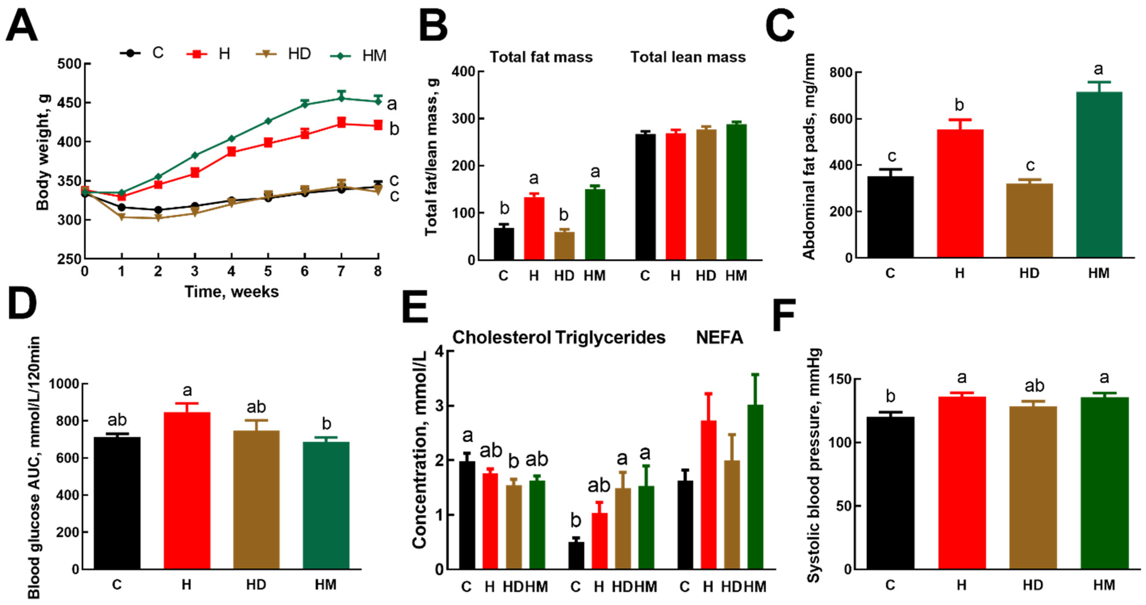
2.2. Metabolic Parameters and Body Composition

High-carbohydrate, high-fat (H) diet-fed rats had increased body weight, whole-body fat mass, abdominal fat pads and systolic blood pressure compared to corn starch (C) diet-fed rats (Figure 1). Treated municipal wastewater-grown macroalgae increased body weight in H rats fed treated municipal wastewater-grown Oedogonium biomass (HM) (Figure 1A; Table 2) with no change in fat or lean mass compared to H rats (Figure 1B). In contrast, ash dam macroalgae prevented increases in body weight in H rats fed ash dam Oedogonium biomass (HD) (Figure 1A; Table 2) and body fat (Figure 1B) so that these parameters were similar to C rats and markedly lower than H rats (Figure 1B); however, the lean mass remained unchanged compared to both C and H rats (Figure 1B). These changes were observed without any changes in food, water or energy intakes compared to H rats, hence decreasing feed efficiency in HD rats to zero while increasing it in HM rats compared to H rats (Table 2). Treated municipal wastewater-grown macroalgae increased total abdominal and retroperitoneal fats with no change in epididymal and omental fats (Figure 1C; Table 2). In contrast, ash dam macroalgae prevented the increase in total abdominal, retroperitoneal, epididymal and omental fats in HD rats. Bone mineral density and bone mineral content were decreased in HD rats but were unaffected in HM rats compared to H rats (Table 2).
Biomass from both water sources reduced basal blood glucose concentrations and blood glucose concentrations at 120 min compared to H rats. The area under the curve for HD rats was intermediate to H and HM rats with HM rats having lower area under the curve compared to H rats (Figure 1D; Table 2). Plasma lipid concentrations were unaffected by dietary macroalgae (Figure 1E).

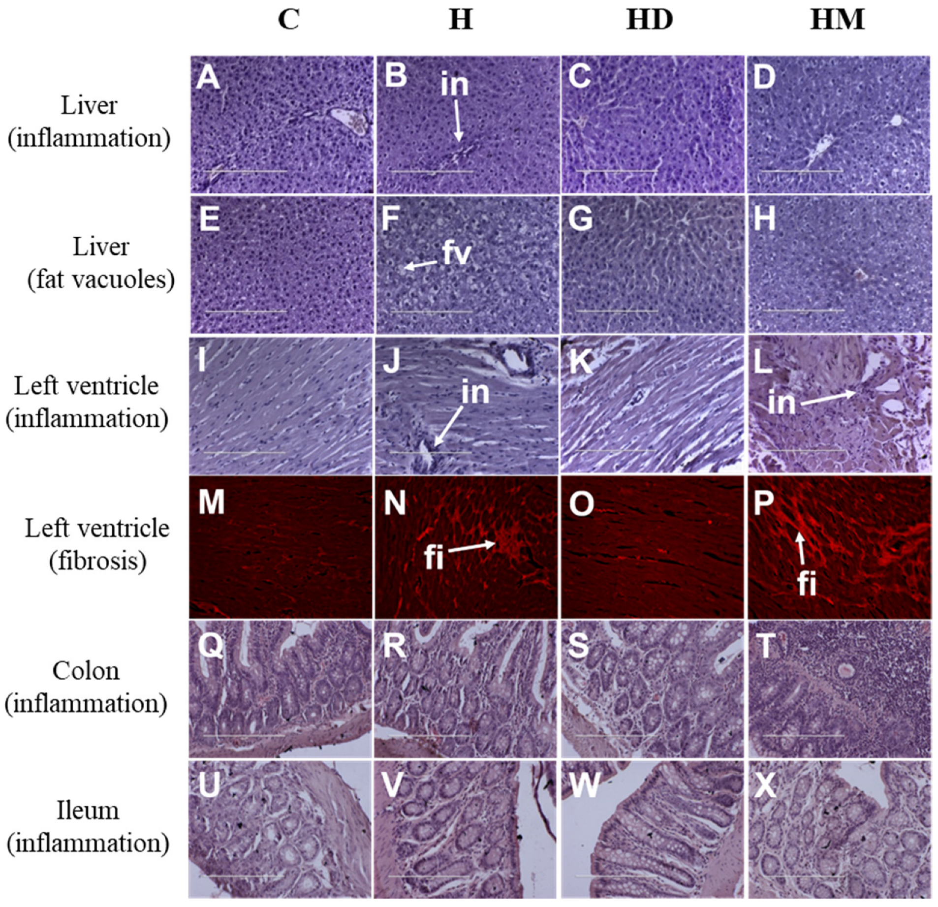
2.3. Liver, Heart and Gastrointestinal Parameters

Liver wet weights of both treated groups were similar to H rats and higher than C rats (Table 2). Plasma activities of alanine transaminase and aspartate transaminase were unchanged in both HM and HD rats (Table 2). Liver B content was very low in H rats compared to C rats and both macroalgae-treated groups had intermediate B content compared to C and H rats (Table 3). Liver V content was similar in C, H and HM rats; ash dam macroalgae markedly increased V content in the liver of HD rats compared to other groups (Table 3). Livers from HD and HM rats showed an absence of inflammation (Figure 2C,D) and steatosis (Figure 2G,H), unlike livers from H rats which showed inflammatory cell infiltration and steatosis (Figure 2B,F).
Systolic blood pressure was unchanged in HM and HD rats compared to H rats (Figure 1F). Left and right ventricular weights were unchanged by supplementation with macroalgae whereas kidney and spleen weights were increased in HM rats compared to H rats (Table 2). HM (Figure 2L,P) rats had higher inflammation and fibrosis in the left ventricle compared to H rats (Figure 2J,N) whereas HD rats (Figure 2K,O) had lower inflammation and fibrosis in the left ventricle compared to H rats (Figure 2J,N). The colon and ileum from HM rats showed higher inflammation than other groups whereas the ileum from HD rats showed no abnormalities (Figure 2S,W) unlike the colon (Figure 2T,X). Table 2 and Table 3 provide other metabolic and tissue variables including metal contents in the liver.

3. Discussion

Malnutrition, including obesity, undernutrition and other dietary risks, is now the leading cause of poor health in the world [30]. Overnutrition and obesity are major challenges in both developed and developing countries [31]. The consumption of energy-rich foods has increased with most of the energy coming from simple sugars and saturated fats, leading to a higher prevalence of obesity [32]. The risk of developing cardiovascular disease, type 2 diabetes and cancers also increases with obesity [33]. The greatest burden of malnutrition in low- and middle-income countries comes from the combination of the high prevalence of stunting (28%), wasting (8.8%) and underweight (17.4%) with increasing overweight in children less than five years of age [30]. The Lancet Commission in 2019 reported economic losses of about $3.5 trillion annually from malnutrition, equivalent to 11% of the GDP in Africa and Asia [30].
Seaweeds have been an essential part of human diets [1], possibly providing the nutritional and energetic requirements for the unique development of a large human brain [34] and are now consumed mainly in East Asia [35,36]. Freshwater macroalgae have received little attention as functional foods for metabolic syndrome compared to seaweeds or microalgae [4]. It is feasible that functional foods from freshwater macroalgae could provide solutions to both over- and under-nutrition. As an example, the freshwater macroalgae Spirogyra varians can provide a viable source of nutrition in human diets [37].
This study showed that the biomass of a freshwater macroalgae, Oedogonium, cultivated in ash dam wastewater containing increased concentrations of metals and metalloids prevented obesity development in rats fed a high-carbohydrate, high-fat diet. This response was manifested as a prevention of increases in body weight, whole-body fat mass and abdominal fat together with improvements in blood glucose tolerance. Ash dam macroalgae prevented inflammation in the heart and colon, steatosis in the liver and fibrosis in the heart. These changes occurred without obvious toxicity, despite the accumulation of metals and metalloids in the macroalgal biomass and in the rat liver after the consumption of the macroalgae. While the consumption of Oedogonium cultivated in treated municipal wastewater also resulted in improvements in blood glucose tolerance and decreased steatosis in the liver, these changes were accompanied by increased body weight and abdominal fat, inflammation in the heart and colon, and fibrosis in the heart. These results clearly distinguish the metabolic responses of the rat model to two different sources of macroalgae with notably differentiated biochemical profiles.
Metabolic syndrome in humans is characterised by extensive changes in the gut microbiota with decreased capacity to metabolise carbohydrates and short-chain fatty acids. This decrease may be reversed with increased dietary prebiotic fibre [38]. The complex polysaccharides in seaweeds may be a viable nutritional source of the prebiotics to reverse changes in the gut microbiota [39]. The prebiotic role of these macroalgal polysaccharides, mostly from seaweed, and their health benefits have been widely discussed but clinical trials are still necessary [40]. While almost all investigations have been carried out on marine macroalgae, the freshwater green macroalgae, Cladophora surera, synthesised sulphated polysaccharides similar to the marine species of this genus [41]. However, the increased insoluble fibre in the treated municipal wastewater biomass (37.4%) compared to ash dam wastewater biomass (21.4%) does not correlate with the greater effectiveness of the ash dam biomass in the prevention of increased abdominal fat deposition.
Trace elements are involved in the development and treatment of obesity. They are chemical micronutrients including Cr, Co, Cu, Fe, Mn, Se and Zn that are needed in very small amounts, yet have a prominent role in the homeostasis of physiological and metabolic processes in our body [42]. Seaweeds may accumulate heavy metals and thus induce toxicity in humans when consumed, albeit rarely [43]. This is consistent with studies with freshwater macroalgae showing that Oedogonium (Genbank Accession Number KF606974) cultured with flue gas reduced the concentrations of all metals in wastewater from a coal-fired power station [25], that Oedogonium westii effectively removed Cd, Ni, Cr and Pb ions from aqueous systems [44], and that the freshwater macroalgae, Cladophora fructa, removed Cu, Zn, Cd and Hg ions from aqueous solutions [45]. However, trace metals found in industrial wastewater including Cr and V may be beneficial to human health and disease [46,47,48,49]. Their importance in reducing diet-induced obesity has been shown by improved insulin signalling and glucose metabolism [46,49,50]. In high-fat diet-fed female Wistar rats, decreased adipose tissue content of Cr and V correlated with an increase in adipose tissue endocrine dysfunction [49]. Decreased content of these trace elements in adipose tissue due to caloric excess has been hypothesised to lead to the development of adipose tissue insulin resistance through disruption of intra-adipocyte insulin signalling, and further adipokine imbalance that leads to obesity [50]. Along with decreased insulin resistance, Cr and Zn also decrease inflammation, which is the link between insulin resistance and obesity that leads to metabolic syndrome [50].
The trace elements in macroalgae when consumed as part of the diet could play a role in the control of metabolic syndrome risk factors in places where these dietary nutrients are missing or where traditional tillage cropping has removed many of the minerals from the soil. Zn produces insulinomimetic effects and improved glucose tolerance and ash dam macroalgae had high amounts of Zn [51]. Na and K were present in high amounts in comparison to other minerals and, though there was no change in systolic blood pressure, these minerals play roles in managing hypertension [52]. Trace elements such as Cr, Se and V when taken in optimal doses can improve the symptoms of metabolic syndrome with their insulinomimetic effects [53]. Cr administration lowered adipose tissue content in obese rats compared to control rats [49], and supplementation of Cr histidinate lowered body weight and serum glucose with increased Nrf2 expression and decreased NF-κB expression [54]. Se deficiency can cause heart failure [55] and its supplementation showed cardioprotective effects [56]. VO (dmpp)2, an oxovanadium, administered to fat rats reduced obesity and hepatic triglycerides [57]. High-fat diet-fed C57BL/6 mice treated with V-rich groundwater had reduced obesity which may be due to the inhibition of preadipocyte differentiation by decreasing the peroxisome proliferator-activated receptor γ (PPAR-γ) and CCAAT-enhancer-binding protein expression [58]. Ash dam biomass had higher contents of these three trace elements than the treated municipal wastewater biomass and supports their role in the prevention of obesity in rats treated with ash dam grown Oedogonium biomass. Inorganic As has been established as a carcinogen for humans with increased risk of certain cancers and other metabolic disease [59,60,61]. Many foods can accumulate high levels of inorganic As and can introduce these into the food cycle [59]. Organic As, on the other hand, has been considered to be non-toxic [62,63]. In this study, higher amounts of trace metals such as As, V and Se were absorbed into the macroalgae (10 to 100 times), but the relative difference between the concentrations in the rat liver was much lower than that between the algae. This may suggest that the excess amount of these metals including As may not be absorbed and accumulated from macroalgae in the rats.
Omega-3 fatty acids and carotenoids are present in macroalgae, although at lower concentrations. These components have proven health benefits associated with them [64,65,66,67], but their lower concentrations in macroalgae may not provide sufficient doses for physiological responses. However, these compounds may provide benefits to complement responses from other components.
One of the limitations of our study is the lack of a macroalgae intervention to corn starch diet-fed rats, as our primary aim was to investigate effects of macroalgae in obesity prevention. Although both interventions showed no signs of toxicity in high-carbohydrate, high-fat diet-fed rats, the high content of metals and metalloids in biomass cultured in ash dam water could exhibit some toxic effects in corn starch diet-fed rats or other basal diets more broadly. Further studies on feeding macroalgae interventions to corn starch diet-fed rats would identify any such effects. Moreover, a reversal protocol of feeding macroalgae to both corn starch diet-fed rats and high-carbohydrate, high-fat diet-fed rats would provide responses that can be compared with our reversal protocol studies with seaweeds showing beneficial responses [8,9,10,11,12].
Thus, our first hypothesis that Oedogonium biomass grown in ash dam water would be toxic has now been shown to be incorrect as this biomass prevented most of the signs of diet-induced metabolic syndrome in rats without signs of gastrointestinal, liver or heart toxicity. Further, our second hypothesis that Oedogonium biomass grown in municipal wastewater would improve the symptoms of diet-induced metabolic syndrome in rats was shown to be valid only for the prevention of diet-induced hyperglycaemia. Based on the results in our study, it would be interesting to analyse how these macroalgae impact gut microbiota.

4. Materials and Methods

4.1. Sources of Freshwater Macroalgae

Two strains of the freshwater macroalgal genus Oedogonium were cultured for this study. The first strain (Oedogonium intermedium, Genbank Accession number: KF606977 [68]) was originally collected from an agricultural irrigation channel at Brandon, QLD, Australia (19°39′ S, 147°24′ E) [69]. This strain was maintained in stock cultures at the Marine and Aquaculture Facilities, James Cook University, Townsville, QLD, Australia prior to culture in the primary treated municipal wastewater from Cleveland Bay Wastewater purification plant in Townsville. Cultures were maintained at James Cook University in 3 × 10,000 L tanks with a water exchange (new for old) of 800 L every day over a 4-week period [27]. The biomass yield over the culture period ranged from 6.8 to 9.9 g DW m−2 d−1 (equivalent to ~30 tonnes per hectare per year). A second strain (Oedogonium sp., Genbank Accession number: KF606974 [70]) was isolated from the 46,000 ML ash dam of Tarong Power station, a 700 MW coal-fired power station located at Tarong, QLD, Australia (26°46′ S, 151°54′ E). This strain was cultured in 3 × 10,000 L tanks on site at the power station using ash dam water amended with flue gas from the combustion of coal, with 5000 L exchanged every three days throughout the 4-week culture period [25]. The biomass yield over the culture period ranged from 2.9 to 8.2 g DW m−2 d−1 (equivalent to ~20 tonnes per hectare per year). Biomass from each water source was collected weekly over the 4-week culture period and a pooled blended sample was used for assays and biochemical analysis. Biomass was air-dried and stored at room temperature until use. Ash dam macroalgae was labelled as D and municipal waste treatment plant macroalgae was labelled as M. The two Oedogonium strains were closely related (Clade Tar1 [KF606974] and Tsv2 [KF606977] with a bootstrap value of 88%; we cannot state that these are of the same species [69]). Dried biomass was used for all biochemical analyses and for diet inclusion in the live rat study.

4.2. Chemical Analysis of Macroalgae

The full biochemical profiles of biomass from each wastewater source were determined by analysis for proximate components (lipid, protein, carbohydrate, ash, moisture and dietary fibre) and element concentrations (C, H, O, N, S and metal/metalloid/halogens), as well as for specific nutritional compounds (antioxidants, vitamins and pigments).
Total lipids were quantified in a 200.0 ± 0.1 mg subsample using solvent extraction [71]. Fatty acids were extracted and transesterified from a separate subsample of biomass (30.0 ± 0.1 mg) following a one-step extraction/transesterification method (methanol/acetyl chloride; 95:5 v/v) [72]. Fatty acid methyl esters were separated and quantified by gas chromatography–mass spectrophotometry on an Agilent 7890c GC/5975c EIMS system equipped with a DB-23 capillary column (cyanopropyl stationary phase [60 m × 0.25 mm id × 0.15 μm], Agilent Technologies, Mulgrave, VIC, Australia) [72]. The quantity of fatty acids was determined by the comparison of peak areas of external standards (Sigma Aldrich, Castle Hill, NSW, Australia) and was corrected for recovery of internal standard (heptadecanoic acid, C17:0).
Protein was determined as the sum of amino acids based on a quantitative amino acid analysis (16 amino acids) performed at Australian Proteome Analysis Facility (APAF) at Macquarie University, Sydney, NSW, Australia, while carbohydrate was calculated by difference as 100 – ∑ (lipid, protein, ash, moisture) where lipids, proteins, ash and moisture are expressed as a percentage of the total mass. Moisture content was measured on a minimum of 1.0 g biomass at 105 °C to constant weight (MS-70 moisture analyser, A&D Company Ltd., Toshima, Tokyo, Japan). The same biomass sample was then weighed to 1 mg precision followed by combustion in air (550 °C, 6 h; SEM muffle furnace, LabTek, Brendale, QLD, Australia) to determine ash content per gram dry weight.
The total dietary fibre content, including soluble and insoluble components that comprise the main forms of carbohydrates, was analysed on a 10 g sample, following standard methods (AOAC Official Method 985.29 total dietary fibre in foods, and AOAC Official Method 993.19 soluble dietary fibre in food and food products) by Grain Growers Ltd. (Sydney, Australia; now Australian Export Grains Innovation Centre (AEGIC)).
A subsample (200 mg) of each biomass source was also analysed for the contents of C, N, H and S (OEA Laboratory Ltd., United Kingdom, http://www.oealabs.com) using GC-TCD. The content of oxygen was calculated by difference as %O = 100 – ∑(C, H, N, S, ash) where C, H, N, S and ash are expressed as a percentage of the total mass. Gross energy content (kJ/g) of the biomass expressed as the higher heating value was then calculated [73] based on CHONS elemental composition and ash content. The content of 24 elements in the biomass was measured by Inductively Coupled Plasma Mass Spectrometry (ICP/MS) (Varian 820-MS, Varian, Belrose, NSW, Australia) at the Advanced Analytical Centre, James Cook University [74].
A further series of subsamples was taken for the following nutritional analyses. Vitamins were quantified by HPLC (ascorbic acid and vitamin K1) and by the Euglena gracilis microbiological assay (vitamin B12) at the National Measurement Institute, Sydney, NSW, Australia. The pigments chlorophyll a and b were extracted in HPLC-grade methanol and quantified spectrophotometrically (SpectroStar nano, BMG LabTech, Mornington, VIC, Australia) [75], while α- and β-carotene were quantified by HPLC at the National Measurement Institute following standard protocols [76].

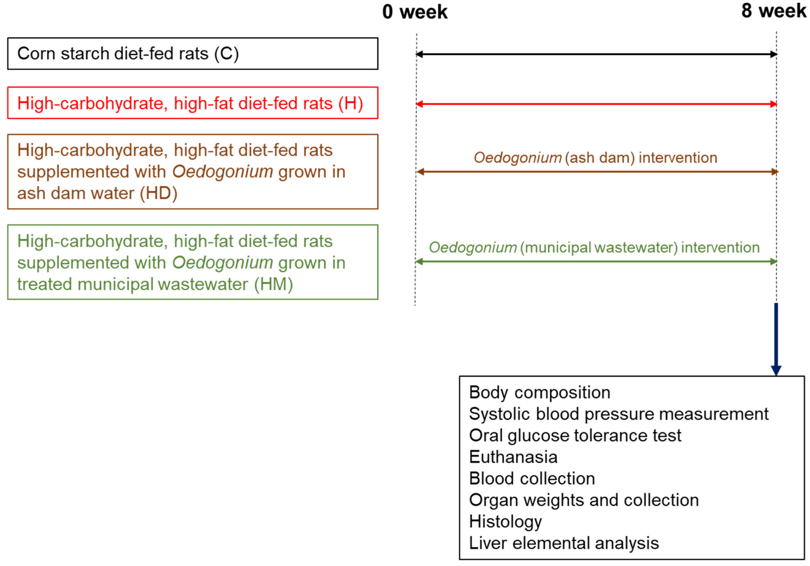
4.3. Studies in Live Rats

Male Wistar rats (8–9 weeks old, 330–340 g, n = 32) were obtained from the Animal Resources Centre, Murdoch, WA, Australia. Rats were randomly divided into four groups (Figure 3). Control groups were fed either a corn starch diet (C; n = 8) or a high-carbohydrate, high-fat diet (H; n = 8) [29]. Two groups were fed a high-carbohydrate, high-fat diet containing 5% dried biomass of freshwater macroalgae grown in ash dam water or treated municipal wastewater (HD and HM) to replace 5% water in these diets (n = 8 in each group). These diets were given to the rats for eight weeks. Drinking water for high-carbohydrate, high-fat diet-fed rats was supplemented with 25% fructose, whereas corn starch diet-fed rats were given drinking water without any additive. The composition of C and H diets is described in our previous studies [29,77]. All the rats were individually housed in temperature-controlled 12-h light/dark conditions and were given ad libitum access to food and water.
Body weight and intakes of food and water were measured daily for all rats. Abdominal circumferences were measured using a standard measuring tape under light sedation with Zoletil (tiletamine 10 mg/kg, zolazepam 10 mg/kg, i.p.; Virbac, Peakhurst, NSW, Australia) [29]. Energy intakes (including the values for D—14.77 kJ/g; M—17.66 kJ/g) and feed efficiency were calculated from daily food intakes [29]. Body composition was measured using dual-energy X-ray absorptiometry at the end of the protocol using a Norland XR36 DXA instrument (Norland Corp., Fort Atkinson, WI, USA) under light anaesthesia with Zoletil (tiletamine 10 mg/kg and zolazepam 10 mg/kg, i.p.) and Ilium Xylazil (xylazine 6 mg/kg, i.p.). Scans were analysed using the manufacturer’s recommended software for use in laboratory animals (Small Subject Analysis Software, version 2.5.3/1.3.1; Norland Corp.) [29].
Systolic blood pressures of rats were measured at 0 and 8 week periods of the protocol under sedation with Zoletil (tiletamine 10 mg/kg, zolazepam 10 mg/kg, i.p.), using an MLT1010 Piezo-Electric Pulse Transducer (ADInstruments, Sydney, NSW, Australia) and an inflatable tail cuff connected to a MLT844 Piezo-Electric Pressure Transducer (ADInstruments) and PowerLab data acquisition unit (ADInstruments) [29].
At eight weeks of the protocol, rats were deprived of food overnight for 12 h for oral glucose tolerance testing. During this food deprivation period, fructose-supplemented drinking water was replaced with normal drinking water. Oral glucose tolerance tests were performed after determining basal blood glucose concentrations in tail vein blood using Medisense Precision Q.I.D. glucose meters (Abbott Laboratories, Bedford, MA, USA). Rats were given a glucose load of 2 g/kg body weight as 40% glucose solution by oral gavage and blood glucose concentrations were measured at 0, 30, 60, 90 and 120 min after oral glucose administration [29]. Blood glucose concentrations over the period of 120 min were used to calculate the area under the curve.

4.4. Studies after Euthanasia

Rats were euthanised with Lethabarb (pentobarbitone sodium, 100 mg/kg, i.p.; Virbac). After the induction of euthanasia, heparin (200 IU; Sigma-Aldrich, Bayswater, Australia) was injected through the right femoral vein. The abdomen was then opened, and blood (~5 mL) was withdrawn from the abdominal aorta and collected into heparinised tubes. Blood was centrifuged at 5000× g for 15 min to obtain plasma. Plasma concentrations of total cholesterol and triglycerides and plasma activities of alanine transaminase and aspartate transaminase were determined using kits and controls supplied by Olympus using an Olympus analyser (AU 400, Tokyo, Japan) [29]. Nonesterified fatty acids in plasma were determined using a commercial kit (Wako, Osaka, Japan) [29].
After blood withdrawal, hearts were perfused to clear the blood and separated into left ventricle (with septum) and right ventricle and weighed. The liver, kidneys, spleen, retroperitoneal fat, epididymal fat and omental fat were removed separately and weighed. These organ weights were normalised against the tibial length at the time of organ removal and expressed as mg/mm of tibial length [29].
The heart, liver, ileum and colon were removed from the rats (n = 5 from each group) within five minutes after euthanasia and these tissues were fixed in 10% neutral buffered formalin. The samples were then dehydrated and embedded in paraffin wax. Thin sections (5 µm) of these tissues were cut and stained with hematoxylin and eosin to study infiltration of inflammatory cells. Heart tissues were also stained with picrosirius red to study collagen deposition. Liver tissues were stained for fat vacuoles with hematoxylin and eosin stain [29].
Livers collected after euthanasia were stored at −20 °C until analysis. Liver samples were thawed and homogenised using an automated homogeniser (Miltenyi Biotec, San Diego, CA, USA). Samples were then dried at 60 °C and used for quantification of 24 elements by the Advanced Analytical Centre at James Cook University using ICP/MS.

4.5. Statistical Analysis

Macroalgae composition values are presented as mean ± standard deviation (SD), n = 3 (replicate subsamples of the biomass used for the rat component were taken to characterise the biochemical properties, not for formal comparison between the two water sources). Values from rat measurements are presented as mean ± standard error of the mean (SEM), n = 8. Differences between the groups were determined by one-way analysis of variance. Variables with statistically significant differences were treated with Newman–Keuls post hoc test to compare all groups of animals. Statistical analyses were performed using GraphPad Prism version 9.4 for Windows. A p value of <0.05 was considered as statistically significant.

5. Conclusions

The freshwater macroalgae, Oedogonium, cultivated in ash dam water had a markedly different biochemical profile to that cultivated in treated municipal wastewater with elevated levels of metals and trace elements, including Al, Fe, V, Zn, Mn and As. These metals and trace elements contributed to the prevention of obesity and the symptoms of metabolic syndrome as measured in rats fed a high-carbohydrate, high-fat diet with Oedogonium cultivated in ash dam water. Biomass cultivated in treated municipal wastewater failed to prevent obesity but helped in improving other metabolic parameters. This study supports the capacity to cultivate freshwater macroalgae with targeted biochemical profiles in selected or modified water sources, such that the biomass provides nutraceutical benefits in mitigating obesity and the symptoms of metabolic syndrome.

Author Contributions

Conceptualisation, L.B., N.A.P. and S.K.P.; methodology, S.K.P., N.K.R.G., M.M., A.C., N.N. and D.R.; formal analysis, S.K.P., N.K.R.G., M.M., N.N. and N.A.P.; investigation, S.K.P., N.K.R.G., N.A.P., M.M., A.C., N.N. and D.R.; resources, L.B. and N.A.P.; writing—original draft preparation, S.K.P. and N.K.R.G.; writing—review and editing, S.K.P., N.A.P., M.M. and L.B.; supervision, L.B., S.K.P. and N.A.P.; project administration, S.K.P.; funding acquisition, L.B., S.K.P. and N.A.P. All authors have read and agreed to the published version of the manuscript.

Funding

This study was supported by Strategic Research Funding received from the University of Southern Queensland Research and Innovation Division (SRF-09) and Pacific Biotechnology (previously MBD Industries Ltd.) Research and Development program.

Institutional Review Board Statement

Experimental protocols were approved by the University of Southern Queensland Animal Ethics Committee under the guidelines of the National Health and Medical Research Council of Australia (approval number 14REA007).

Informed Consent Statement

Not applicable.

Data Availability Statement

The original data generated for the study are included in this article.

Acknowledgments

The authors would like to thank Brian Bynon, School of Veterinary Sciences, The University of Queensland, Gatton, for the plasma analyses. The authors also thank Pacific Biotechnologies Australia for providing the Oedogonium biomass from the Tarong Power station for this study and the MBD program (now Pacific Biotechnologies Australia) for the biomass composition and liver analysis.

Conflicts of Interest

The authors declare no conflict of interest. The funders had no role in the design of the study, in the collection, analyses, or interpretation of data, in the writing of the manuscript, or in the decision to publish the results.

References

  1. Mouritsen, O.G.; Rhatigan, P.; Cornish, M.L.; Critchley, A.T.; Pérez-Lloréns, J.L. Saved by seaweeds: Phyconomic contributions in times of crises. J. Appl. Phycol. 2021, 33, 443–458. [Google Scholar] [CrossRef] [PubMed]
  2. Pérez-Lloréns, J.L.; Mouritsen, O.G.; Rhatigan, P.; Cornish, M.L.; Critchley, A.T. Seaweeds in mythology, folklore, poetry, and life. J. Appl. Phycol. 2020, 32, 3157–3182. [Google Scholar] [CrossRef]
  3. Raja, K.; Kadirvel, V.; Subramaniyan, T. Seaweeds, an aquatic plant-based protein for sustainable nutrition—A review. Future Foods 2022, 5, 100142. [Google Scholar] [CrossRef]
  4. Wells, M.L.; Potin, P.; Craigie, J.S.; Raven, J.A.; Merchant, S.S.; Helliwell, K.E.; Smith, A.G.; Camire, M.E.; Brawley, S.H. Algae as nutritional and functional food sources: Revisiting our understanding. J. Appl. Phycol. 2017, 29, 949–982. [Google Scholar] [CrossRef]
  5. Brown, L.; Poudyal, H.; Panchal, S.K. Functional foods as potential therapeutic options for metabolic syndrome. Obes. Rev. 2015, 16, 914–941. [Google Scholar] [CrossRef]
  6. Granato, D.; Barba, F.J.; Bursać Kovačević, D.; Lorenzo, J.M.; Cruz, A.G.; Putnik, P. Functional foods: Product development, technological trends, efficacy testing, and safety. Annu. Rev. Food Sci. Technol. 2020, 11, 93–118. [Google Scholar] [CrossRef] [PubMed] [Green Version]
  7. O’Neill, S.; O’Driscoll, L. Metabolic syndrome: A closer look at the growing epidemic and its associated pathologies. Obes. Rev. 2015, 16, 1–12. [Google Scholar] [CrossRef] [Green Version]
  8. Kumar, S.A.; Magnusson, M.; Ward, L.C.; Paul, N.A.; Brown, L. Seaweed supplements normalise metabolic, cardiovascular and liver responses in high-carbohydrate, high-fat fed rats. Mar. Drugs 2015, 13, 788–805. [Google Scholar] [CrossRef] [PubMed]
  9. du Preez, R.; Majzoub, M.E.; Thomas, T.; Panchal, S.K.; Brown, L. Caulerpa lentillifera (sea grapes) improves cardiovascular and metabolic health of rats with diet-induced metabolic syndrome. Metabolites 2020, 10, 500. [Google Scholar] [CrossRef]
  10. du Preez, R.; Magnusson, M.; Majzoub, M.E.; Thomas, T.; Praeger, C.; Glasson, C.R.K.; Panchal, S.K.; Brown, L. Brown seaweed Sargassum siliquosum as an intervention for diet-induced obesity in male Wistar rats. Nutrients 2021, 13, 1754. [Google Scholar] [CrossRef]
  11. du Preez, R.; Paul, N.; Mouatt, P.; Majzoub, M.E.; Thomas, T.; Panchal, S.K.; Brown, L. Carrageenans from the red seaweed Sarconema filiforme attenuate symptoms of diet-induced metabolic syndrome in rats. Mar. Drugs 2020, 18, 97. [Google Scholar] [CrossRef] [PubMed] [Green Version]
  12. Wanyonyi, S.; du Preez, R.; Brown, L.; Paul, N.A.; Panchal, S.K. Kappaphycus alvarezii as a food supplement prevents diet-induced metabolic syndrome in rats. Nutrients 2017, 9, 1261. [Google Scholar] [CrossRef] [PubMed] [Green Version]
  13. John, O.D.; du Preez, R.; Panchal, S.K.; Brown, L. Tropical foods as functional foods for metabolic syndrome. Food Funct. 2020, 11, 6946–6960. [Google Scholar] [CrossRef]
  14. Food and Agriculture Organization of the United Nations. The State of the World’s Fisheries and Aquaculture 2020. Sustainability in Action; Food and Agriculture Organization of the United Nations: Rome, Italy, 2020; p. 244. Available online: https://www.fao.org/3/ca9229en/ca9229en.pdf (accessed on 22 August 2022).
  15. Duarte, C.M.; Gattuso, J.-P.; Hancke, K.; Gundersen, H.; Filbee-Dexter, K.; Pedersen, M.F.; Middelburg, J.J.; Burrows, M.T.; Krumhansl, K.A.; Wernberg, T.; et al. Global estimates of the extent and production of macroalgal forests. Glob. Ecol. Biogeogr. 2022, 31, 1422–1439. [Google Scholar] [CrossRef]
  16. Yun, J.-H.; Smith, V.H.; deNoyelles, F.J.; Roberts, G.W.; Stagg-Williams, S.M. Freshwater macroalgae as a biofuels feedstock: Mini-review and assessment of their bioenergy potential. Indus. Biotechnol. 2014, 10, 212–220. [Google Scholar] [CrossRef]
  17. Lawton, R.J.; de Nys, R.; Paul, N.A. Selecting reliable and robust freshwater macroalgae for biomass applications. PLoS ONE 2013, 8, e64168. [Google Scholar] [CrossRef]
  18. Arumugam, N.; Chelliapan, S.; Kamyab, H.; Thirugnana, S.; Othman, N.; Nasri, N.S. Treatment of wastewater using seaweed: A review. Int. J. Environ. Res. Public Health 2018, 15, 2851. [Google Scholar] [CrossRef] [PubMed] [Green Version]
  19. Deniz, F.; Ersanli, E.T. An ecofriendly approach for bioremediation of contaminated water environment: Potential contribution of a coastal seaweed community to environmental improvement. Int. J. Phytoremediat. 2018, 20, 256–263. [Google Scholar] [CrossRef]
  20. Kidgell, J.T.; de Nys, R.; Hu, Y.; Paul, N.A.; Roberts, D.A. Bioremediation of a complex industrial effluent by biosorbents derived from freshwater macroalgae. PLoS ONE 2014, 9, e94706. [Google Scholar] [CrossRef] [Green Version]
  21. Rajasulochana, P.; Preethy, V. Comparison on efficiency of various techniques in treatment of waste and sewage water—A comprehensive review. Resour.-Effic. Technol. 2016, 2, 175–184. [Google Scholar] [CrossRef]
  22. Han, W.; Clarke, W.; Pratt, S. Composting of waste algae: A review. Waste Manag. 2014, 34, 1148–1155. [Google Scholar] [CrossRef] [PubMed]
  23. Michalak, I.; Chojnacka, K.; Saeid, A. Plant growth biostimulants, dietary feed supplements and cosmetics formulated with supercritical CO2 algal extracts. Molecules 2017, 22, 66. [Google Scholar] [CrossRef]
  24. Neveux, N.; Nugroho, A.A.; Roberts, D.A.; Vucko, M.J.; de Nys, R. Selecting extraction conditions for the production of liquid biostimulants from the freshwater macroalga Oedogonium intermedium. J. Appl. Phycol. 2020, 32, 539–551. [Google Scholar] [CrossRef]
  25. Roberts, D.A.; Paul, N.A.; Bird, M.I.; de Nys, R. Bioremediation for coal-fired power stations using macroalgae. J. Environ. Manag. 2015, 153, 25–32. [Google Scholar] [CrossRef] [PubMed]
  26. Cole, A.J.; Neveux, N.; Whelan, A.; Morton, J.; Vis, M.; de Nys, R.; Paul, N.A. Adding value to the treatment of municipal wastewater through the intensive production of freshwater macroalgae. Algal Res. 2016, 20, 100–109. [Google Scholar] [CrossRef]
  27. Neveux, N.; Magnusson, M.; Mata, L.; Whelan, A.; de Nys, R.; Paul, N.A. The treatment of municipal wastewater by the macroalga Oedogonium sp. and its potential for the production of biocrude. Algal Res. 2016, 13, 284–292. [Google Scholar] [CrossRef]
  28. Roberts, D.A.; Shiels, L.; Tickle, J.; de Nys, R.; Paul, N.A. Bioremediation of aluminium from the waste water of a conventional water treatment plant using the freshwater macroalga Oedogonium. Water 2018, 10, 626. [Google Scholar] [CrossRef] [Green Version]
  29. Panchal, S.K.; Poudyal, H.; Iyer, A.; Nazer, R.; Alam, M.A.; Diwan, V.; Kauter, K.; Sernia, C.; Campbell, F.; Ward, L.; et al. High-carbohydrate, high-fat diet-induced metabolic syndrome and cardiovascular remodeling in rats. J. Cardiovasc. Pharmacol. 2011, 57, 611–624. [Google Scholar] [CrossRef]
  30. Swinburn, B.A.; Kraak, V.I.; Allender, S.; Atkins, V.J.; Baker, P.I.; Bogard, J.R.; Brinsden, H.; Calvillo, A.; De Schutter, O.; Devarajan, R.; et al. The global syndemic of obesity, undernutrition, and climate change: The Lancet Commission report. Lancet 2019, 393, 791–846. [Google Scholar] [CrossRef]
  31. NCD Risk Factor Collaboration (NCD-RisC). Trends in adult body-mass index in 200 countries from 1975 to 2014: A pooled analysis of 1698 population-based measurement studies with 19.2 million participants. Lancet 2016, 387, 1377–1396. [Google Scholar] [CrossRef]
  32. Kearney, J. Food consumption trends and drivers. Philos. Trans. R. Soc. Lond. B Biol. Sci. 2010, 365, 2793–2807. [Google Scholar] [CrossRef] [PubMed] [Green Version]
  33. Bray, G.A.; Kim, K.K.; Wilding, J.P.H.; World Obesity, F. Obesity: A chronic relapsing progressive disease process. A position statement of the World Obesity Federation. Obes. Rev. 2017, 18, 715–723. [Google Scholar] [CrossRef] [PubMed] [Green Version]
  34. Cunnane, S.C.; Crawford, M.A. Energetic and nutritional constraints on infant brain development: Implications for brain expansion during human evolution. J. Hum. Evol. 2014, 77, 88–98. [Google Scholar] [CrossRef] [PubMed]
  35. Lange, K.W.; Hauser, J.; Nakamura, Y.; Kanaya, S. Dietary seaweeds and obesity. Food Sci. Hum. Wellness 2015, 4, 87–96. [Google Scholar] [CrossRef] [Green Version]
  36. Wan-Loy, C.; Siew-Moi, P. Marine algae as a potential source for anti-obesity agents. Mar. Drugs 2016, 14, 222. [Google Scholar] [CrossRef] [Green Version]
  37. Tipnee, S.; Ramaraj, R.; Unpaprom, Y. Nutritional evaluation of edible freshwater green macroalga Spirogyra varians. Emer. Life Sci. Res. 2015, 1, 1–7. [Google Scholar]
  38. Santos-Marcos, J.A.; Perez-Jimenez, F.; Camargo, A. The role of diet and intestinal microbiota in the development of metabolic syndrome. J. Nutr. Biochem. 2019, 70, 1–27. [Google Scholar] [CrossRef]
  39. Lopez-Santamarina, A.; Miranda, J.M.; Mondragon, A.D.C.; Lamas, A.; Cardelle-Cobas, A.; Franco, C.M.; Cepeda, A. Potential use of marine seaweeds as prebiotics: A review. Molecules 2020, 25, 1004. [Google Scholar] [CrossRef] [Green Version]
  40. Gotteland, M.; Riveros, K.; Gasaly, N.; Carcamo, C.; Magne, F.; Liabeuf, G.; Beattie, A.; Rosenfeld, S. The pros and cons of using algal polysaccharides as prebiotics. Front. Nutr. 2020, 7, 163. [Google Scholar] [CrossRef]
  41. Arata, P.X.; Alberghina, J.; Confalonieri, V.; Errea, M.I.; Estevez, J.M.; Ciancia, M. Sulfated polysaccharides in the freshwater green macroalga Cladophora surera not linked to salinity adaptation. Front. Plant Sci. 2017, 8, 1927. [Google Scholar] [CrossRef] [Green Version]
  42. Bhattacharya, P.T.; Misra, S.R.; Hussain, M. Nutritional aspects of essential trace elements in oral health and disease: An extensive review. Scientifica 2016, 2016, 5464373. [Google Scholar] [CrossRef] [PubMed]
  43. Chen, Q.; Pan, X.D.; Huang, B.F.; Han, J.L. Distribution of metals and metalloids in dried seaweeds and health risk to population in southeastern China. Sci. Rep. 2018, 8, 3578. [Google Scholar] [CrossRef] [PubMed] [Green Version]
  44. Shamshad, I.; Khan, S.; Waqas, M.; Asma, M.; Nawab, J.; Gul, N.; Raiz, A.; Li, G. Heavy metal uptake capacity of fresh water algae (Oedogonium westti) from aqueous solution: A mesocosm research. Int. J. Phytoremediat. 2016, 18, 393–398. [Google Scholar] [CrossRef] [PubMed]
  45. Ji, L.; Xie, S.; Feng, J.; Li, Y.; Chen, L. Heavy metal uptake capacities by the common freshwater green alga Cladophora fracta. J. Appl. Phycol. 2012, 24, 979–983. [Google Scholar] [CrossRef]
  46. Lewicki, S.; Zdanowski, R.; Krzyzowska, M.; Lewicka, A.; Debski, B.; Niemcewicz, M.; Goniewicz, M. The role of chromium III in the organism and its possible use in diabetes and obesity treatment. Ann. Agric. Environ. Med. 2014, 21, 331–335. [Google Scholar] [CrossRef]
  47. Rehder, D. The future of/for vanadium. Dalton Trans. 2013, 42, 11749–11761. [Google Scholar] [CrossRef]
  48. Rehder, D. The role of vanadium in biology. Metallomics 2015, 7, 730–742. [Google Scholar] [CrossRef] [Green Version]
  49. Tinkov, A.A.; Popova, E.V.; Polyakova, V.S.; Kwan, O.V.; Skalny, A.V.; Nikonorov, A.A. Adipose tissue chromium and vanadium disbalance in high-fat fed Wistar rats. J. Trace Elem. Med. Biol. 2015, 29, 176–181. [Google Scholar] [CrossRef]
  50. Tinkov, A.A.; Sinitskii, A.I.; Popova, E.V.; Nemereshina, O.N.; Gatiatulina, E.R.; Skalnaya, M.G.; Skalny, A.V.; Nikonorov, A.A. Alteration of local adipose tissue trace element homeostasis as a possible mechanism of obesity-related insulin resistance. Med. Hypotheses 2015, 85, 343–347. [Google Scholar] [CrossRef]
  51. Chimienti, F. Zinc, pancreatic islet cell function and diabetes: New insights into an old story. Nutr. Res. Rev. 2013, 26, 1–11. [Google Scholar] [CrossRef] [Green Version]
  52. Koliaki, C.; Katsilambros, N. Dietary sodium, potassium, and alcohol: Key players in the pathophysiology, prevention, and treatment of human hypertension. Nutr. Rev. 2013, 71, 402–411. [Google Scholar] [CrossRef] [PubMed]
  53. Panchal, S.K.; Wanyonyi, S.; Brown, L. Selenium, vanadium, and chromium as micronutrients to improve metabolic syndrome. Curr. Hypertens. Rep. 2017, 19, 10. [Google Scholar] [CrossRef] [PubMed]
  54. Tuzcu, M.; Sahin, N.; Orhan, C.; Agca, C.A.; Akdemir, F.; Tuzcu, Z.; Komorowski, J.; Sahin, K. Impact of chromium histidinate on high fat diet induced obesity in rats. Nutr. Metab. 2011, 8, 28. [Google Scholar] [CrossRef] [PubMed] [Green Version]
  55. Alexanian, I.; Parissis, J.; Farmakis, D.; Pantziou, C.; Ikonomidis, I.; Paraskevaidis, I.; Ioannidou, S.; Sideris, A.; Kremastinos, D.; Lekakis, J.; et al. Selenium contributes to myocardial injury and cardiac remodeling in heart failure. Int. J. Cardiol. 2014, 176, 272–273. [Google Scholar] [CrossRef]
  56. Chaabane, M.; Tir, M.; Hamdi, S.; Boudawara, O.; Jamoussi, K.; Boudawara, T.; Ghorbel, R.E.; Zeghal, N.; Soudani, N. Improvement of heart redox states contributes to the beneficial effects of selenium against penconazole-induced cardiotoxicity in adult rats. Biol. Trace Elem. Res. 2016, 169, 261–270. [Google Scholar] [CrossRef] [PubMed]
  57. Metelo, A.M.; Perez-Carro, R.; Castro, M.M.; Lopez-Larrubia, P. VO(dmpp)2 normalizes pre-diabetic parameters as assessed by in vivo magnetic resonance imaging and spectroscopy. J. Inorg. Biochem. 2012, 115, 44–49. [Google Scholar] [CrossRef] [Green Version]
  58. Park, S.J.; Youn, C.K.; Hyun, J.W.; You, H.J. The anti-obesity effect of natural vanadium-containing Jeju ground water. Biol. Trace Elem. Res. 2013, 151, 294–300. [Google Scholar] [CrossRef]
  59. Nigra, A.E.; Olmedo, P.; Grau-Perez, M.; O’Leary, R.; O’Leary, M.; Fretts, A.M.; Umans, J.G.; Best, L.G.; Francesconi, K.A.; Goessler, W.; et al. Dietary determinants of inorganic arsenic exposure in the Strong Heart Family Study. Environ. Res. 2019, 177, 108616. [Google Scholar] [CrossRef]
  60. Spratlen, M.J.; Grau-Perez, M.; Best, L.G.; Yracheta, J.; Lazo, M.; Vaidya, D.; Balakrishnan, P.; Gamble, M.V.; Francesconi, K.A.; Goessler, W.; et al. The association of arsenic exposure and arsenic metabolism with the metabolic syndrome and its individual components: Prospective evidence from the Strong Heart Family Study. Am. J. Epidemiol. 2018, 187, 1598–1612. [Google Scholar] [CrossRef] [Green Version]
  61. Moon, K.A.; Oberoi, S.; Barchowsky, A.; Chen, Y.; Guallar, E.; Nachman, K.E.; Rahman, M.; Sohel, N.; D’Ippoliti, D.; Wade, T.J.; et al. A dose-response meta-analysis of chronic arsenic exposure and incident cardiovascular disease. Int. J. Epidemiol. 2018, 47, 1013. [Google Scholar] [CrossRef]
  62. Khairul, I.; Wang, Q.Q.; Jiang, Y.H.; Wang, C.; Naranmandura, H. Metabolism, toxicity and anticancer activities of arsenic compounds. Oncotarget 2017, 8, 23905–23926. [Google Scholar] [CrossRef] [PubMed]
  63. Taylor, V.; Goodale, B.; Raab, A.; Schwerdtle, T.; Reimer, K.; Conklin, S.; Karagas, M.R.; Francesconi, K.A. Human exposure to organic arsenic species from seafood. Sci. Total Environ. 2017, 580, 266–282. [Google Scholar] [CrossRef]
  64. Shahidi, F.; Ambigaipalan, P. Omega-3 polyunsaturated fatty acids and their health benefits. Annu. Rev. Food Sci. Technol. 2018, 9, 345–381. [Google Scholar] [CrossRef] [PubMed]
  65. Kumar, M.; Pal, N.; Sharma, P.; Kumawat, M.; Sarma, D.K.; Nabi, B.; Verma, V.; Tiwari, R.R.; Shubham, S.; Arjmandi, B.; et al. Omega-3 fatty acids and their interaction with the gut microbiome in the prevention and amelioration of type-2 diabetes. Nutrients 2022, 14, 1723. [Google Scholar] [CrossRef] [PubMed]
  66. Eggersdorfer, M.; Wyss, A. Carotenoids in human nutrition and health. Arch. Biochem. Biophys. 2018, 652, 18–26. [Google Scholar] [CrossRef]
  67. Rivera-Madrid, R.; Carballo-Uicab, V.M.; Cárdenas-Conejo, Y.; Aguilar-Espinosa, M.; Siva, R. Overview of carotenoids and beneficial effects on human health. In Carotenoids: Properties, Processing and Applications; Galanakis, C.M., Ed.; Academic Press: Cambridge, MA, USA, 2020; pp. 1–40. [Google Scholar] [CrossRef]
  68. Lawton, R.J. Oedogonium sp. Tsv2 Internal Transcribed Spacer 1, Partial Sequence; 5.8S Ribosomal RNA Gene, Complete Sequence; and Internal Transcribed Spacer 2, Partial Sequence. GenBank: KF606977.1. 2014. Available online: https://www.ncbi.nlm.nih.gov/nuccore/KF606977 (accessed on 14 October 2022).
  69. Lawton, R.J.; de Nys, R.; Skinner, S.; Paul, N.A. Isolation and identification of Oedogonium species and strains for biomass applications. PLoS ONE 2014, 9, e90223. [Google Scholar] [CrossRef] [Green Version]
  70. Lawton, R.J. Oedogonium sp. Tar1 Internal Transcribed Spacer 1, Partial Sequence; 5.8S Ribosomal RNA Gene, Complete Sequence; and Internal Transcribed Spacer 2, Partial Sequence. GenBank: KF606974.1. 2014. Available online: https://www.ncbi.nlm.nih.gov/nuccore/KF606974 (accessed on 14 October 2022).
  71. Folch, J.; Lees, M.; Sloane Stanley, G.H. A simple method for the isolation and purification of total lipides from animal tissues. J. Biol. Chem. 1957, 226, 497–509. [Google Scholar] [CrossRef]
  72. Gosch, B.J.; Magnusson, M.; Paul, N.A.; de Nys, R. Total lipid and fatty acid composition of seaweeds for the selection of species for oil-based biofuel and bioproducts. GCB Bioenergy 2012, 4, 919–930. [Google Scholar] [CrossRef] [Green Version]
  73. Channiwala, S.A.; Parikh, P.P. A unified correlation for estimating HHV of solid, liquid and gaseous fuels. Fuel 2002, 81, 1051–1063. [Google Scholar] [CrossRef]
  74. Saunders, R.J.; Paul, N.A.; Hu, Y.; de Nys, R. Sustainable sources of biomass for bioremediation of heavy metals in waste water derived from coal-fired power generation. PLoS ONE 2012, 7, e36470. [Google Scholar] [CrossRef]
  75. Ritchie, R.J. Consistent sets of spectrophotometric chlorophyll equations for acetone, methanol and ethanol solvents. Photosynth. Res. 2006, 89, 27–41. [Google Scholar] [CrossRef] [PubMed]
  76. Weast, R.C. CRC—Handbook of Chemistry and Physics, 56th ed.; Chemical Rubber Publishing Co.: Cleveland, OH, USA, 1975–1976. [Google Scholar]
  77. Poudyal, H.; Panchal, S.K.; Waanders, J.; Ward, L.; Brown, L. Lipid redistribution by α-linolenic acid-rich chia seed inhibits stearoyl-CoA desaturase-1 and induces cardiac and hepatic protection in diet-induced obese rats. J. Nutr. Biochem. 2012, 23, 153–162. [Google Scholar] [CrossRef] [PubMed]
Figure 1. Effects of Oedogonium grown in ash dam water and treated municipal wastewater on (A) body weight, (B) whole-body fat and lean mass, (C) total abdominal fat pads, (D) blood glucose AUC during glucose tolerance test, (E) plasma lipid profile and (F) systolic blood pressure. Values are presented as means ± SEM (n = 8 rats per group). Means without a common letter are significantly different (a, b or c; p < 0.05). C, corn starch diet-fed rats; H, high-carbohydrate, high-fat diet-fed rats; HD, high-carbohydrate, high-fat diet-fed rats supplemented with Oedogonium grown in ash dam water; HM, high-carbohydrate, high-fat diet-fed rats supplemented with Oedogonium grown in treated municipal wastewater; AUC, area under the curve; NEFA, non-esterified fatty acids.
Ijms 23 13811 g001
Figure 2. Effects of Oedogonium grown in ash dam water and treated municipal wastewater on inflammation and fibrosis in the heart, colon and ileum structure of rats (n = 4 rats per group). Hematoxylin and eosin staining of the liver showing inflammatory cells (in, arrow head) and fat vacuoles (fv, arrow head) in C (A,E); H (B,F); HD (C,G) and HM (D,H) rats. Hematoxylin and eosin staining and picrosirius red staining of the left ventricle showing inflammatory cells (in, arrow head) and fibrosis (fi, arrow head), respectively, in C (I,M), H (J,N), HD (K,O) and HM (L,P) rats. Hematoxylin and eosin staining of the colon and ileum showing inflammatory cells in C (Q,U), H (R,V), HD (S,W) and HM (T,X) rats. Scale bar, 100 μm. C, corn starch diet-fed rats; H, high-carbohydrate, high-fat diet-fed rats; HD, high-carbohydrate, high-fat diet-fed rats supplemented with Oedogonium grown in ash dam water; and HM, high-carbohydrate, high-fat diet-fed rats supplemented with Oedogonium grown in treated municipal wastewater.
Ijms 23 13811 g002
Figure 3. Study design to characterise responses to Oedogonium biomass in rats with diet-induced metabolic syndrome.
Ijms 23 13811 g003
Table 1. Elemental and macronutrient composition of macroalgae.
Oedogonium Cultivated in Ash Dam Water (D)Oedogonium Cultivated in Treated
Municipal Wastewater (M)
Elements and proximate composition (% dry weight)
C32.41 ± 0.2938.90 ± 0.05
H5.31 ± 0.146.09 ± 0.01
O19.64 ± 0.02424.03 ± 1.68
N5.15 ± 0.025.37 ± 0.11
S0.33 ± 0.040.30 ± 0.015
F0.035 ± 0.00020.0006 ± 0.0
Cl0.209 ± 0.0020.814 ± 0.012
Br0.0032 ± 0.00010.0021 ± 0.00005
I0.005 ± 0.0001<0.0016
Moisture5.16 ± 0.356.41 ± 1.38
Ash32.00 ± 0.0618.90 ± 0.20
Protein (sum of amino acids)21.4423.87
Total lipids3.40 ± 0.254.82 ± 0.24
Carbohydrate (by difference)37.9946.01
Metals, non-metals and metalloids (mg/kg dry weight)
Al1315.0 ± 35.4276.5 ± 2.1
As99.95 ± 1.491.26 ± 0.014
B9.61 ± 0.167.63 ± 0.09
Ba209.5 ± 0.7177.85 ± 0.50
Ca9010 ± 14114,450 ± 212
Cd2.20 ± 0.020.11 ± 0.009
Co5.75 ± 0.0990.84 ± 0.008
Cr16.35 ± 0.0711.32 ± 0.04
Cu48.70 ± 0.8513.35 ± 0.21
Fe14,500 ± 141423.5 ± 6.4
Hg<0.5<0.5
K10,550 ± 7128,400 ± 1839
Mg2485 ± 356165 ± 35
Mn737.0 ± 9.9184.0 ± 2.8
Mo10.10 ± 0.000.82 ± 0.38
Na1550 ± 141990 ± 14
Ni42.60 ± 0.572.05 ± 0.02
P10,900 ± 013,900 ± 141
Pb0.83 ± 0.0162.15 ± 0.134
S3975 ± 2193080 ± 141
Se13.60 ± 0.28<1
Sr124.50 ± 0.71180.5 ± 0.71
V723.0 ± 4.240.82 ± 0.02
Zn258.0 ± 4.2442.15 ± 1.91
Fatty acids (% of total fatty acids)
C14:09.08 ± 0.350.91 ± 0.01
C15:00.44 ± 0.020.12 ± 0.01
C16:050.84 ± 0.2726.50 ± 0.09
C16:1n-722.90 ± 0.384.69 ± 0.13
C16:2n-60.11 ± 0.060.94 ± 0.04
C17:00.26 ± 0.090.32 ± 0.04
C16:3n-30.59 ± 0.3818.28 ± 0.30
C18:01.23 ± 0.040.80 ± 0.01
C18:1n-95.24 ± 0.211.75 ± 0.04
C18:2n-61.90 ± 0.074.59 ± 0.04
C18:3n-6ND2.97 ± 0.10
C18:3n-35.63 ± 0.0530.82 ± 0.18
C18:4n-3ND3.81 ± 0.05
C20:1n-90.62 ± 0.05ND
C20:2n-61.20 ± 0.180.45 ± 0.10
C20:3n-6ND1.99 ± 0.07
C22:0ND1.05 ± 0.06
Total SFA61.85 ± 0.5429.71 ± 0.06
Total MUFA28.75 ± 0.416.44 ± 0.12
Total PUFA9.40 ± 0.4963.85 ± 0.17
Total n-3 PUFA6.22 ± 0.3552.90 ± 0.40
Total n-6 PUFA3.18 ± 0.2610.95 ± 0.26
n-3:n-65.63 ± 0.0530.82 ± 0.18
Dietary fibre (% dry weight)
Total dietary fibre23.835.3
Insoluble fibre21.434.5
Vitamins
α-carotene (μg/100 g)<5<5
Ascorbic acid (mg/100 g)<1<1
β-carotene (μg/100 g)3206400
Vitamin K1 (μg/100 g)3712
Vitamin B12 (μg/100 g)218634
Chlorophyll (% dry weight)
Chlorophyll a0.20 ± 0.010.59 ± 0.01
Chlorophyll b0.50 ± 0.011.43 ± 0.04
Amino acids (mg/g dry weight)
Histidine3.74.6
Serine10.512.3
Arginine14.514.3
Glycine12.613.0
Aspartic acid24.828.7
Glutamic acid28.232.1
Threonine11.913.2
Alanine15.116.5
Proline10.312.0
Lysine12.915.0
Tyrosine8.39.6
Methionine4.04.1
Valine14.315.9
Isoleucine10.911.3
Leucine19.921.9
Phenylalanine12.514.2
Values are mean ± SD, n = 3 for each group where replicate samples were analysed. Three subsamples of both macroalgae biomass used in the rat feeding trial were taken for biochemical analysis. Biomass was milled and mixed thoroughly prior to analysis and use in the feeding trial. Standard deviation for each analysis is provided to inform the consistency of the variable. No statistical comparisons were made between the biomass as the subsamples were not independent. SFA, saturated fatty acids; MUFA, monounsaturated fatty acids; PUFA, polyunsaturated fatty acids; ND, not detected.
Table 2. Effects of macroalgae on metabolic, tissue and biochemical variables.
VariablesCHHDHM
Body weight gain, g17.0 ± 7.0 c81.0 ± 5.0 b−1.0 ± 5.6 c124.0 ± 6.6 a
Food intake, g/day36.7 ± 1.3 a21.7 ± 0.4 b22.8 ± 1.5 b22.6 ± 0.4 b
Water intake, g/day28.9 ± 2.520.3 ± 1.624.2 ± 1.826.4 ± 1.3
Energy intake, kJ/day411.6 ± 14.6 b465.7 ± 7.6 a515.6 ± 28.5 a524.8 ± 10.0 a
Feed efficiency, g/kJ0.04 ± 0.02 c0.17 ± 0.01 b0.00 ± 0.01 d0.24 ± 0.01 a
Abdominal circumference, cm18.2 ± 0.3 c21.1 ± 0.2 b18.6 ± 0.3 c22.4 ± 0.2 a
Bone mineral density, g/cm20.163 ± 0.003 ab0.170 ± 0.004 a0.158 ± 0.003 b0.172 ± 0.002 a
Bone mineral content, g10.0 ± 0.3 b11.8 ± 0.3 a9.3 ± 0.3 b12.6 ± 0.2 a
Basal blood glucose concentrations (week 8), mmol/L4.1 ± 0.3 b5.2 ± 0.2 a4.5 ± 0.2 b4.3 ± 0.1 b
Blood glucose 120 min (week 8), mmol/L4.7 ± 0.2 b6.2 ± 0.4 a4.7 ± 0.4 b4.5 ± 0.2 b
Retroperitoneal fat, mg/mm154.0 ± 13.3 c253.8 ± 26.7 b130.1 ± 10.6 c338.9 ± 24.9 a
Epididymal fat, mg/mm89.7 ± 8.1 c131.2 ± 10.5 b70.4 ± 3.6 c182.5 ± 15.3 a
Omental fat, mg/mm108.3 ± 11.2 b169.6 ± 8.4 a120.6 ± 9.1 b194.4 ± 9.9 a
Left ventricle + septum, mg/mm18.6 ± 0.7 b20.0 ± 0.8 ab19.2 ± 0.9 b22.1 ± 0.7 a
Right ventricle, mg/mm4.25 ± 0.194.08 ± 0.463.40 ± 0.224.38 ± 0.32
Liver, mg/mm223 ± 10 b279 ± 4 a323 ± 19 a323 ±13 a
Kidneys, mg/mm47.5 ± 1.8 c55.2 ± 1.1 b58.4 ± 3.2 ab63.6 ± 1.8 a
Spleen, mg/mm14.6 ± 0.7 b16.9 ± 0.9 b15.9 ± 0.6 b20.2 ± 1.0 a
Plasma alanine transaminase activity, U/L31.5 ± 6.639.5 ± 3.247.0 ± 3.031.0 ± 2.0
Plasma aspartate transaminase activity, U/L82.0 ± 11.275.5 ± 6.872.0 ± 3.065.0 ± 4.0
Values are mean ± SEM and n = 8 for each group. Mean values within a row with unlike superscript letters are significantly different (a, b, c or d; p < 0.05). C, corn starch diet-fed rats; H, high-carbohydrate, high-fat diet-fed rats; HD, high-carbohydrate, high-fat diet-fed rats supplemented with Oedogonium grown in ash dam water; HM, high-carbohydrate, high-fat diet-fed rats supplemented with Oedogonium grown in treated municipal wastewater.
Table 3. Effects of Oedogonium grown in ash dam water and treated municipal wastewater on metal composition in the liver.
Metals (mg/kg Dry Weight)CHHDHM
Al1.99 ± 0.89<0.50.53 ± 0.241.93 ± 0.86
As4.94 ± 2.212.60 ± 1.1617.34 ± 7.762.53 ± 1.13
B28.20 ± 12.61 a0.24 ± 0.11 b11.62 ± 5.20 ab4.36 ± 1.95 ab
Ba<0.10.08 ± 0.030.06 ± 0.030.11 ± 0.05
Ca135 ± 60129 ± 58108 ± 48108 ± 48
Cd<0.05<0.05<0.05<0.05
Co0.05 ± 0.02<0.10.21 ± 0.100.07 ± 0.03
Cr0.25 ± 0.110.18 ± 0.080.16 ± 0.070.14 ± 0.06
Cu15.24 ± 6.8216.55 ± 7.4011.92 ± 5.3313.92 ± 6.23
Fe781 ± 349543 ± 243762 ± 341526 ± 235
Hg2.67 ± 1.191.75 ± 0.781.62 ± 0.721.81 ± 0.81
K10,378 ± 464110,416 ± 46588622 ± 38568197 ± 3666
Mg717 ± 321714 ± 319611 ± 273655 ± 293
Mn6.24 ± 2.795.49 ± 2.465.57 ± 2.496.37 ± 2.85
Mo0.87 ± 0.391.03 ± 0.461.00 ± 0.450.96 ± 0.43
Na3052 ± 13652800 ± 12521373 ± 6141143 ± 511
Ni0.08 ± 0.030.05 ± 0.020.08 ± 0.040.07 ± 0.03
P9048 ± 40469342 ± 41787026 ± 31427590 ± 3394
Pb0.08 ± 0.040.07 ± 0.030.07 ± 0.030.07 ± 0.03
S5232 ± 23405146 ± 23014060 ± 18164218 ± 1886
Se1.76 ± 0.791.81 ± 0.812.99 ± 1.342.27 ±1.01
Sr0.12 ± 0.050.18 ± 0.080.22 ± 0.100.19 ± 0.08
V0.06 ± 0.03 b0.09 ± 0.04 b4.07 ± 1.82 a0.18 ± 0.08 b
Zn66.5 ± 29.769.7 ± 31.257.3 ± 25.666.3 ± 29.7
Values are mean ± SEM and n = 8 for each group. Mean values within a row with unlike superscript letters are significantly different (a or b; p < 0.05). C, corn starch diet-fed rats; H, high-carbohydrate, high-fat diet-fed rats; HD, high-carbohydrate, high-fat diet-fed rats supplemented with Oedogonium grown in ash dam water; HM, high-carbohydrate, high-fat diet-fed rats supplemented with Oedogonium grown in treated municipal wastewater.
Publisher’s Note: MDPI stays neutral with regard to jurisdictional claims in published maps and institutional affiliations.

