Article

Detection of the Omicron SARS-CoV-2 Lineage and Its BA.1 Variant with Multiplex RT-qPCR

by
Nikita D. Yolshin
1,
Andrey B. Komissarov
1,
Kirill V. Varchenko
1,
Tamila D. Musaeva
1,
Artem V. Fadeev
1 and
Dmitry A. Lioznov
1,2,*
1
Smorodintsev Research Institute of Influenza, 197376 Saint Petersburg, Russia
2
Department of Infectious Diseases and Epidemiology, First Pavlov State Medical University, 197022 Saint Petersburg, Russia
*
Author to whom correspondence should be addressed.
Int. J. Mol. Sci. 2022, 23(24), 16153; https://doi.org/10.3390/ijms232416153
Submission received: 22 November 2022 / Revised: 14 December 2022 / Accepted: 15 December 2022 / Published: 18 December 2022
(This article belongs to the Special Issue Genomic Variation and Epidemiology of SARS-CoV-2)

Abstract

:
Whole genome sequencing (WGS) is considered the best instrument to track both virus evolution and the spread of new, emerging variants. However, WGS still does not allow the analysis of as many samples as qPCR does. Epidemiological and clinical research needs to develop advanced qPCR methods to identify emerging variants of SARS-CoV-2 while collecting data on their spreading in a faster and cheaper way, which is critical for introducing public health measures. This study aimed at designing a one-step RT-qPCR assay for multiplex detection of the Omicron lineage and providing additional data on its subvariants in clinical samples. The RT-qPCR assay demonstrated high sensitivity and specificity on multiple SARS-CoV-2 variants and was cross-validated by WGS.

1. Introduction

The global pandemic caused by severe acute respiratory syndrome coronavirus 2 (SARS-CoV-2) started in 2019. More than 640 million people have been infected and have suffered from the coronavirus disease 2019 (COVID-19) to date (November 2022) [1]. The virus, which killed more than 6,600,000 people in two years, continues to evolve and spread throughout the globe [2] despite various non-pharmaceutical interventions and the availability of plenty of vaccines.
Several months after SARS-CoV-2 appeared, new distinct lineages were detected. One after the other, lineages emerged and replaced previous variants. Practically worldwide, the “wave” of the Delta variant put an end to the Alpha variant dominance, only to be replaced by Omicron BA.1 in December 2021–January 2022. However, BA.1 domination did not persist and was replaced by BA.2 in March 2022. New variants causing “epidemic waves” often demonstrate higher transmission rates and decreased susceptibility to neutralization by pre-existing antibodies against SARS-CoV-2 due to previous infection or vaccination [3]. Omicron (B.1.1.529) was designated a variant of concern (VOC) by WHO on 26 November 2021. Omicron shares some mutations with the Delta variant, including D614G and T478K, which facilitate ACE2 binding and fusion with human cells [4,5]. Omicron has also acquired new mutations that contribute to immune evasion by masking highly immunogenic sites. Thus, the Omicron variant poses an increased risk of reinfection with SARS-CoV-2 as compared to other VOCs because it is able to resist neutralization by antibodies [6,7] or sera from vaccinated people [8]. The Omicron lineage includes several subvariants with strikingly different genetic characteristics. The most widespread subvariants are BA.1 and BA.2, with more than 2.3 and 1.1 million genomes, respectively, shared via the Global Initiative on Sharing All Influenza Data (GISAID) [9] by May 2022.
Quantitative (q) PCR plays a key role in the rapid and easily scalable assessment of SARS-CoV-2 variants’ spread, e.g., in outbreak scenarios. Although next-generation sequencing (NGS) is performed routinely for surveillance purposes, it is limited in sample number and, hence, less informative. It is of special concern in the case of low- and middle-income countries and other limited resource settings where sequencing capacity is low [10]. In the first assessment of the Alpha variant (B.1.1.7), population-wide frequency was performed by analyzing so-called S gene target failure (SGTF) in widely used commercial qPCR assays; false negatives for the S gene target occasionally proved to be a good proxy estimate of the B.1.1.7 spread. SGTF was found to be a reliable marker of Alpha after validation with whole-genome sequencing (WGS) [11]. SGTF was also used for Omicron detection [12]. However, it is not a specific test since it identifies other lineages carrying the 69/70 deletion and some Omicron variants do not carry this mutation.
qPCR assays for tracking the emerging variants of SARS-CoV-2 make surveillance cheaper and faster, which is critical for promptly implementing public health measures. qPCR relies on detecting unique mutations as a sign of emerging variants. The surveillance of SARS-CoV-2 evolution has been focused on the detection of nucleotide substitutions. qPCR assays could be targeted at single nucleotide polymorphisms (SNP), but this application would require highly specific enzymes and chemistry that would be too expensive for epidemiological studies. SNPs can be detected by high-resolution melting (HRM) technology. However, the high mutability of viral genomes makes HRM too sensitive to emerging mutations, thus making it hard to interpret [13].
Insertion-deletion polymorphisms (indels) would make a more feasible qPCR target, allowing for less laborious assay development and optimization. Increased numbers of indels seem to mediate immune escape and growing population resistance due to vaccination and previous infections. Indels occur most frequently in the Spike protein but are also found in others, especially those involved in interactions with the host immune system [14]. The Omicron lineage (BA*) was found to have a unique deletion that proved to be a good target for its detection, while the BA.1 variant sequence also contains unique lineage-specific insertion.
Here, we report on a novel multiplex RT-qPCR test which includes (1) a generic assay detecting all known SARS-CoV-2 variants, (2) an assay targeting all subvariants of the Omicron lineage, (3) an assay specific to BA.1, labeling the non-BA.1 variants as drop-outs, and (4) internal control reaction. The multiplex RT-qPCR assay simultaneously detects any SARS-CoV-2 and discriminates the Omicron lineage and its BA.1 SARS-CoV-2 subvariant in RNA samples in a single reaction, thus simplifying testing and reducing test costs.

2. Results

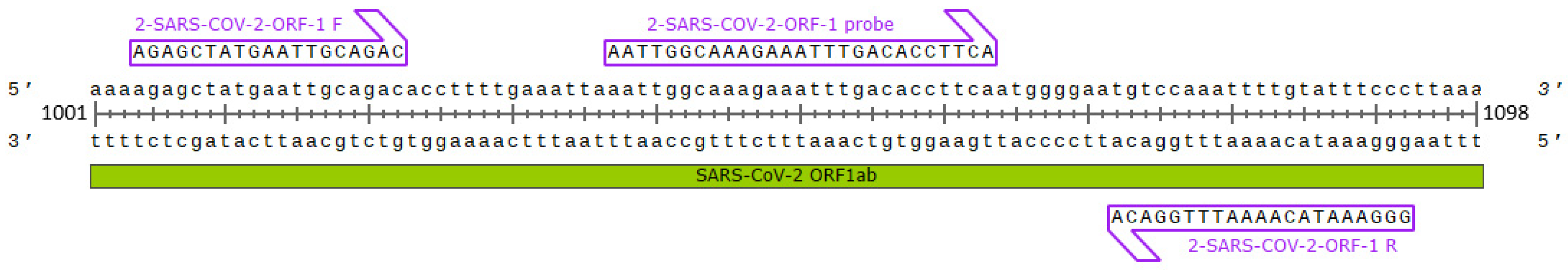
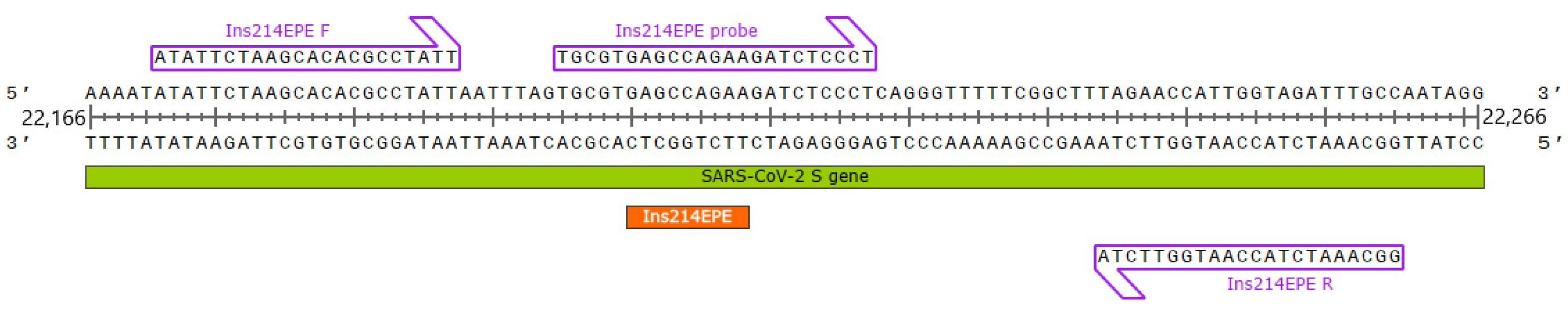
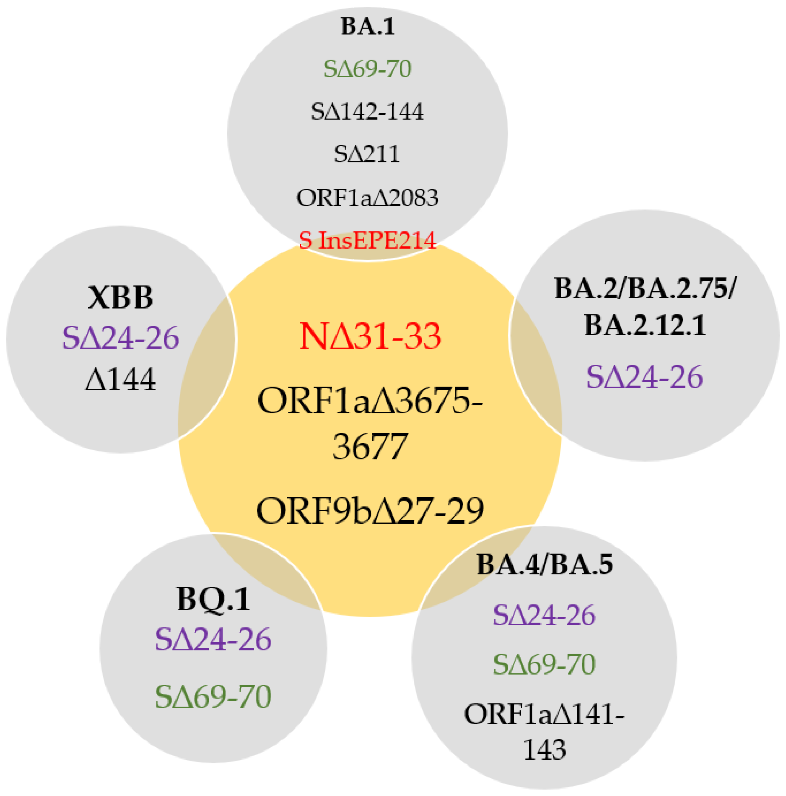
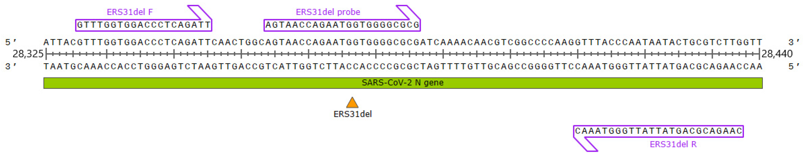
2.1. Primer and Probe Design

Oligonucleotide primers and probes targeting the conservative region in ORF1 (Figure 1) were designed to detect any SARS-CoV-2 RNA and were used in this multiplex assay as an indicator of SARS-CoV-2 in a clinical sample (2-SARS-CoV-2-ORF-1). The primer and probe sequences were confirmed to have perfect matches within all SARS-CoV-2 genome sequences available from GISAID to the moment.
A unique ins214EPE insertion is located in the area termed an “insertion hotspot” in the Spike protein-encoding gene of BA.1 [15]. Hence, this mutation was incorporated in the RT-qPCR design to detect the Omicron BA.1 lineage (Figure 2). Long insertions are thought to be generated via the template-switching mechanism associated with the synthesis of subgenomic RNAs, while short inserts seem to result from RdRP slippage on short runs of A or U [16].
All Omicron lineages (BA*) carry a three amino acid deletion ERS31del in the N gene encoding the Nucleocapsid protein (Figure 3). According to metadata, this mutation is found in 93% of BA* genomes submitted to the GISAID EpiCoV repository by the end of April 2022. Therefore, it was used to design a qPCR assay discriminating Omicron from other SARS-CoV-2 variants (Figure 4). ERS31del is considered lineage-defining and is found in all BA* genomes. However, it is not annotated in all the genomes, presumably because algorithms struggle with processing NGS data if it contains insertions and deletions since accurate indel calling is known to be an issue [17]. Assays were designed to amplify DNA from RNA templates derived from different SARS-CoV-2 lineages while detecting insertions and deletions with the probes complementary to a sequence containing the corresponding variant-specific mutation (Figure 3 and Figure 4).

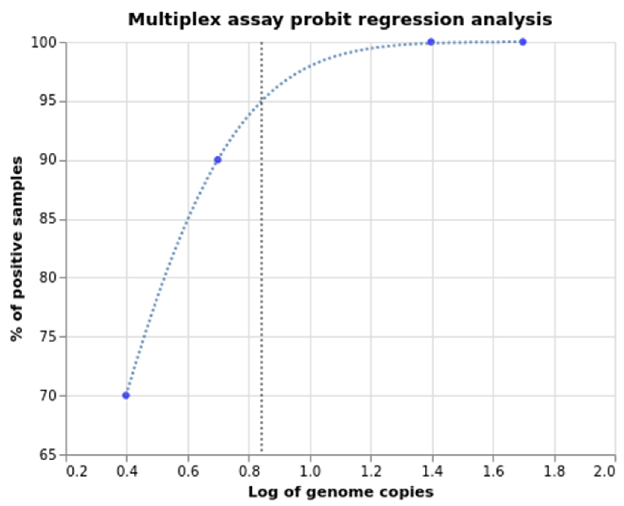
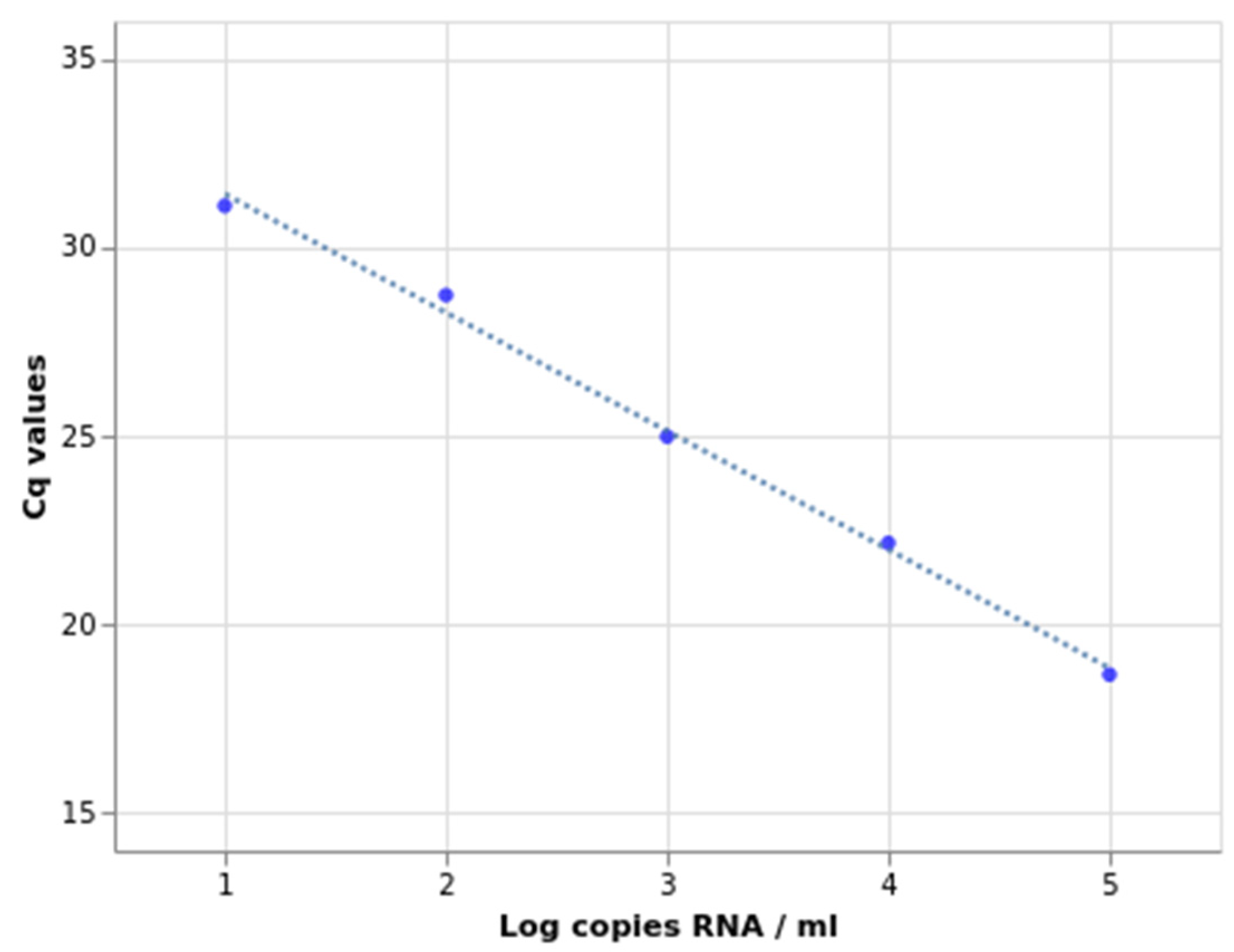
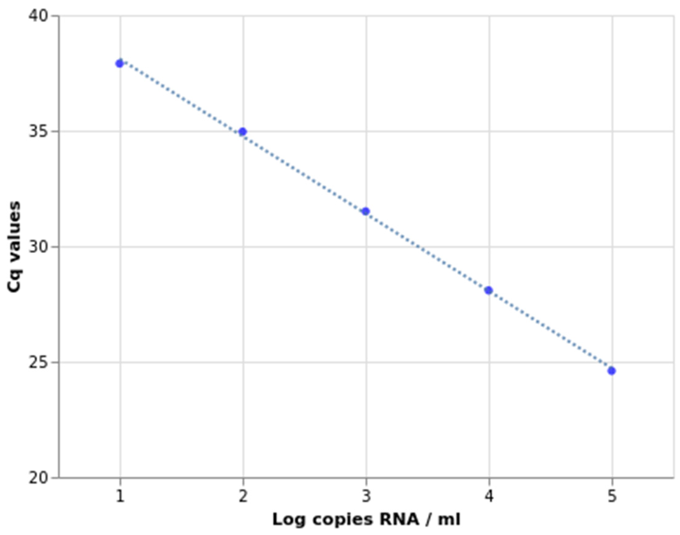
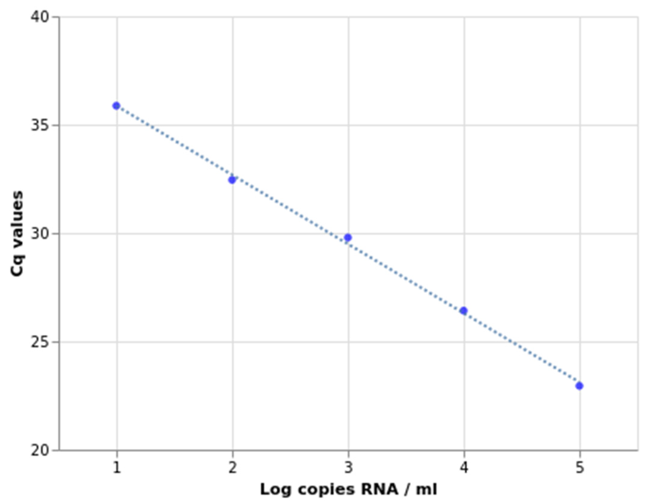
2.2. Sensitivity, Specificity, and Amplification Efficiency Assessment

The limit of detection (LOD) was determined by serial dilution of full-length SARS-CoV-2 RNA and probit regression analysis (Table 1, Figure 5). Standard SARS-CoV-2 RNA concentration was calculated with digital (d) PCR (QIAcuity Digital PCR System) using SARS-CoV-2 detection kit (ModularDx, TIB Molbiol). The concentration amounted to 49,860 copies per microliter. All primer-probe sets detected SARS-CoV-2 at 1000 virus copies per ml at the lowest.
The developed multiplex RT-qPCR assay demonstrates high specificity. It was preliminarily tested on 28 clinical samples of RNA extracted from oropharyngeal swabs, which contained 11 SARS-CoV-2 lineages according to WGS data (Table 2). Specific signal was detected only in samples with SARS-CoV-2 Omicron lineage RNA.
The specificity of the multiplex RT-qPCR assay was further tested on another 96 samples containing variants previously defined by WGS: 32 non-BA.1 Omicron, 32 BA.1, 22 non-Omicron variants, and 10 negative controls. The experiment yielded 100% true positives and 100% true negatives (Table 3). Detailed data is provided in Appendix A, Table A1.
Ten-fold serial dilutions of Omicron RNA were used to assess PCR efficiency. The PCR efficiency of 2-SARS-CoV-2, Ins214EPE, and ERS31del assays reached 108%, 98.9%, and 105%, respectively. The graphs are presented in Appendix B (Figure A1, Figure A2 and Figure A3).
Additionally, the specificity was tested on clinical samples that tested positive for other respiratory viruses from the collection of Smorodintsev Research Institute of Influenza, such as influenza and parainfluenza viruses, human seasonal coronaviruses (OC43, NL63, 229E, HKU1), hRSV, rhinoviruses, bocaviruses, and metapneumovirus (33 in total). The test yielded no false-positive results. Data is shown in Appendix C (Table A2).
After combining the four reactions into a single multiplex, its accuracy and specificity were found to be similar to those of a single RT-qPCR (Appendix D, Table A3). Thus, the developed multiplex RT-qPCR assay provides diagnostic performance comparable to currently used singleplex RT-qPCR tests while requiring fewer reagents and less time. Interpretation of the RT-qPCR results is presented in Table 4.

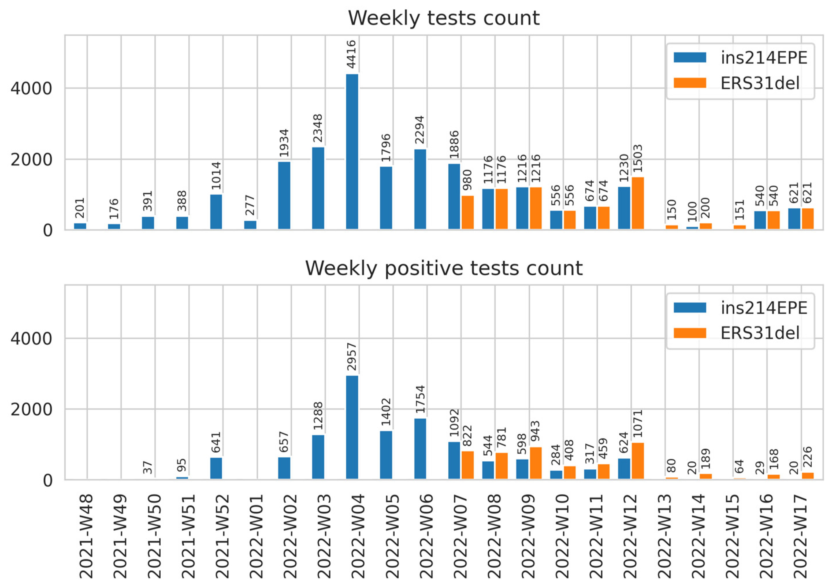
2.3. Implementing the Developed Assays in SARS-CoV-2 Surveillance in Russia

More than 30,000 specimens were tested with the multiplex RT-qPCR assay while screening clinical specimens collected in various Russian Federation regions from December 2021 to May 2022. The multiplex RT-qPCR allowed us to monitor Omicron variant frequencies from its first appearance and initial spread to the highest epidemic peaks since the beginning of the COVID-19 pandemic in Russia (Figure 6).
Thus, we developed a sensitive and specific multiplex RT-qPCR, which detects the SARS-CoV-2 Omicron lineage and specifically identifies the BA.1 subvariant, and verified it with WGS on plenty of clinical samples.
The Ins214EPE assay protocol was shared with the scientific community on protocols.io in December 2021 [18]. Afterward, the multiplex assay was developed and the protocol was also made available on protocols.io in February 2022 [19]. The described RT-qPCR assay is patented in Russia (#2779025) and commercialized by the BioLabMix company.

3. Discussion

Epidemiological and clinical research requires collecting data on the spreading of emerging SARS-CoV-2 variants. Some emerging mutations are good targets for detecting particular viral genomes. RT-qPCR can detect SNPs but demands highly specific enzymes and chemistry, such as PACE-RT chemistry [20]. HRM technology is a suitable technique to detect SNPs in some applications. However, its robustness is jeopardized if the target is genetically diverse or mutable [21]: the presence of more than one mutation site can result in complex melt curves which may be difficult to interpret. Hence, HRM is not perfect in the case of the S and N genes of SARS-CoV-2.
SGTF assay [11], the so-called “drop-out” technique, also has an inherent disadvantage in this kind of test. It may fail to detect the S target in swab samples with lower virus concentration, resulting in false negatives.
Other RT-qPCR assays for Omicron detection are known. One of the assays identifies the Omicron lineage [22] based on oligonucleotide complementarity to eight nucleotide polymorphisms in the SARS-CoV-2 Omicron genome. The assay uses the SNP cluster in the S gene as a marker of the Omicron lineage. However, some of these point mutations, such as E484Q and N501Y, are present in non-Omicron SARS-CoV-2 variants as well, while others are absent in some Omicron subvariants, such as Q493R in BA.4&5. The main limitation of this method is the necessity to run another SARS-CoV-2 assay in parallel for Cq comparing since there is also a weak signal for the Wuhan Hu-1, the Alpha, Beta, Delta, and Gamma variants. The authors also noticed that the temperature profile of amplification is critical to assay specificity. It could also hinder the reproducibility of this test if different RT-qPCR reagents are used. Another described Omicron detection technique relies on Sanger sequencing following amplification [23]. However, this method is laborious, expensive, difficult to scale up, and thus could hardly be used for screening.
Nevertheless, SARS-CoV-2 emerging variants often carry not only SNPs but also lineage-defining indels. Targeting these with PCR could be a less laborious approach to develop and optimize. Some published assays for Omicron lineage detection are based on identified 69/70 deletion [24,25], but widespread BA.2 subvariants of Omicron lack this mutation. The assay developed in our laboratory avoids the abovementioned disadvantages and carries only limitations specific to the qPCR method, such as sensitivity to virus genetic variability at the points of oligonucleotide annealing.
Two indels unique to the Omicron variant, Ins214EPE and ERS31del, were chosen for RT-qPCR assays designed to detect the Omicron lineage and its BA.1 subvariant. The developed multiplex assay for Omicron and its BA.1 variant detection has been in use for more than 9 months. Despite the rapid virus evolution, ERS31del in the N gene is still present in all circulating variants, including new immune-evasive variants with global spread BQ and XBB, and Ins214EPE is still a unique marker of the BA.1 variant to the moment of article submission (November 2022). The assay is cheap, fast, robust, and simple to be introduced and implemented in any PCR laboratory.
The developed assays, including the multiplex RT-qPCR, demonstrate high specificity and sensitivity. The assays were verified, broadly tested, and used to screen thousands of samples. The final multiplex platform included the ERS31del assay for the Omicron lineage (BA*) detection, Ins214EPE assay against the BA.1 variant, ORF1 SARS-CoV-2 for the detection of any type of SARS-CoV-2, and the RP assay for human RNA amplification as a control for RNA extraction, sample storage, and PCR conditions. For kits based on these assays, RNA controls were also obtained.
ERS31del is typical for all Omicron subvariants detected by July 2022: BA.1, BA.2, BA.3, BA.4, BA.5, BA.2.12.1, and BA.2.75. Thus, the ERS31del assay can detect all the variants that are spreading in 2022. However, some SNP-specific assays are needed to discriminate Omicron subvariants since no specific indels are found (except for BA.1). Similarly to SGTF, which has already been proved useful twice during the pandemic, the Ins214EPE and ERS31del assays could be useful for targeting novel SARS-CoV-2 variants, too. One or several SARS-CoV-2 variants are highly likely to become a future seasonal infection; thus, the new RT-qPCR assays are a promising approach to seasonal SARS-CoV-2 identification.

4. Materials and Methods

4.1. Sample Collection and RT-PCR Testing

De-identified samples used in this study were collected during the ongoing surveillance of SARS-CoV-2 variability routinely conducted by the Smorodintsev Research Institute of Influenza under the Coronavirus Russian Genetic Initiative (CORGI). Written informed consent was obtained from all subjects following the order of the Ministry of Health of the Russian Federation of 21 July 2015 #474 n. This study was reviewed and deemed exempt by the Local Ethics Committee of the Smorodintsev Research Institute of Influenza (protocol No. 152, 18 June 2020).
Nasopharyngeal and throat swabs were collected in virus transport media. Total RNA was extracted using the Auto-Pure 96 Nucleic Acid Purification System (Allsheng, Hangzhou, China) and the NAmagp DNA/ RNA extraction kit (Biolabmix, Novosibirsk, Russia). Extracted RNA was immediately tested for SARS-CoV-2 using Biolabmix SARS-CoV-2 RT-PCR Detection System (Biolabmix, Novosibirsk, Russia), following the Hong Kong University protocol with modifications [26].
To assess the specificity of the assays, a panel of respiratory viruses was obtained by routine testing of nasopharyngeal swabs from patients with respiratory infection diseases using PCR assay “ARVI-screen” (Amplisens, Moscow, Russia).

4.2. RT-qPCR Assay Design

In silico specificity tests were carried out using NCBI BLAST. Target regions of oligonucleotide sets were searched against the nucleotide sequence database with excluded SARS-CoV-2 genomes. Additionally, Ins214EPE and ERS31del oligonucleotides’ complementarity with BA.1 and BA* genomes submitted to the GISAID repository by the end of January was checked in silico, and the oligonucleotides aligned successfully with the majority of the corresponding genomes.
Sequences of oligonucleotides used in the multiplex RT-qPCR assay are presented in Table 5.
The expected amplicon size of ORF1 is 88 bp, S gene–89 bp, N gene–99 bp. Human RNA polymerase gene amplification assay used as an internal control was developed by the Center for Disease Control and Prevention [27]. All primers and probes were designed manually following basic primer design rules [28] and manufactured by ALCOR BIO company (Russia, Saint-Petersburg). The assays were designed to amplify a DNA fragments, including indels, from RNA templates derived from different SARS-CoV-2 variants. The detecting probes were complementary to a sequence containing the corresponding variant-specific mutation. The slope of the standard curves for assays 2SARS-CoV-2-ORF-1, Ins214EPE, and ERS31del was −3.14, −3.34 and −3.20, respectively. The amplification efficiency exceeded 95% for all primer–probe sets (Appendix B).
Oligonucleotides for the multiplex RT-qPCR were premixed as shown in Table 6.
PCR master mix contained 12.5 μL of PCR buffer, 1.2 μL of oligonucleotide premix (Table 6), 1 μL of enzyme mix, and 5.3 μL of water per reaction. Then, 5 μL of RNA extracted from a clinical sample was added. The thermocycler program included reverse transcription for 15 min at 45 °C, initial denaturation for 5 min at 95 °C, and 40 cycles of denaturation (10 s at 95 °C) followed by annealing/elongation (30 s at 62 °C). Bio-Rad CFX96 machine and Biolabmix reagents for PCR (RT-qPCR kit) were used in this work.

4.3. Digital PCR

dPCR was run with the QIAcuity One 5-Plex from QIAGEN (Hilden, DE) using QIAcuity One-Step Viral RT-PCR kit with SARS-CoV-2 detection kit (ModularDx, TIB Molbiol). BA.1 pooled sample was used as template RNA (GenBank: OP810428.1). A total of 5 μL template RNA was added to master-mix containing 3 μL One-Step Viral RT-PCR Master Mix, 0.12 μL 100x Multiplex RT mix, 0.5 TIB oligonucleotides mix, and 3.4 μL of water per reaction. dPCR settings were as follows: 40 min for reverse transcription at 50 °C, 2 min at 95 °C for polymerase activation followed by 40 cycles of 5 s at 95 °C for denaturation, and 30 s at 60 °C for annealing and extension.

4.4. Genome Assembly and Consensus Correction

WGS data were obtained by Illumina MiSeq and MinIon (Oxford Nanopore Technology, Oxford, UK) and used to confirm PCR results and create the SARS-CoV-2 lineage panel. Libraries for Illumina sequencing were prepared using the Nextera XT library preparation kit (Illumina, San Diego, CA, USA) and then sequenced on a MiSeq instrument (Illumina, San Diego, CA, USA) with a MiSeq Sequence kit v3. FastQC software was used for sequence data quality assessment. Trimmomatic was applied for quality data trimming. Reads were mapped onto reference sequences using BWA. SAMtools-mpileup v1.10.68 [29] was used to produce draft consensus sequences which were then corrected. Libraries for Oxford Nanopore sequencing were prepared using an SQK-LSK109 DNA Ligation Sequence kit (Oxford Nanopore, Oxford, UK). Sequencing was performed using a MinIon instrument (Oxford Nanopore, Oxford, UK) with a R9.4.1 flowcell. Guppy software was used for base-calling and data quality trimming. Reads were mapped onto the reference sequence Wuhan-Hu-1 SARS-CoV-2 using Minimap2 [30]. Lineage was defined using PANGO algorithms.

4.5. Molecular Cloning

Positive control RNA for RT-qPCR assays was produced via in vitro transcription (IVT) from pDNA templates, obtained by cloning the corresponding viral cDNA fragments to pJet using CloneJet Kit (Thermo Scientific, Waltham, USA). Bacterial clones were grown in LB media with ampicillin 100 μg/mL and plasmid DNA was extracted with Evrogen (Russia) plasmid midi kit.

5. Patents

The following three patents registered in Russian provide the details about the described RT-qPCR assays: Nº2761481, Nº2772362, and Nº2779025 for 2-SARS-CoV-2 test, Ins214EPE test, and multiplex RT-qPCR, respectively.

Author Contributions

N.D.Y.: experiment design, qPCR experiments, methodology, writing—original draft preparation; A.B.K.: conceptualization, writing—review and editing, supervision, resources; K.V.V.: visualization, sequence annotation analyses; T.D.M.: ARI panel creation, processing of clinical samples, assay validation; A.V.F.: whole genome sequencing data analysis; D.A.L.: project administration and funding acquisition. All authors have read and agreed to the published version of the manuscript.

Funding

The Ministry of Health of the Russian Federation supported this study as research work within the state assignment #121110800170-8 (2022). Part of this study was funded by the Russian Ministry of Science and Higher Education as a part of the World-class Research Center program: Advanced Digital Technologies (contract No. 075-15-2022-313, dated 20 April 2022).

Institutional Review Board Statement

This study was reviewed and deemed exempt by the Local Ethics Committee of Smorodintsev Research Institute of Influenza (protocol No. 152, 18 June 2020).

Informed Consent Statement

Written informed consent was obtained from all subjects in accordance with order #474 n of the Ministry of Health of the Russian Federation of 21 July 2015.

Data Availability Statement

Sequences of all used genomes were deposited to GISAID, protocols shared on protocols.io: Ins214EPE assay protocol https://www.protocols.io/view/one-step-rt-pcr-ins214epe-assay-for-omicron-b-1-1-3byl4b2e8vo5/v1 (accessed on 18 November 2022). Omicron SARS-CoV-2 lineage and its BA.1 variant multiplex RT-qPCR https://www.protocols.io/view/sars-cov-2-omicron-detection-rt-qpcr-assay-with-b-kqdg3p9nql25/v1 (accessed on 18 November 2022). Other data will be provided upon reasonable request.

Acknowledgments

We acknowledge World Health Organization for providing the SARS-CoV-2 detection kit (ModularDx, TIB Molbiol). We gratefully acknowledge all data contributors, i.e., the authors and their originating laboratories responsible for obtaining the specimens, and their submitting laboratories for generating the SARS-CoV-2 genetic sequence and metadata and sharing via the GISAID Initiative. We would also like to acknowledge Vladimir Gushchin (Pathogenic Microorganisms Variability Laboratory, Gamaleya Institute, Moscow, Russia) for providing RNA extracts from the first confirmed cases of Omicron VOC in Russia (hCoV-19/Russia/MOW-Moscow_PMVL-O11/2021.

Conflicts of Interest

The authors declare no conflict of interest. The funders had no role in the design of the study, in the collection, analyses, or interpretation of data, in the writing of the manuscript, or in the decision to publish the results.

Appendix A

Table A1. Test of the multiplex RT-qPCR assay specificity on 96 samples containing variants previously defined by WGS: 32 non-BA.1 Omicron, 32 BA.1, 22 non-Omicron variants, and 10 negative controls. “Nd”—Not detected.
GISAID Accession NumberPango
Variant
2-SARS-CoV-2-ORF1 AssayERS31del AssayIns214EPE AssayRP Assay
EPI_ISL_15919776CL.127.6526.25Nd29.26
EPI_ISL_15919773CL.127.2225.7Nd26.96
EPI_ISL_15919771BA.5.2.625.0224.91Nd30.1
EPI_ISL_15919779CL.120.719.25Nd31.08
EPI_ISL_15919777BA.5.2.624.6924.82Nd30.52
EPI_ISL_15919787CL.124.1422.65Nd26.27
EPI_ISL_15919784BA.5.2.3027.0326.92Nd29.38
EPI_ISL_15919785BA.5.224.9824.8Nd30.39
EPI_ISL_15919782BA.5.219.8719.64Nd29.05
EPI_ISL_15919780CL.123.622.37Nd25.07
EPI_ISL_15919781BA.5.2.124.324.19Nd25.02
EPI_ISL_15920157BA.5.219.9419.64Nd27.27
EPI_ISL_15920158BA.5.221.3521.39Nd27.96
EPI_ISL_15920173XBB24.7723.9Nd29
EPI_ISL_15920176BF.529.7329.58Nd32.5
EPI_ISL_15920178BA.5.220.4320.64Nd26.32
EPI_ISL_15920160BA.5.1.1020.0518.63Nd24.2
EPI_ISL_15920162BA.5.2.720.3620.17Nd24.92
EPI_ISL_15920168BA.5.222.4122.29Nd25.2
EPI_ISL_15983536BA.5.2.618.4618.1Nd25.53
EPI_ISL_15983633BA.5.225.0825.13Nd26.2
EPI_ISL_15983624BA.5.224.7324.63Nd30.28
EPI_ISL_15983623BA.5.226.626.42Nd30.2
EPI_ISL_15983622CL.124.5823.24Nd26.31
EPI_ISL_15983538BA.5.2.125.3231.43Nd25.43
EPI_ISL_15983599BA.5.226.3426.27Nd25.38
EPI_ISL_15983534BA.5.221.0120.9Nd28.43
EPI_ISL_15983596BA.5.225.8525.81Nd29.7
EPI_ISL_15983595BA.5.2.627.5827.43Nd28.65
EPI_ISL_15983592CL.123.8722.3Nd25.03
EPI_ISL_15983594BA.5.2.3433.0433.94Nd29.34
EPI_ISL_15983593CL.128.8327.65Nd30.17
EPI_ISL_11503185BA.124.322.8525.2230.12
EPI_ISL_11503189BA.125.2123.6826.0331.63
EPI_ISL_12043917BA.127.0425.6927.6235.07
EPI_ISL_12043919BA.128.527.1528.8430.65
EPI_ISL_12043923BA.130.7729.4530.9729.68
EPI_ISL_12043930BA.127.6526.2728.0229.08
EPI_ISL_9338420BA.118.0216.3118.2831.63
EPI_ISL_9338432BA.122.5621.2422.7623.9
EPI_ISL_9338433BA.128.7827.1928.5528
EPI_ISL_9338434BA.124.0922.8124.2626.93
EPI_ISL_9338498BA.127.1825.8127.4627.17
EPI_ISL_9338506BA.121.3520.0422.4625.32
EPI_ISL_9338534BA.134.6234.0735.3829.87
EPI_ISL_9338538BA.124.1822.7124.7727.53
EPI_ISL_9338588BA.122.220.7322.2730.59
EPI_ISL_9338592BA.119.4517.8519.3927.42
EPI_ISL_9338686BA.129.0728.0128.9227.9
EPI_ISL_9338701BA.124.2622.9324.2727.95
EPI_ISL_9338747BA.128.1426.7927.9826.34
EPI_ISL_9338818BA.121.2820.0721.1224.44
EPI_ISL_9861967BA.126.7425.426.5228.24
EPI_ISL_9862115BA.127.6326.4827.4825.9
EPI_ISL_9862127BA.127.3125.9927.729.04
EPI_ISL_9862130BA.130.9429.6231.2534.54
EPI_ISL_9862144BA.125.824.2626.1528.42
EPI_ISL_9862153BA.123.9522.5723.9629.04
EPI_ISL_9862158BA.122.8421.522.8927.67
EPI_ISL_9862159BA.129.6528.3329.4733.28
EPI_ISL_9862161BA.126.0524.5926.129.05
EPI_ISL_9862162BA.126.5625.1626.8528.19
EPI_ISL_9862172BA.123.3221.8223.2527.38
EPI_ISL_9862177BA.123.3622.1722.9125.54
EPI_ISL_3122915B.1.617.224.87NdNd26.64
EPI_ISL_3122916B.1.617.233.94NdNd28.28
EPI_ISL_3122917AY.12225.77NdNd29.62
EPI_ISL_3122923AY.12225.72NdNd27.69
EPI_ISL_3122927AY.12226.36NdNd27.12
EPI_ISL_3122928AY.12222.99NdNd26.28
EPI_ISL_3122931AY.12230.06NdNd29.54
EPI_ISL_3122933AY.12222.81NdNd26.56
EPI_ISL_3122971AY.12231.27NdNd26.36
EPI_ISL_3122972AY.12229.13NdNd32.05
EPI_ISL_3122973AY.12223.03NdNd27.8
EPI_ISL_3122974AY.12230.12NdNd28.77
EPI_ISL_3122975B.1.617.226.55NdNd24.21
EPI_ISL_3122976AY.12236.43NdNd28.2
EPI_ISL_3122977B.1.1.728.96NdNd28.26
EPI_ISL_3122978AY.12230.76NdNd31.7
EPI_ISL_3122979AY.12234.45NdNd32.33
EPI_ISL_3122980AY.12225.22NdNd28.57
EPI_ISL_3122981AY.12232.53NdNd26.87
EPI_ISL_3122982B.1.617.231.29NdNd26.91
EPI_ISL_3122986AY.12222.96NdNd29.73
EPI_ISL_3122987AY.12223.33NdNd26.07
Negative control-NdNdNdNd

Appendix B

Figure A1. The effectiveness of the SARS-CoV-2 RT-qPCR is 108%. The slope of the standard curve is −3.14.
Ijms 23 16153 g0a1
Figure A2. The effectiveness of the Ins214EPE RT-qPCR is 99%. The slope of the standard curve is −3.34.
Ijms 23 16153 g0a2
Figure A3. The effectiveness of the ERS31del qRT-PCR is 105%. The slope of the standard curve is −3.20.
Ijms 23 16153 g0a3

Appendix C

Table A2. Analytical specificity of the multiplex qPCR assay tested on 33 clinical samples positive for other respiratory viruses. The RP assay (human RNAse P assay) developed by US CDC was used to check for the presence of human RNA in clinical samples. “Nd”—Not detected.
Clinical Sample Positive forRP Assay, CqSARS-CoV-2 Assay, CqIns214EPE, Cq
RSV A28.76Nd Nd
RSV A30.56Nd Nd
RSV A27.70NdNd
RSV B31.49NdNd
RSV B30.98NdNd
RSV B32.33NdNd
NL6332.20NdNd
NL6330.42NdNd
NL6324.95NdNd
Oc4330.34NdNd
Oc4330.69NdNd
Oc4328.64NdNd
HKU130.06NdNd
HKU128.30NdNd
HKU130.73NdNd
229E29.11NdNd
229E32.52NdNd
229E29.37NdNd
BoV32.26NdNd
BoV30.75NdNd
BoV27.25NdNd
Rv32.85NdNd
Rv33.76NdNd
Rv27.75NdNd
Piv128.63NdNd
Piv224.72NdNd
Piv327.01NdNd
Piv423.90NdNd
Adv29.47NdNd
MPV30.12NdNd
HIV A29.13NdNd
HIV A28.45NdNd
HIV A28.1639.06Nd
SARS-CoV-2 B.1.1.52934.1526.6128.44

Appendix D

Table A3. Comparison of qPCR results of the same SARS-CoV-2 BA.1 sample tested in 6 dilutions with multiplex and single-plex RT-qPCR assays (a single reaction in one tube, 4 tubes for each dilution of the sample). “Nd”—Not detected.
Multiplex RT-qPCR Results
dilution2-SARS-CoV-2ERS31delIns214EPERP
1x19.0121.0019.3829.38
10x22.2224.1422.4433.19
100x25.5627.7826.1334.51
1000x28.9631.0529.3737.74
10,000x31.4434.0432.25Nd
100,000x34.5537.6935.52Nd
Singleplex assay results
2-SARS-CoV-2ERS31delIns214EPERP
1x19.0319.4718.9429.46
10x22.0422.9522.6133.15
100x25.3426.4226.0136.11
1000x28.4429.7929.3238.42
10,000x31.4932.4531.87Nd
100,000x34.1635.8735.64Nd

References

  1. Worldometers. Available online: https://www.worldometers.info/coronavirus/ (accessed on 12 November 2022).
  2. WHO. Weekly Epidemiological Update on COVID-19. Available online: https://www.who.int/emergencies/diseases/novel-coronavirus-2019/situation-reports (accessed on 12 November 2022).
  3. Mistry, P.; Barmania, F.; Mellet, J.; Peta, K.; Strydom, A.; Viljoen, I.M.; James, W.; Gordon, S.; Pepper, M.S. SARS-CoV-2 Variants, Vaccines, and Host Immunity. Front Immunol. 2022, 12, 809244. [Google Scholar] [CrossRef] [PubMed]
  4. Zhang, J.; Xiao, T.; Cai, Y.; Lavine, C.L.; Peng, H.; Zhu, H.; Anand, K.; Tong, P.; Gautam, A.; Mayer, M.L.; et al. Membrane fusion and immune evasion by the spike protein of SARS-CoV-2 Delta variant. Science 2021, 374, 1353–1360. [Google Scholar] [CrossRef] [PubMed]
  5. Gobeil, S.M.-C.; Janowska, K.; McDowell, S.; Mansouri, K.; Parks, R.; Manne, K.; Stalls, V.; Kopp, M.F.; Henderson, R.; Edwards, R.J.; et al. D614G Mutation Alters SARS-CoV-2 Spike Conformation and Enhances Protease Cleavage at the S1/S2 Junction. Cell Rep. 2021, 34, 108630. [Google Scholar] [CrossRef] [PubMed]
  6. Hoffmann, M.; Krüger, N.; Schulz, S.; Cossmann, A.; Rocha, C.; Kempf, A.; Nehlmeier, I.; Graichen, L.; Moldenhauer, A.-S.; Winkler, M.S.; et al. The Omicron variant is highly resistant against antibody-mediated neutralization: Implications for control of the COVID-19 pandemic. Cell 2022, 185, 447–456.e11. [Google Scholar] [CrossRef]
  7. Schmidt, F.; Muecksch, F.; Weisblum, Y.; Da Silva, J.; Bednarski, E.; Cho, A.; Wang, Z.; Gaebler, C.; Caskey, M.; Nussenzweig, M.C.; et al. Plasma Neutralization of the SARS-CoV-2 Omicron Variant. N. Engl. J. Med. 2022, 386, 599–601. [Google Scholar] [CrossRef]
  8. Lu, L.; Mok, B.W.Y.; Chen, L.L.; Chan, J.M.C.; Tsang, O.T.Y.; Lam, B.H.S.; Chuang, V.W.M.; Chu, A.W.H.; Chan, W.M.; Ip, J.D.; et al. Neutralization of Severe Acute Respiratory Syndrome Coronavirus 2 Omicron Variant by Sera From BNT162b2 or CoronaVac Vaccine Recipients. Clin. Infect. Dis. 2021, 75, e822–e826. [Google Scholar] [CrossRef]
  9. Shu, Y.; McCauley, J. GISAID: Global initiative on sharing all influenza data—From vision to reality. Eurosurveillance 2017, 22, 30494. [Google Scholar] [CrossRef] [Green Version]
  10. Knyazev, S.; Chhugani, K.; Sarwal, V.; Ayyala, R.; Singh, H.; Karthikeyan, S.; Deshpande, D.; Baykal, P.I.; Comarova, Z.; Lu, A.; et al. Unlocking capacities of genomics for the COVID-19 response and future pandemics. Nat. Methods 2022, 19, 374–380. [Google Scholar] [CrossRef]
  11. Volz, E.; Mishra, S.; Chand, M.; Barrett, J.C.; Johnson, R.; Geidelberg, L.; Ferguson, N.M.; Hinsley, W.R.; Laydon, D.J.; Dabrera, G.; et al. Transmission of SARS-CoV-2 lineage B.1.1.7 in England: Insights from linking epidemiological and genetic data. MedRxiv 2021. [Google Scholar] [CrossRef]
  12. Subramoney, K.; Mtileni, N.; Bharuthram, A.; Davis, A.; Kalenga, B.; Rikhotso, M.; Maphahlele, M.; Giandhari, J.; Naidoo, Y.; Pillay, S.; et al. Identification of SARS-CoV-2 Omicron variant using spike gene target failure and genotyping assays, Gauteng, South Africa, 2021. J. Med. Virol. 2022, 94, 3676–3684. [Google Scholar] [CrossRef]
  13. Kalthoff, D.; Beer, M.; Hoffmann, B. High resolution melting analysis: Rapid and precise characterisation of recombinant influenza A genomes. Virol. J. 2013, 10, 284. [Google Scholar] [CrossRef] [PubMed] [Green Version]
  14. Alisoltani, A.; Jaroszewski, L.; Iyer, M.; Iranzadeh, A.; Godzik, A. Increased Frequency of Indels in Hypervariable Regions of SARS-CoV-2 Proteins—A Possible Signature of Adaptive Selection. Front. Genet. 2022, 13, 875406. [Google Scholar] [CrossRef] [PubMed]
  15. Gerdol, M.; Dishnica, K.; Giorgetti, A. Emergence of a recurrent insertion in the N-terminal domain of the SARS-CoV-2 spike glycoprotein. Virus Res. 2022, 310, 198674. [Google Scholar] [CrossRef] [PubMed]
  16. Garushyants, S.K.; Rogozin, I.B.; Koonin, E.V. Template switching and duplications in SARS-CoV-2 genomes give rise to insertion variants that merit monitoring. Commun. Biol. 2021, 4, 1343. [Google Scholar] [CrossRef]
  17. Albers, C.A.; Lunter, G.; MacArthur, D.G.; McVean, G.; Ouwehand, W.H.; Durbin, R. Dindel: Accurate indel calls from short-read data. Genome Res. 2011, 21, 961–973. [Google Scholar] [CrossRef] [Green Version]
  18. Yolshin, N.; Varchenko, K.; Komissarova, K.; Danilenko, D.; Komissarov, A.; Lioznov, D. One-step RT-PCR Ins214EPE assay for Omicron (B.1.1.529) variant detection. Protocols.Io 2021. [Google Scholar] [CrossRef]
  19. Yolshin, N.; Varchenko, K.; Komissarov, A. SARS-CoV-2 Omicron detection RT-qPCR assay with BA.1 and BA.2/BA.3 differentitation. Protocols.Io 2022. [Google Scholar] [CrossRef]
  20. Harper, H.; Burridge, A.; Winfield, M.; Finn, A.; Davidson, A.; Matthews, D.; Hutchings, S.; Vipond, B.; Jain, N.; The COVID-19 Genomics UK (COG-UK) Consortium; et al. Detecting SARS-CoV-2 variants with SNP genotyping. PLoS ONE 2022, 16, e0243185. [Google Scholar] [CrossRef]
  21. Gene-Quantification. Available online: https://www.gene-quantification.de/ab-hrm-guide.pdf (accessed on 12 November 2022).
  22. Corbisier, P.; Petrillo, M.; Marchini, A.; Querci, M.; Buttinger, G.; Bekliz, M.; Spiess, K.; Polacek, C.; Fomsgaard, A.; Eede, G.V.D. A qualitative RT-PCR assay for the specific identification of the SARS-CoV-2 B.1.1.529 (Omicron) Variant of Concern. J. Clin. Virol. 2022, 152, 105191. [Google Scholar] [CrossRef]
  23. Bloemen, M.; Rector, A.; Swinnen, J.; Van Ranst, M.; Maes, P.; Vanmechelen, B.; Wollants, E. Fast detection of SARS-CoV-2 variants including Omicron using one-step RT-PCR and Sanger sequencing. J. Virol. Methods 2022, 304, 114512. [Google Scholar] [CrossRef]
  24. Phan, T.; Boes, S.; McCullough, M.; Gribschaw, J.; Marsh, J.; Harrison, L.H.; Wells, A. Development of a One-Step Qualitative RT-PCR Assay to Detect the SARS-CoV-2 Omicron (B.1.1.529) Variant in Respiratory Specimens. J. Clin. Microbiol. 2022, 60, e0002422. [Google Scholar] [CrossRef] [PubMed]
  25. Chassalevris, T.; Chaintoutis, S.C.; Koureas, M.; Petala, M.; Moutou, E.; Beta, C.; Kyritsi, M.; Hadjichristodoulou, C.; Kostoglou, M.; Karapantsios, T.; et al. SARS-CoV-2 wastewater monitoring using a novel PCR-based method rapidly captured the Delta-to-Omicron ΒA.1 transition patterns in the absence of conventional surveillance evidence. Sci. Total Environ. 2022, 844, 156932. [Google Scholar] [CrossRef] [PubMed]
  26. Chu, D.K.W.; Pan, Y.; Cheng, S.M.S.; Hui, K.P.Y.; Krishnan, P.; Liu, Y.; Ng, D.Y.M.; Wan, C.K.C.; Yang, P.; Wang, Q.; et al. Molecular Diagnosis of a Novel Coronavirus (2019-nCoV) Causing an Outbreak of Pneumonia. Clin. Chem. 2020, 66, 549–555. [Google Scholar] [CrossRef] [Green Version]
  27. CDC. Available online: https://www.cdc.gov/coronavirus/2019-ncov/lab/rt-pcr-panel-primer-probes.html (accessed on 12 November 2022).
  28. Rodríguez, A.; Rodríguez, M.; Córdoba, J.J.; Andrade, M.J. Design of primers and probes for quantitative real-time PCR methods. In Methods in Molecular Biology (Clifton, N.J.); Humana Press: New York, NY, USA, 2015; Volume 1275, pp. 31–56. [Google Scholar] [CrossRef]
  29. Li, H. A statistical framework for SNP calling, mutation discovery, association mapping and population genetical parameter estimation from sequencing data. Bioinformatics 2011, 27, 2987–2993. [Google Scholar] [CrossRef] [Green Version]
  30. Li, H. Minimap2: Pairwise alignment for nucleotide sequences. Bioinformatics 2018, 34, 3094–3100. [Google Scholar] [CrossRef] [PubMed] [Green Version]
Figure 1. 2-SARS-CoV-2-ORF-1 oligonucleotides binding scheme.
Ijms 23 16153 g001
Figure 2. Ins214EPE oligonucleotides binding scheme.
Ijms 23 16153 g002
Figure 3. Diagram of the deletions shared by Omicron subvariants.
Ijms 23 16153 g003
Figure 4. ERS31del oligonucleotides binding scheme.
Ijms 23 16153 g004
Figure 5. Probit regression model of the multiplex assay LOD.
Ijms 23 16153 g005
Figure 6. Samples collected in different regions of Russia during Omicron spreading were tested in Smorodintsev Research Institute of Influenza using the developed multiplex RT-qPCR.
Ijms 23 16153 g006
Table 1. Serial dilution analysis with the multiplex RT-qPCR assay.
Copies per ReactionLog% PositivesSignal Ratio
501.69910020/20
251.39810020/20
50.6999018/20
2.50.3987014/20
LOD (C95) = 0.84 logLOD (C95) = 7 copies per reaction
Table 2. Assessment of the analytical specificity of the assays to different SARS-CoV-2 variants. “Nd”—“Not detected”.
GISAID #PANGO
Lineage
RP Assay, CqSARS-CoV-2 Assay, CqERS31del Assay, CqIns214EPE Assay, Cq
P.135.3515.45NdNd
EPI_ISL_1257814B.1.35139.91NdNdNd
EPI_ISL_415710B.1Nd15.69NdNd
EPI_ISL_1652610B.133.8426.69NdNd
EPI_ISL_1919527B.124.2626.98NdNd
EPI_ISL_2698439B.1.1.725.2720.64NdNd
EPI_ISL_2523457B.1.1.730.5428.05NdNd
EPI_ISL_2450515B.1.1.726.9931.25NdNd
EPI_ISL_3454801AT.130.3033.07NdNd
EPI_ISL_3454797AT.130.0231.33NdNd
EPI_ISL_3454782AT.128.0033.00NdNd
EPI_ISL_2698481B.1.1.52329.1227.09NdNd
EPI_ISL_2698509B.1.1.52327.3923.46NdNd
EPI_ISL_2523540B.1.1.52330.0227.09NdNd
EPI_ISL_2698496B.1.1.31725.2225.31NdNd
EPI_ISL_2523528B.1.1.31727.5822.71NdNd
EPI_ISL_2450468B.1.1.31728.7023.91NdNd
EPI_ISL_4563892B.1.617.226.6634.03NdNd
EPI_ISL_5263415B.1.617.225.7228.43NdNd
EPI_ISL_4563887AY.12926.7825.42NdNd
EPI_ISL_6831894AY.12223.9628.95NdNd
EPI_ISL_6831897AY.12231.1635.05NdNd
EPI_ISL_6831903AY.12224.1934.93NdNd
EPI_ISL_11503033BA.127.9822.2023.5722.08
EPI_ISL_10931393BA.127.0824.9327.1624.65
EPI_ISL_10931401BA.125.1927.4829.8325.57
EPI_ISL_9338305BA.226.5019.6421.07Nd
EPI_ISL_9338650BA.224.7020.8122.70Nd
Table 3. Multiplex RT-qPCR specificity test on 96 samples panel.
Omicron BA* Non-BA.1 (32)Omicron BA.1 (32)Non-Omicron (22)NTC (10)
2-SARS-CoV-2 ORF1 assayTrue positive (32)
False negative (0)
True positive (32)
False negative (0)
True positive (22)
False negative (0)
True negative (10)
False positive (0)
ERS31del assayTrue positive (32)
False negative (0)
True positive (32)
False negative (0)
True negative (22)
False positive (0)
True negative (10)
False positive (0)
Ins214EPE assayTrue negative (32)
False positive (0)
True positive (32)
False negative (0)
True negative (22)
False positive (0)
True negative (10)
False positive (0)
Table 4. Interpretation of the multiplex RT-qPCR assay results.
2-SARS-CoV-2 AssayERS31del AssayIns214EPE AssayRP Assay
FAMHEXROXCy5
++SARS-CoV-2, non-Omicron
+++SARS-CoV-2 Omicron lineage, non-BA.1
++++SARS-CoV-2 Omicron lineage, BA.1
+No SARS-CoV-2 RNA in the sample
No RNA in the reaction/
RNA extraction failed
Table 5. Primer and probe sequences for the multiplex RT-qPCR assay for the Omicron lineage detection with BA.1 determination.
TargetNameSequence
ORF12-SARS-CoV-2-ORF-1-FAGAGCTATGAATTGCAGAC
ORF12-SARS-CoV-2-ORF-1-RGGGAAATACAAAATTTGGACA
ORF12-SARS-CoV-2-ORF-1-PFAM-AATTGGCAAAGAAATTTGACACCTTCA-BHQ1
ERS31delN31-33del FGTTTGGTGGACCCTCAGATT
ERS31delN31-33del RCAAGACGCAGTATTATTGGGTAAAC
ERS31delN31-33del PHEX-AGTAACCAGAATGGTGGGGCGCG-BHQ1
Ins214EPEIns214EPE FATATTCTAAGCACACGCCTATT
Ins214EPEIns214EPE RGGCAAATCTACCAATGGTTCTA
Ins214EPEIns214EPE PROX-TGCGTGAGCCAGAAGATCTCCCT-BHQ2
Human RPRP-CB-FAGATTTGGACCTGCGAGCG
Human RPRP-CB-RGAGCGGCTGTCTCCACAAGT
Human RPRP-CB-PCy5-TTCTGACCTGAAGGCTCTGCGCG-BHQ2
Table 6. Oligonucleotides’ premixing protocol for 100 reactions.
ReagentsμLConcentrationQuantity
2-SARS-CoV-2-ORF-1-F7.5100 pmol/μL300 nM
2-SARS-CoV-2-ORF-1-R7.5100 pmol/μL300 nM
2-SARS-CoV-2-ORF-1-P5100 pmol/μL200 nM
N31-33del F2.5100 pmol/μL100 nM
N31-33del R2.5100 pmol/μL100 nM
N31-33del P1.25100 pmol/μL50 nM
Ins214EPE F10100 pmol/μL400 nM
Ins214EPE R10100 pmol/μL400 nM
Ins214EPE P5100 pmol/μL200 nM
RP F5100 pmol/μL200 nM
RP R5100 pmol/μL200 nM
RP P5100 pmol/μL200 nM
water53.75
Total volume120
Publisher’s Note: MDPI stays neutral with regard to jurisdictional claims in published maps and institutional affiliations.

