Review

DNA Damage Response Mechanisms in Head and Neck Cancer: Significant Implications for Therapy and Survival

by
Chara Papalouka
1,
Maria Adamaki
1,
Panagiota Batsaki
2,
Panagiotis Zoumpourlis
1,
Antonis Tsintarakis
1,
Maria Goulielmaki
2,
Sotirios P. Fortis
2,
Constantin N. Baxevanis
2 and
Vassilis Zoumpourlis
1,*
1
Biomedical Applications Unit, Institute of Chemical Biology, National Hellenic Research Foundation (NHRF), 11635 Athens, Greece
2
Cancer Immunology and Immunotherapy Center, Saint Savas Cancer Hospital, 11522 Athens, Greece
*
Author to whom correspondence should be addressed.
Int. J. Mol. Sci. 2023, 24(3), 2760; https://doi.org/10.3390/ijms24032760
Submission received: 28 December 2022 / Revised: 27 January 2023 / Accepted: 29 January 2023 / Published: 1 February 2023
(This article belongs to the Special Issue Advances in Molecular Mechanism of Head and Neck Cancer)

Abstract

:
Head and neck cancer (HNC) is a term collectively used to describe a heterogeneous group of tumors that arise in the oral cavity, larynx, nasopharynx, oropharynx, and hypopharynx, and represents the sixth most common type of malignancy worldwide. Despite advances in multimodality treatment, the disease has a recurrence rate of around 50%, and the prognosis of metastatic patients remains poor. HNCs are characterized by a high degree of genomic instability, which involves a vicious circle of accumulating DNA damage, defective DNA damage repair (DDR), and replication stress. Nonetheless, the damage that is induced on tumor cells by chemo and radiotherapy relies on defective DDR processes for a successful response to treatment, and may play an important role in the development of novel and more effective therapies. This review summarizes the current knowledge on the genes and proteins that appear to be deregulated in DDR pathways, their implication in HNC pathogenesis, and the rationale behind targeting these genes and pathways for the development of new therapies. We give particular emphasis on the therapeutic targets that have shown promising results at the pre-clinical stage and on those that have so far been associated with a therapeutic advantage in the clinical setting.

1. Introduction

Head and neck cancer (HNC) is a term collectively used to describe a heterogeneous group of neoplasms which include cancers of the lip and oral cavity, the sinus and nasal cavity, the pharynx and larynx, the salivary gland and the thyroid gland, and is associated with poor prognosis in advanced stages [1]. The most common type of HNC is squamous cell carcinoma (HNSCC), accounting for approximately 90% of all HNCs, as most subtypes are derived from squamous cells of the mucosal epithelium [1]. In 2017, the Global Burden of Disease (GBD) study estimated that HNC represents 5.3% of all cancers (excluding nonmelanoma skin cancers), with the lip and oral cavity (LOC) cancers being the most frequent, followed by larynx cancers [2,3]. It has also been estimated that approximately 600,000 new HNSCC cases are diagnosed annually throughout the world, with mortality rates reaching as high as 50% [4,5,6].
The onset of HNC depends on an interplay between genetic factors, such as Fanconi anemia (FA), Bloom syndrome and xeroderma pigmentosum, and environmental factors, such as age, geographic region and various lifestyle contributors [7]. A major risk lifestyle factor is tobacco smoking, while smoking electronic cigarettes, despite being considered less harmful than conventional cigarettes, has also been associated with the development of HNSCC due to their high content of substances that can cause oral mucosal lesions [2]. Another major lifestyle risk factor is alcohol consumption, even in individuals who have never been smokers, while combined use of alcohol and tobacco has been shown to have a synergistic effect on tumor development [2,8,9]. Notably, a significant association, albeit to a lesser extent compared to smoking and alcohol consumption, has also been observed between Human papillomavirus (HPV) infection and the development of various HNCs, especially oropharyngeal (OPC), nasopharyngeal (NPC), oral cavity (OC), and larynx cancers [6]. To date, thirteen types of HPV have been characterized as carcinogenic by the International Agency for Research on Cancer, with the HPV16 subtype being regarded as the most potent contributor to the development of HNC [2,10].
The heterogeneity and diversity of anatomical sites in HNC make its treatment a challenging task. Current approaches include a combination of surgery, radiotherapy, and chemotherapy [11,12]. Radiotherapy (RT) combined with platinum compounds such as cisplatin, with or without 5-fluorouracil, has been the standard of care for many years but has, however, been associated with a modest survival advantage and significant treatment-related mortality [13,14]. More recent chemotherapeutics include taxols and the anti-epidermal growth factor receptor (anti-EGFR) antibody cetuximab, which, despite having been specifically approved for recurrent and metastatic HNC, has also been associated with an improvement in response rates and increased survival in locoregional HNSCC [15,16]. Of note, cancer immunotherapy has emerged in recent years as an additional treatment approach to HNCs, especially in the form of immune checkpoint inhibitors (ICIs), based on the rationale that these monoclonal antibodies have the ability to inhibit immune checkpoint receptors and, therefore, to prevent the inactivation of T-cell function and subsequent cancer immune escape [17]. In this regard, the ICIs nivolumab and pembrolizumab, which specifically bind to immune checkpoint receptor programmed cell death protein 1 (PD-1), have been approved for patients with recurrent or metastatic disease previously treated with platinum chemotherapy, conferring a survival benefit in 20–30% of patients [16,18,19]. However, despite advances in multimodality treatment and promising results from administering therapeutic combinations in clinical trials, relapse rates remain very high, and 5-year survival is still estimated at 66% [19]. This undoubtedly highlights the need for devising new strategies that will target resistant clones and prolong survival.
As with most solid tumors, HNCs are characterized by genomic instability, which may manifest both as chromosomal instability (CIN), in the form of chromosomal rearrangements or changes in chromosomal ploidy, as nucleotide instability (NIN), in the form of gene amplification, deletion, inversion, and base substitution or mutation, or as microsatellite instability (MIN), in the form of loss or addition of oligonucleotide repeats [13,20,21,22]. The mechanism behind these aberrations seems to involve a vicious circle of accumulating DNA damage, defective DNA damage repair (DDR), and replication stress [13,21,23,24]. The latter can induce the collapse of replication forks and the occurrence of DNA double-strand breaks (DSBs), which appear to be a prominent feature of HNCs [21,25,26]. The inability to appropriately repair these DSBs translates to inability to effectively inhibit cell cycle progression for DNA repair or apoptosis, leading to an ever-increasing accumulation of DNA lesions and the creation of a highly unstable genetic environment that is driven by mutagenic stress [13]. Interestingly, a positive correlation has been identified between the number of chromosomal arms bearing allelic loss and tumor grade, neck nodal status, disease recurrence, metastasis, and overall survival in patients with HNC [20,27]. The extent of DNA damage and the efficiency of DNA repair mechanisms are also significantly associated with response to treatment, and may play an important role in the development of novel therapies.
Even though malignant transformation results from a defective DDR mechanism and subsequent development of genomic instability, the induction of DSBs by chemotherapeutic agents and radiotherapy relies on defective cellular DNA repair processes for a successful response to treatment [28]. Overall, DNA repair mechanisms for DSBs fall into two main types: (i) the homology recombination repair (HRR or HR) mechanism, which uses extensive homology and DNA synthesis is facilitated through a template, usually the sister chromatid, in order to regenerate the lost sequences, and (ii) the non-homologous end joining (NHEJ) mechanism, which rejoins DSBs without any homology requirement and is therefore considered an error-prone DNA repair mechanism [28,29,30]. HR occurs largely during the late S and G2 phases, when the double helix on one sister chromatid can provide sequence information to repair the damaged chromatid, while NHEJ occurs predominantly during the G1 phase of the cell cycle, when sister chromatids are unavailable to allow homology-directed repair, and less during S/G2; in other words, NHEJ exhibits decreased fidelity in repairing DSBs compared to HR [30,31]. Even though defects in both mechanisms have been shown to confer a pro-carcinogenic effect, the chemo- and radioresistant properties of certain tumors appear to rely on effective DDR signaling to escape the cell-cycle arrest and apoptotic pathways induced by DSBs [28,32,33,34]. Recent evidence also highlights the existence of tumors with impaired or inactivated DDR pathways, as well as tumors with mutated DNA repair genes that act as tumor suppressors, which may be specifically targeted to improve therapeutic outcomes and patient survival [35,36,37,38].
This review gives an overview of the main DNA repair mechanisms involved in the pathogenesis of HNC, including the different genes and proteins that appear to be aberrantly expressed or mutated in DDR pathways, and how these aberrations are differentially implicated in cancer development and therapeutic resistance. We discuss the rationale behind targeting these genes and pathways for the development of novel therapeutic interventions and give a detailed account of those that have shown promising results in pre-clinical and clinical studies and the potential to confer a therapeutic advantage or survival benefit, either used as monotherapy or in combination with other anti-cancer therapies.

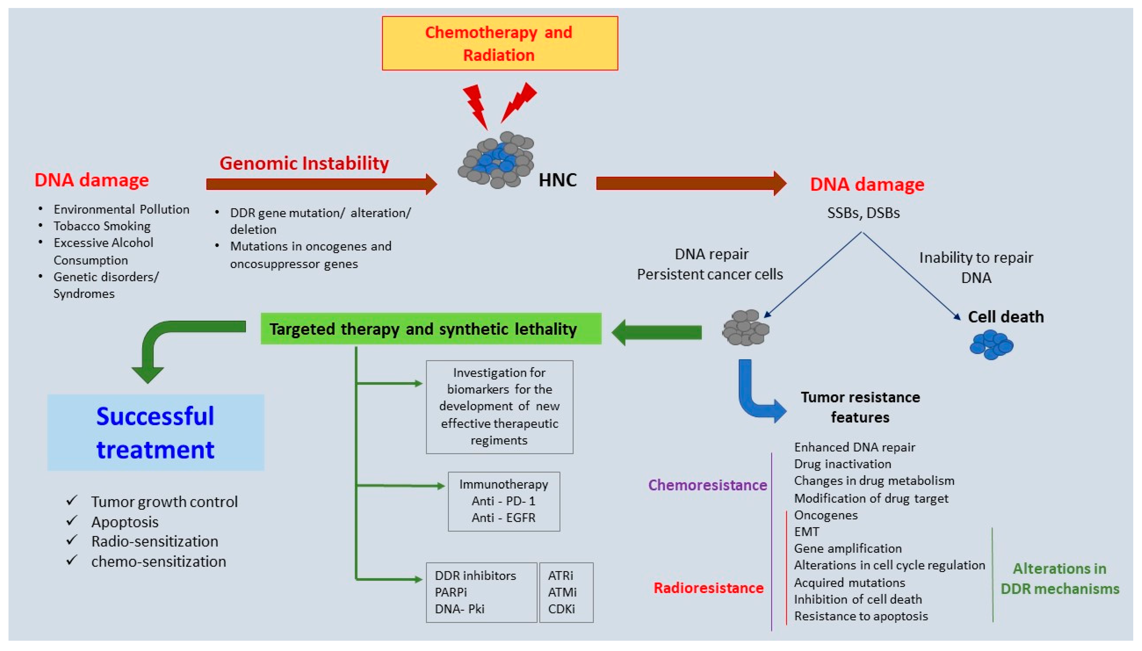
2. Genomic Instability in HNC

It has long become evident that genomic instability is implicated in tumor initiation, progression, and survival [21,39], and that it may manifest as one of three main types: chromosomal instability, nucleotide instability (ΝΙΝ), and microsatellite instability [21,22,40]. CΙΝ is the high rate of change in the structure and number of chromosomes in cancer cells in relation to normal cells [21,22]; MIN is the addition or loss of a number of oligonucleotide repeats due to defects in DNA mismatch repair (MMR) [40,41]; whereas NIN is the increased rate of point mutations, including base substitutions, deletions, and insertions, and is believed to occur as a result of base excision repair (BER) and nucleotide excision repair (NER) pathway malfunctioning [40]. Even though heritable germline mutations naturally increase the risk of cancer occurrence, hereditary cancers are also characterized by acquired somatic mutations [41,42]. Additionally, regardless of the type of gene that is affected by the hereditary mutation, i.e., whether the mutation occurs in an oncogene, an onco-suppressor gene, or a gene that is related to DNA repair, the prevailing hypothesis states that genomic instability is present in precancerous lesions and leads to tumor growth by increasing the rate of mutations, thereby supporting the notion of a continuous mutator phenotype (the so-called “mutator hypothesis”) [21,43,44]. Examples of hereditary HNCs include the Gorlin–Goltz and FA syndromes, with the former arising from mutations in the patched tumor suppressor gene (PTCH) [45,46] and the latter from mutations in genes implicated in the Fanconi anemia pathway [47], both being characterized by increased genomic instability [48]. On the other hand, in sporadic cancers, the mutations are acquired spontaneously and randomly; however, they also lead to replication stress and increased genomic instability [21,42,49,50].
Replication stress may be induced by both endogenous and exogenous sources. In addition to the overexpression or constitutive activation of oncogenes such as RAS, MYC, and EGFR families, and cyclin E [51], replication stress may also arise from replication barriers such as the presence of DNA–RNA hybrids, collisions between replication and transcription complexes [52], altered chromatin compaction and hypo-acetylation, shortened telomeres [53], repetitive sequences, early replicating fragile sites (ERFSs), and common fragile sites (CFSs) [54,55,56]. Additional barriers include DNA lesions and misincorporation of ribonucleotides, secondary DNA structures, and dormant replication origins due to external or internal factors of DNA damage that have not been repaired [54]. Exogenous sources of DNA damage include environmental agents such as chemicals, ultraviolet light, ionizing radiation, toxins, and pollutants, while major sources of endogenous DNA damage include oxidative DNA damage from the presence of reactive oxygen species (ROS), oxygen, and lipid peroxidation products (e.g., MDA-induced DNA damage derived from lipid peroxidation, propano, and etheno adducts), hydrolytic deamination and carbonyl stress, and several estrogen metabolites, among others [57].
Εxposure to carcinogens that are produced by environmental pollution or through lifestyle habits such as tobacco smoking and excessive alcohol consumption, and its association with an increased risk of developing HNCs, have been widely studied [58,59,60,61]. Nitrosamines and polycyclic aromatic hydrocarbons (PAHs) that are formed during tobacco smoking, as well as acetaldehyde production and cytochrome p450 2E1 (CYP2E1) enzyme induction by alcohol consumption, contribute to the formation of persistent DNA adducts, which disrupt the DNA double helix and lead to the formation of hypermutations and chromosomal instability [62,63,64,65,66,67]. Smoking has also been implicated in the dysregulation of antitumor immune mechanisms, and particularly in the impairment of T-cell-mediated immune responses in patients with esophageal squamous cell carcinoma (ESCC) [68]. In addition, betel quid chewing, due to its alkaloid ingredients, has been associated with an increased risk of HNC development [69,70]. Current research suggests that the betel quid ingredient, arecoline, may suppress the expression of ATM and BRCA1 genes, thereby interrupting DDR and repair processes, ultimately leading to carcinogenesis [71].
The consequences of genomic instability in HNCs are reflected in the high heterogeneity of these tumors, which may influence the outcome of targeted therapy and disease prognosis [72]. Specifically, intra-tumoral heterogeneity is associated with poor prognosis in HNC patients [73]. HPV positive (HPV(+)) HNSCCs are significantly different in mutation patterns [29,30], gene expression [74,75,76,77,78], protein abundance profiles, and genome methylation [79,80], as compared to HPV negative (HPV(−)) HNSCCs [81]. For example, most HPV(−) tumors present with mutations in the p53 gene, which is rarely mutated in HPV(+) tumors, and are also characterized by significantly higher rates of allelic loss and chromosomal alterations [49,82,83,84]. On the other hand, HPV(+) tumors seem to be driven by E6 and E7 viral oncogenes, which may be sufficient to deregulate onco-suppressor factors leading to carcinogenesis [49]. Several studies have revealed differences in the expression of cell cycle-related genes in HPV(+) tumors, which may further explain the faster cell proliferation observed in these tumors in comparison with HPV(−) HNCs [49,85,86]. However, the high complexity and heterogeneity in the landscape of HNC are observed in both HPV(−) and HPV(+) HNSCCs.
HPV-associated HNCs present with altered DNA methylation status, with genes in HPV(+) HNSCCs being either hypo or hypermethylated as compared to HPV(−) HNSCCs [87]. However, the majority of HPV(+) HNCs exhibit higher DNA methylation levels than HPV(−) HNCs, and these have been associated with immune responses on virally infected cells and viral invasion processes [88]. Demethylation treatment with 5-azacytidine-generating DSBs seems to be effective in these cancers due to a defective FA-dependent homologous recombination mechanism [89]. The Fanconi anemia pathway is involved in interstrand crosslink (ICL) repair and seems to play an important role in HNSCC prognosis and treatment response [90].

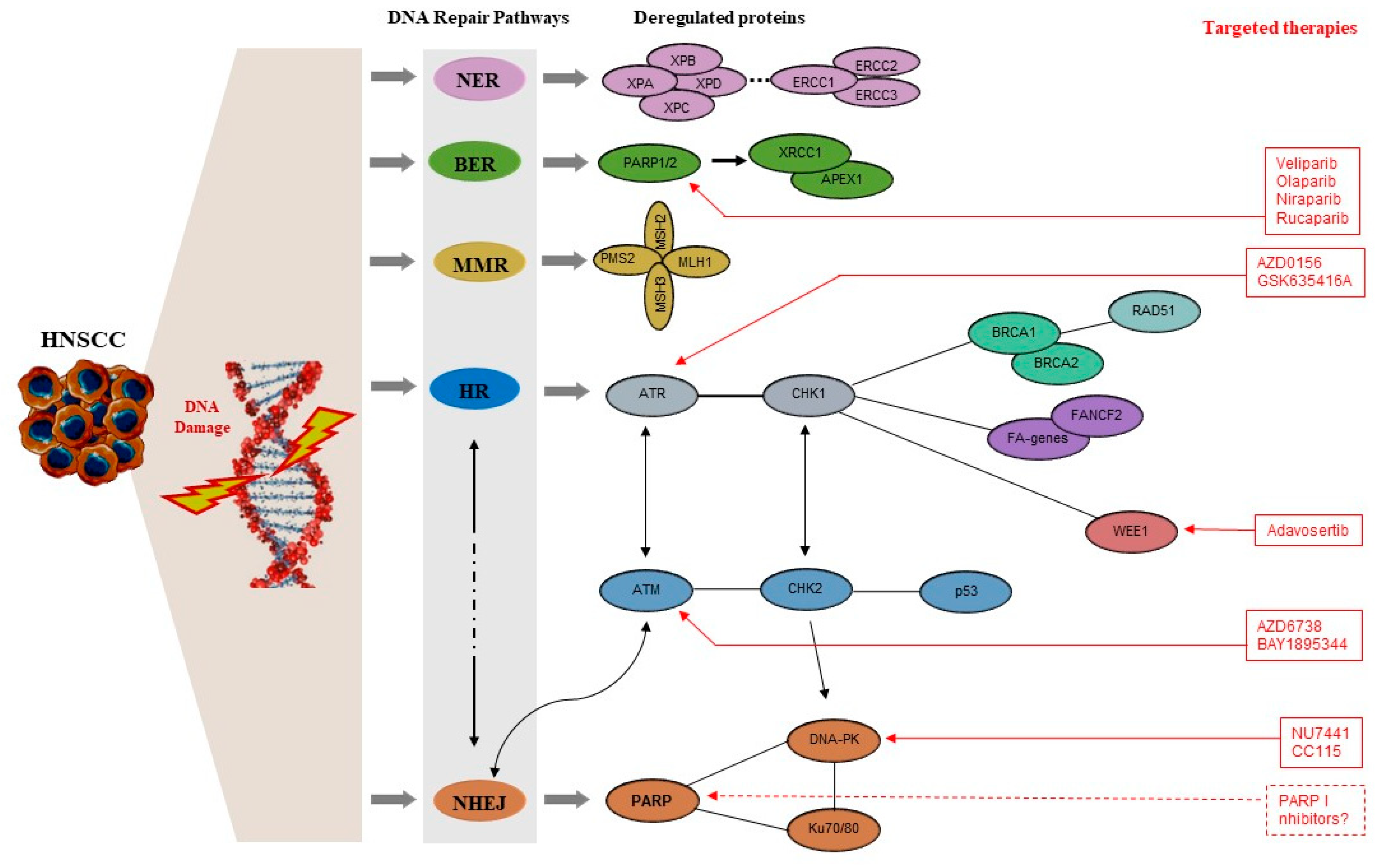
3. DDR Mechanisms—A Brief Overview

The cellular response to DNA damage and replication stress is the activation of the DDR mechanisms, which signal the presence of DNA damage and promote its repair, or, in some cases, lead the cell to apoptosis in order to prevent the spread of damage. Apart from NHEJ and HR, several other DNA repair pathways are also included in the DDR armamentarium. These are as follows:
(i)
Alternative end joining (A-EJ) is a recently described mechanism that repairs DSBs through a subset of A-EJ pathways and relies on microhomology-mediated repair. The best characterized Alt-EJ pathway is the microhomology end joining (MMEJ) pathway; this requires 2 to 20 nucleotides of homologous sequence and shares the same first steps of DDR with HR, i.e., the repair process begins with end resection and involves many of the factors that contribute to the HR end resection machinery. These mechanisms are considered to be error-prone and highly mutagenic as they are associated with deletions flanking the original DSB, leading to chromosomal rearrangements and genomic instability [91,92,93,94].
(ii)
Nucleotide excision repair (NER) is a mechanism that repairs bulky DNA lesions that distort the DNA double helix and are usually caused by external mutagens, such as UV light and chemical carcinogenic substances [91,95]. NER is divided into two sub-pathways: global genome NER (GG-NER), which takes place throughout the genome, independently of transcriptional processes, and transcription-coupled NER, which only occurs in order to repair the damage on the transcribed strand [96,97,98].
(iii)
Mismatch repair (MMR) is a mechanism that repairs base–base mismatches, deletions, or insertions predominantly generated during DNA replication and recombination processes or mismatches that escape DNA polymerase proofreading activity. MMR is considered a highly conserved biological pathway and plays an important role in genome maintenance [91,99].
(iv)
Base excision repair (BER) is a mechanism that tends to repair DNA damage (small base lesions) that originate from endogenous sources, such as those attributed to ROS, alkylation, deamination, and methylation, and which do not create structural distortions of the DNA double helix [91,100]. BER is initiated by a DNA glycosylase that recognizes and removes the damaged base, leaving an empty space that is further processed by short-patch repair or long-patch repair to complete the repair mechanism. A study in a Pakistani population suggested that the deregulation of genes in the BER pathway may drive HNSCC progression [101].
Below, we discuss proteins related to DDR mechanisms that have been found mutated or aberrantly expressed in HNC cell lines or patients, and their implication in the development, prognosis, and therapy of HNCs.

4. DDR Molecules with Evidence of Implication in the Pathogenesis of HNC

4.1. DDR Signaling Kinases

In mammalian cells, the main upstream kinases orchestrating the DDR signaling pathway are the ATM (ataxia telangiectasia mutated) and ATR (ATM- and Rad3-related) serine/threonine kinases, while the DNA-PKcs (DNA-dependent protein kinase catalytic subunit) is a DDR sensor regulating a smaller number of targets and plays a primary role in NHEJ [102,103]. However, the upstream molecules of DDR not only play a crucial part in the repair of DNA lesions, but they are also implicated in the regulation of the cell cycle, cell cycle arrest, and apoptosis. For this reason, DDR plays a critical role in tumor initiation and progression, and in the response of HNCs to radiotherapy. In this respect, cell cycle checkpoint mechanisms constitute an important part of the DNA damage response, as they either pause the cell cycle to repair the DNA damage or, in cases where the damaged DNA cannot be repaired, they signal programmed cell death in order to minimize the transmission of DNA errors [104].

4.1.1. Ataxia Telangiectasia Mutated (ATM)

ATM is a well-characterized serine-threonine kinase, primarily activated upon DSBs, which are considered the most deleterious DNA lesions invoked by replication stress [102]. A key regulator of ATM activation upon the formation of DSBs is the MRN complex (MRE11-RAD50-NBS1), which is usually responsible for the rapid localization of ATM to DSBs [102]. Although there is enough evidence to support that MRN acts as a DSB sensor, the way that it activates the ATM and ATR kinases has not been completely elucidated [102]. ATM is able to autophosphorylate, a property that promotes its association with Mdc1 and its accumulation at DSBs [102,105,106]. The activation of ATM is followed by phosphorylation of downstream molecules such as H2AX, BRCA1, Chk2, p53, and Chk2, among others, promoting DNA repair via HR, or leading the cell to apoptosis [102,107,108,109,110]. Germline inactivation of ATM leads to Ataxia–Telangiectasia Syndrome, an inherited disorder characterized by DNA repair defects and subsequent genomic instability [111]. These patients have been associated with an increased risk (one in three affected individuals) of developing HNSCC and gynaecological squamous cell carcinomas, with the onset of the disease usually occurring during early adulthood [112,113]. In addition, in certain tumors, ATM gene aberrations usually involve somatic mutations [114], while ATM promoter hypermethylation is also evident in a significant proportion of HNCs (25%), with the resulting downregulation of ATM expression being associated with poor prognosis [71,115,116]. Recent evidence also suggests that an impaired ATM-orchestrated DNA damage response, irrespectively of ATM expression levels, might be responsible for the radiosensitivity of HPV(+) HNSCCs, even though the exact molecular mechanism remains unknown [100]. Indeed, the interaction between ATM and the E7 viral oncoprotein has been described to induce the phosphorylation and activation of the DNA damage protein kinase CHK2 in HPV(+) cells, which is required for HPV differentiation and genome amplification [117].

4.1.2. ATM- and Rad3-Related (ATR)

In contrast to ATM, ATR responds to a wide variety of DNA damage, in addition to DSBs [102,111]. ATR, along with its partner, ATRIP, are localized to sites of single-stranded DNA (ssDNA), and this may constitute the trigger that induces the ATR response in DSBs generated during the S and G2 phases of the cell cycle, i.e., resected DSBs or DSBs associated with stalled replication forks [102,111,118]. Additionally, ATR appears to be implicated in a mechanism that prevents the premature onset of mitosis, while it is activated by replication origins arising during the S phase [119]. Due to the plausible role of ATR in mammalian cell DNA integrity, mutation rates in this gene are quite low, with the exception of a rare genetic syndrome, the Seckel syndrome, which is characterized by biallelic ATR mutations [111]. Nonetheless, it has been estimated that 4.36% of patients with HNC present with ATR gene mutations [120].

4.1.3. Chk1 and Chk2

Control of the cell cycle mainly involves two checkpoint kinases, Chk1 and Chk2, both of which are serine/threonine kinases. Chk2 is involved in all three main cell cycle checkpoints, i.e., the G1/S checkpoint, the intra-S checkpoint, and the G2/M checkpoint [121]. It is mainly activated by ATM upon DNA double-strand breakage, and activation involves autophosphorylation and dimerization [121]. Chk1 is not as stable as Chk2; its activation is restricted to the S and G2 phases [122] with the involvement of ATR in single-strand breaks (SSBs) [123]. However, ATM is also required for the activation of Chk1 in response to DSBs and for repair through homologous recombination [123].

4.1.4. p53-Binding Protein 1 (53BP1)

53BP1 also participates in the activation of Chk1 and Chk2 and in the repair process [124]. Specifically, 53BP1 is rapidly recruited to DSBs and mediates their repair in mammalian cells. In addition, it is actively implicated in the protection of the replication forks during cellular replication stress responses, as defective ATR-Chk1-p53 signaling and caspase 3-mediated cell death are observed in the absence of 53BP1 [125]. During the G1 phase, 53BP1 has been reported to promote the repair of DSBs through the NHEJ mechanism and, in this way, to counteract repair via HR, i.e., through DNA end resection. Rap1-interacting factor 1 (RIF1) and Pax transactivation domain-interacting protein (PTIP) have been shown to interact with 53BP1 in an ATM-dependent manner and to act as key effectors that allow 53BP1 to block DSB resection in G1 and to promote NHEJ [126,127,128]. In contrast, during the G2-S phase, BRCA1 and CtIP have been shown to antagonize the actions of 53BP1-RIF1 and 53BP1-PTIP in order to promote HR repair; BRCA1 appears to recruit CtIP to DSBs and to promote DNA end resection, but the exact mechanism has not been elucidated yet [129,130]. Overall, it is generally accepted that 53BP1-RIF1 and 53BP1-PTIP protein complexes guide the decision toward the NHEJ repair process, whereas the BRCA1-CtIP protein complex guides the decision toward the initiation of the HR process [130].
Research on human HNC samples has also revealed a perturbed activation of DDR, or rather a defect in the synchronization of the cell cycle during DDR activation, as evidenced by elevated expression of Ki-67, a protein marker for cell proliferation, and localization of 53BP1 foci in the nucleus [131]. The researchers proposed 53BP1 expression as a potential biomarker for estimating the genomic instability invoked by DSBs or by abnormally activated DDR in oral squamous epithelial lesions [131]; this is in agreement with the results of another study highlighting the potential usefulness of 53BP1 IF analysis as a diagnostic tool for Thyroid Follicular Tumors [132]. Preclinical data have also demonstrated that 53BP1 can regulate cell cycle arrest through the modulation of TP53, CHK1, and CHK2 gene expression in ESCC; specifically, targeted downregulation of the 53BP1 protein has been shown to cause a reduction in p53 gene expression, but to be negatively correlated with CHK1 and CHK2 gene expression [133].

4.2. Molecules of the Homologous Recombination (HR) Mechanism

In patients with HR-related defects, such as BRCA1 and BRCA2 deficiency, or in patients with FA syndrome, chromosomal instability is usually observed [48,134]. When the HR process is impaired, chromosomal translocations appear when two DSBs from different chromosomes (i.e., four DNA-free ends) are aberrantly joined via NHEJ, leading to genomic rearrangements that are associated with high genomic instability and cancer [135]. Additionally, chromothripsis, which results from multiple DSBs in a single chromosome, may be followed by chromosomal segments rejoined via NHEJ, driving carcinogenesis through the loss of tumor suppressor genes or the activation of oncogenes. Thus, the error-prone nature of NHEJ and HR impairment can lead to genomic rearrangements and carcinogenesis [135]. However, there is substantial evidence to suggest that certain genetic deficiencies are associated with a higher frequency of HR, and can therefore lead to genomic instability and increase the possibility of cancer occurrence [136].

4.2.1. FA Genes

Fanconi Anemia is a genetic disorder caused by the germline inactivation of one or more genes, and patients may present with bone marrow failure and a high predisposition to cancer [137]. The FA pathway is involved in ICL repair through HR-mediated mechanisms [90]. In this context, FA proteins act as DNA integrity factors through the stabilization of replication forks and the regulation of cytokinesis, in addition to their canonical role in ICL repair [90]. In FA patients, HNSCC is considered the most common type of solid cancer, associated with a 700-fold increased risk [137].
Defects in HR repair are not only implicated in the initiation of tumorigenesis, but are also associated with secondary and subsequent stages of carcinogenesis and tumor progression [136]. In sporadic HNSCC, this is evidenced by the downregulation of FA-related genes and FANCF gene silencing. Indeed, functional defects in DNA crosslink repair have been observed in a significant proportion of HNSCC cell lines, in terms of MMC-hypersensitivity, G2-blockade, and olaparib (PARP-inhibitor) hypersensitivity [138]. The mono-ubiquitylation of FANCD2 protein, a necessary prerequisite for the activation of the FA pathway, also appears to be compromised in certain HNC cell lines, while FA/HR-related gene variants are present in 19% of HNSCC tumors and in 24% of cell lines, and defects are associated with worse prognosis [138].
The carcinogen acetaldehyde has been shown to activate the ATR-Chk1-dependent damage response mechanisms leading to S and G2/M cell cycle arrest in esophageal epithelial cells (keratinocytes), followed by FA pathway activation-directed repair [139]. In this context, depletion of FANCD2 protein has been associated with excessive DNA damage and apoptotic cell death, suggesting that it is an important contributor to the repair of acetaldehyde-induced DNA damage in these cells [139]. Such data highlight potential mechanisms of excessive alcohol consumption-related tumorigenesis, as well as mechanisms implicated in genomic instability that increase the risk of developing SCCs in FA patients. Future studies will determine whether these mechanisms also apply to HNSCC tumors.

4.2.2. BRCA1 and BRCA2

As mentioned earlier, BRCA1 and BRCA2 have essential functions in the HR repair pathway, and breast and ovarian tumors with defects in these genes can be successfully treated with PARP inhibitors [140]. Molecular profiling of HNSCC patient samples has identified BRCA1 and BRCA2 mutations with a frequency of 5.75% and 9.2%, respectively, which seem to co-exist with TP53 and PI3KCA pathway mutations [141]. However, the clinical significance of most detected variants remains to be determined.

4.2.3. RAD51

The RAD51 protein has essential roles in successful DSB repair via homologous recombination; it binds to ssDNA near the repair sites and exhibits DNA-dependent ATPase activity [142,143]. By catalyzing the recognition of homology and strand exchange, RAD51 contributes to re-synthesizing the lost sequence [144]. Xrcc3 (X-ray repair cross-complementing group 3; 14q32.3) is responsible for the accumulation of RAD51 at sites of DSBs in the cell nucleus and for the enzymatic resolution of the resulting Holliday junction [145].
Two RAD51 single nucleotide polymorphisms (SNPs) have been identified as potential regulators of mRNA stability, altered translational efficiency, and the potential to induce carcinogenesis: 135GC (rs1801320) and 172GT (rs1801321) [146]. A case study revealed a possible association of RAD51 (135GC genotype) and XRCC3 (722CT and 722TT genotypes) gene polymorphisms with an increased risk of HNSCC occurrence in the Polish population [147]. The RAD51 135GC polymorphism, in particular, already associated with an increased risk of developing breast cancer and acute myeloid leukemia, has also been linked to an increased risk of developing HNC in other populations, especially among smokers and drinkers [148]. Interestingly, a meta-analysis revealed that whereas the RAD51 135GC polymorphism is associated with a high susceptibility to HNC and esophageal cancer among Caucasians, the G172T polymorphism appears to confer a potential protective effect against HNC among the same population, as it is associated with a decreased risk of HNC occurrence [146,149].
In OSCC patients, RAD51 protein expression levels are significantly increased compared to healthy individuals [150]. Notably, among OSCC patients, those with lymphatic metastases appear to have significantly higher RAD51 gene expression, suggesting that the respective protein may be involved in lymph node metastasis. Additionally, RAD51 expression has been related to the stage of malignancy, with significantly higher levels being observed in poorly differentiated tissues, as compared to moderately or well-differentiated tissues, further highlighting its potential to be used as a biomarker for OSCC staging [150].
Increased RAD51 mRNA expression levels are correlated with worse prognosis in HPV(+) HNSCC tumors; the combination of a RAD51 inhibitor with a Wee1 inhibitor has been shown to significantly inhibit tumor growth in mice with HPV(+) HNSCC tumors, as compared to mice with HPV(−) HNSCC, suggesting that HPV(+) tumors may be more dependent on DDR mechanisms in order to counteract the effects of increased replication stress (due to the transformation by the viral oncogenes E6 and E7) [151]. In the same context, the inhibitors B02 and AZD1775 have been shown to act as radiosensitizers to HPV(−) HNSCC tumors in vitro and in vivo [151].

4.3. Molecules of the Non-Homologous End Joining (NHEJ) Mechanism

4.3.1. DNA-Dependent Protein Kinase (DNA-PK)

DNA-PK is a serine/threonine-protein kinase that possesses a catalytic subunit (DNA-PKcs) and a Ku heterodimer, consisting of the Ku70 and Ku80 subunits; it is implicated in DDR mechanisms and other processes that are associated with the maintenance of DNA integrity, such as cell cycle progression, transcription, and telomere maintenance [152]. As with the ATM kinase, DNA-PK mainly responds to DSBs and mediates their repair. In particular, the Ku heterodimer recognizes and binds to the DSB and recruits and activates the DNA-PKcs to form the active DNA-PK complex, in this way promoting the NHEJ DNA repair mechanism [135]. Recent evidence suggests that DNA-PK is also involved in a complex regulatory network for the pathway choice between HR and NHEJ [152,153], while phosphorylation of T2609 and T3950 residues is implicated in radioresistance [153]. Interestingly, while mutational inactivation of DNA-PKcs has been shown to impair NHEJ, subsequently leading to DNA repair through HR, drug inhibition of DNA-PKcs has been shown to impair both pathways [152,154]. Overall, DNA-PKcs is implicated in cancer progression and metastasis [155]. In the context of HNCs, DNA-PK overexpression appears to be significantly associated with decreased survival in NPC patients, suggesting that it may be used as a prognostic biomarker for this particular HNC subtype [156]. In addition, DNA-PKcs overexpression has been associated with poor outcomes in patients with NPC undergoing intensity-modulated radiotherapy, suggesting an additional role in predicting response to therapy [157]. Notably, DNA-PK expression has been shown to increase following irradiation of OSCC, with the rate of increase being directly correlated with therapeutic resistance [158], and for this reason, several DNA-PK inhibitors are currently being investigated for their radiosensitizing properties [159].

4.3.2. Ku Protein

When HR mechanisms are not available, as for example outside of the S and G phases of the cell cycle or due to defects in key repair proteins, DSBs are predominantly repaired via the NHEJ mechanism. The Ku protein constitutes an essential player in this process, being a nuclear DNA-binding protein that recognizes DSBs and facilitates their repair [160]. Ku consists of two subunits, one that is approximately 70 kDa and another that is 80 kDa (Ku70 and Ku80, respectively) [161]. Overexpression of Ku80 is associated with an adverse prognosis in ESCC [162], a cancer type that is not classified as HNC but which is often accompanied by head and neck second primary tumors (in approximately 7% of ESCCs patients) [163]. In vitro experiments on both radiosensitive and radioresistant HNC cell lines have revealed that ku70/80 gene overexpression is associated with radioresistance and may therefore have some utility as a potential predictive marker of radioresistance in HNCs [160]. Additionally, siRNA-silencing of β-catenin in the radiosensitive AMC-HN-9 cell line has been shown to impair the radiation-induced overexpression of Ku80 in an AMPK signaling pathway-dependent manner [164]. Ku80 mRNA expression has also been negatively associated with the expression of checkpoint molecule PD-L1 (programmed cell death-1 ligand) in many cancer types, including HNCs, following irradiation [165]. Such observations provide substantial evidence that the NHEJ mechanism is implicated in the repair of DNA damage invoked by radiation and emphasize the necessity of discovering new NHEJ targets for chemotherapeutic radiosensitization of head and neck tumors.

4.3.3. PARP Molecules

PARP1 (Poly (ADP-ribose) polymerase-1) is another sensor known to detect DSBs; however, despite being involved in many cellular processes, such as cell proliferation, differentiation, the repair process of the SSBs and DSBs through different repair pathways, the stabilization of DNA replication forks and in the modification of chromatin structure, its implication in NHEJ is not fully understood [161]. Nonetheless, it has been observed that PARP1 and the Ku70/80 complex localize to DNA lesions considerably earlier than other DSB sensors, that PARP1 competes with the Ku70/80 complex at the DSBs in the S/G2 phases, and that the Ku complex can be replaced by PARP1 at DSB sites [166]. On the contrary, the Ku70/80 complex appears to occupy the DSB sites exclusively in the G1 phase, thereby suggesting that each sensor recognizes DSBs at different stages of the cell cycle; in addition, it has been postulated that PARP 1 may act as a regulator of the Ku70/80 complex in DSBs in the S/G2 phases, as it has been shown to enzymatically remove the Ku complex from these sites [166].
In addition, PARP5A and PARP5B are PARP molecules that also catalyze poly (ADP- ribose) PAR chains, but their role in cancer is still not fully deciphered. While PARP5B downregulation has been observed in prostate, breast, and HNCs [167,168], recent data demonstrate that null PARP5B tumor cells are characterized by an impairment of the NHEJ mechanism, and that treatment with a PARP5B inhibitor combined with low doses of etoposide drives SCC human cell lines into senescence and apoptosis [169]. The latter seems to be dependent on the actions of the NBS1 protein, which constitutes an essential component of the MRN (MRE11-RAD50-NBS1) complex; by accumulating the MRN complex at damaged sites, NBS1 amplifies ATM activation, which in turn activates and orchestrates DSB repair [170]. In another study, the consequential reduction in NSB1 expression following PARP5B inhibition was associated with the activation of ATR signaling, which in turn led to senescence and apoptosis, highlighting PARP5B as a potential target for chemotherapeutic intervention in HNSCC [169].
Preclinical data from HNSCC cell lines and mouse models have demonstrated that NSB1 gene downregulation is associated with disruption of HR and increased sensitivity to PARP inhibitors; in addition, co-inhibition of the MRN complex and PARP has been shown to induce DSB accumulation, telomere shortening, and ultimately cell death [167]. Such observations indicate that simultaneous inhibition of genes/molecules that participate in two or more DNA repair pathways may induce a cytotoxic response that has been described as synthetic lethality [167,171]. The latter only happens in the presence of another specific defect, in this case, a defect in a DNA repair gene, and is best exemplified with the use of PARP inhibitors in cancer cells carrying defects in BRCA1 or BRCA2 genes [172]. In the context of BRCA1/BRCA2 deficient cells, PARP inhibition tends to accumulate SSBs arising during normal cellular activity and to result in DSBs that are formed through the collision of SSBs with a DNA replication fork; these DSBs require HR repair that cannot take place in the absence of fully functional BRCA1/BRCA2 proteins and ultimately induce cell death [172,173]. Interestingly, the observation that PARP inhibition in HR deficient cells can still lead to synthetic lethality but may not be necessarily accompanied by SSB accumulation has led to the conclusion that PARP1 must also be actively implicated in the Alt-NHEJ repair process, which is believed to take place in HR- and/or NHEJ-defective cancer cells [94]. This could have tremendous implications for the development of therapeutic synthetic lethality approaches in malignancies carrying defects in the two major DSB repair pathways, including HNCs [174,175].

4.4. Molecules of the Nucleotide-Excision Repair (NER), Base-Excision Repair (BER), and Mismatch Repair (MMR) Mechanisms

4.4.1. NER-Associated Genes

These are genes that appear to be downregulated in HNSCC patients and include ERCC1, ERCC2/XPD, XPA, and XPC [11]. Low expression levels of ERCC3 and XPA in lymphoblastoid cells and low levels of XPB in lymphocytes are commonly observed in HNSCC patients and have been associated with an increased risk of developing the disease [176]. In addition, NER-associated genes have been proposed as biomarkers with potential value in predicting treatment efficacy and outcome in HNSCC [177]. However, while certain studies have found an association between ERCC1 and XPA upregulation with poor OS in patients with OSCC, others have noted better OS [178]. Notably, these genes have been proposed as a biomarker for the prediction of 5-FU and cisplatin-based chemotherapy in HNSCC patients [179,180].

4.4.2. BER-Associated Genes

APEX1 and XRCC-1 have recently been found to be downregulated in patients with HNSCC [11]. Overexpression of these genes has been correlated with better clinical staging and negative lymph node metastasis in patients with OTSCC [179,181].

4.4.3. MMR-Associated Genes

Downregulation of MLH1, MSH2, MLH3, and PMS2 genes, all key molecules of the MMR mechanism, has been associated with reduced DNA repair capacity, development of malignant lesions in OSCC, and tobacco-induced methylation in smokers [179].
Interestingly, a prognostic 13-DNA-repair-gene signature has recently been identified and shown to accurately and independently predict the clinical outcome of patients with HNSCC [182]; the signature includes the MORF4L2, COPS2, USP10, WAS, UVSSA, PRRX1, ZBTB1, DCLRE1C, MSH5, DOT1L, ZBTB7A, POLR2C, and MORF4L1 genes, known to be involved in a variety of DNA repair pathways, including NER, BER, MMR, and DSB repair [182,183].
Figure 1 schematically represents the DNA repair pathways that are implicated in the pathogenesis of HNC and the proteins that have been found to be deregulated in these pathways.

4.5. Genes That Have Been Functionally Related to HPV(+) HNSCCs

HPV oncogenes E6 and E7 are responsible for tumor initiation and immortalization, and are known to deregulate the onco-suppressor gene TP53 and retinoblastoma protein (pRb), thereby leading to deregulation of the cell cycle and inhibition of apoptosis [184,185,186]. Relevant information from the TCGA database indicates that a great proportion of oropharyngeal and tonsil tumors are HPV(+) without TP53 gene mutations, while HNSCCs in other sites are more prone to developing TP53 mutations and appear to have lower rates of HPV infection [82]. This inverse relationship may reflect the important role of the viral oncoproteins E6 and E7 in oropharyngeal and tonsil HPV-related tumorigenesis [82]. On the other hand, TP53 gene mutations are implicated in HPV(−) HNC tumorigenesis, as evidenced through experiments in HNC cell lines and animal models; E7 has been shown to increase the radiation-induced DNA damage, to decrease the sub-lethal DNA damage repair, and to lead to overexpression of the HR-related protein RAD51 [187].
In addition, current evidence suggests that HPV-associated tumors exhibit higher radiosensitivity as compared to HPV(−) cell lines through a DDR-dependent manner. Specifically, Jonathan E. Leeman et al. demonstrated that the HPV16 E7 oncoprotein leads to the suppression of cNHEJ and promotes the MMEJ mechanism, which is considered to be error-prone and highly mutagenic [78]. In addition, HPV(+) HNSCCs appear to be deficient in TGF-β signaling, which is associated with a perturbed HR function and increased reliance on Alt-NHEJ for repair [188].
The presence of HPV infection in HNCs has also been correlated with overexpression of the p16 gene, and this may result from pRb inactivation via the E7 HPV protein [189]. P16 protein constitutes an important cell cycle regulator, acting as a CDK inhibitor, and for this reason, alterations in p16 gene expression are frequently observed in several malignancies [189]. Nonetheless, HPV(−) malignancies characterized by p16 overexpression have also been identified [190]. Currently, the evaluation of p16 expression is used as a surrogate marker for HPV infection in HNCs, but the data regarding its diagnostic value are rather contradictive. While a great proportion of oropharyngeal squamous cell carcinomas (OPSCCs), especially tonsillar and tongue base carcinomas (TSCC/BOTSCC), appear to contain the HPV genome and evaluation of p16 gene expression has successfully been used for the estimation of prognosis and recurrence of the disease [191], some researchers propose the cautious use of p16 expression status for the diagnosis of HPV(+) HNSCCs in non-tonsillar, non-base of tongue oropharyngeal cancers [192]. In OSCCs such as oral tongue cancers, for example, HPV infection and p16 overexpression do not seem to coincide [193]. Other studies indicate that HPV infection in OSCCs does not affect the survival outcome of the disease [194] and that p16 overexpression may be the result of other pathways or cellular mechanisms and not the consequence of HPV infection [193]. However, additional large-scale studies need to be performed in order to draw conclusions on the diagnostic role of p16 expression in the different types of HNCs.
Table 1 summarizes the genes involved in the DDR pathways and how these are deregulated in the pathogenesis of HNC.

5. Novel Therapeutic Targets: Preclinical and Clinical Evidence

The significant inter- and intratumoral heterogeneity of HNSCCs make targeted therapy a real challenge. Current treatment approaches include surgery, radiation therapy, chemotherapy, targeted therapy, and immunotherapy or combinations of these treatment modalities [196]. One of the most common treatment options is photon-based radiotherapy (XRT), either alone or with chemotherapy agents such as methotrexate, platinum compounds with or without 5-fluorouracil, and cisplatin [196,197,198]. However, treatment approaches often have severe side effects with unfavorable patient outcomes (i.e., the 5-year overall survival rate after XRT treatment does not exceed 40% for HPV(−) OSCC stages III to IV) [199]. Successful treatment is usually impeded by tumor-related mechanisms which repair the radiotherapy- and/or chemotherapy-induced DNA damage, leading to radioresistance and/or chemoresistance of the tumor [196]. Consequently, there is a need for new effective treatment strategies with the lowest possible toxicity. When it comes to DNA damage response mechanisms, efforts for successful therapy have concentrated on the design of new targeted drugs and on the identification of appropriate drug and radiotherapy combinations, based on specific molecular characteristics of the tumor, in order to generate lethal DNA damage, such as DSBs, and prevent their repair, leading to cell death. Table 2 includes a list of DDR inhibitors that are currently being tested in clinical trials, whereas Table 3 includes the DDR inhibitors that are being investigated at the pre-clinical stage.

5.1. PARP Inhibitors

The most attractive therapeutic targets are poly (ADP-ribose) polymerases (PARPs), as these are implicated in the regulation of more than one DNA repair pathway (BER, NER, Alt-NHEJ, NHEJ, MMEJ) [200]. As mentioned earlier, PARP inhibition was first described in cancer cells with defective BRCA1/BRCA2 protein function, and then clinical trials confirmed the effectiveness of PARP inhibitors as monotherapy in patients with BRCA1(−/−) and BRCA2(−/−) tumors [196]. PARP inhibition was the first targeted therapy of synthetic lethality, based on the observation that inhibition of the BER mechanism leads to the formation and accumulation of SSBs and to the collapse of replication forks in highly replicating cancer cells, with subsequent development of DSBs that cannot be repaired through the high-fidelity HR mechanism in homozygous BRCA-defective cells, leading to apoptosis [195,196,201]. However, more recent studies indicate a potential role of PARPs in NHEJ, and propose that it is the deregulation of NHEJ that leads to synthetic lethality in BRCA2-deficient tumors, as experimental inhibition of key BER proteins in these tumors did not appear to have the same effect on PARP inhibition [202,203,204,205]. In addition to tumors with BRCA mutations, cells bearing HR protein deficiencies, for example ATM deficiency, also appear sensitive to PARP inhibition [196]. This observation supports the notion that PARP inhibitors may be effective in tumors bearing a variety of deficiencies in HR and other DDR mechanisms, which are estimated to constitute approximately 40% of HNC tumors [196].
Interestingly, PARP inhibitors are currently being tested in combination with chemotherapeutic agents, as well as with other inhibitors (i.e., EGFR inhibitors, as a great percentage of HNCs accumulate EGFR mutations) and with radiotherapy. Preclinical testing of the PARP inhibitor veliparib (ABT-888) in HNC cell lines and an HPV(+) patient xenograft has produced encouraging results; HPV(+) HNSCC cell lines that were deficient for BRCA2 and DNA-PK proteins, both key molecules of HR and NHEJ, exhibited sensitivity to the PARP inhibitor, as evidenced by a decreased survival rate in vitro and a delay in tumor growth in vivo [206]. Recently, the results of a phase I trial evaluating the maximum tolerated dose (MTD) and safety of veliparib in combination with carboplatin and paclitaxel induction chemotherapy (IC) for locoregionally advanced HNSCC demonstrated that veliparib is well tolerated, with promising survival data following an adequate follow-up interval [207].
On the other hand, HNSCC cell lines with SMAD4 homozygous deletion have been shown to exhibit sensitivity to olaparib, a well-studied PARP inhibitor, which appears to increase DNA damage and cell death in combination with RT in vivo [208]. In a phase I clinical trial, five out of six SMAD4-negative HNSCCs and four out of eight SMAD4-positive HNSCCs responded to standard treatment plus olaparib [208]. A recent study has highlighted the importance of PARP-1 in regulating the repair of complex DNA damage (CDD) induced by high-LET radiation by demonstrating that high-LET proton-induced CDD and concomitant inhibition of PARP-1 (with olaparib) leads to cell death in the HPV(−) HNSCC cell line UMSCC74A [209].
Another study on two HPV(+) and two HPV(−) HNSCC cell lines revealed that the PARP inhibitor niraparib enhanced their sensitivity to both photon and proton radiotherapy and increased the relative biological effectiveness (RBE) of protons, possibly by inhibiting DDR [210]. In addition, the combination of niraparib with the Chk1 inhibitor MK-8776 has been shown to enhance the radiosensitivity of HPV(+) HNSCC cells, whereas co-administration with the Wee1 inhibitor MK-1775 seems to further enhance the radiosensitivity of HPV(−) HNSCC cells in vitro [211]. An ongoing phase II study is currently evaluating the combination of niraparib with PD-1 blockade (dostarlimab) in patients with recurrent/metastatic HNSCC, with primary endpoints including stable disease, partial or complete response, and secondary endpoints including adverse events, progression-free survival (PFS) and overall survival (OS) [212].

5.2. Immune Checkpoint Inhibitors

PD-1 and its associated ligand, PD-L1 (Programmed death ligand 1), both constitute immune checkpoint molecules expressed on the surface of T-cells and cancer cells, respectively. The binding of PD-1 to PD-L1 produces a negative signal that leads to the inactivation of T cells and allows immune evasion of tumor cells, leading to treatment failure; therefore, inhibition of these molecules with specific antibodies (ICIs), represents an attractive therapeutic strategy [213]. A recent meta-analysis revealed that the use of anti-PD-1 monoclonal antibodies is more effective in smokers and in HPV(−) patients, whilst anti-PD-L1-based therapy appears to be more efficient in female patients with locally recurrent HNC and in HPV(+) patients [214]. Despite the fact that this type of immunotherapy appears to offer a therapeutic advantage in terms of tumor progression control and OS compared to chemotherapy, only a small subset of HNSCC patients really benefit from ICI treatment [214]. Current data demonstrate that despite the PD-L1 protein being over-expressed in over 50% of HNSCC tissues, only 15% of patients will respond to PD-1/PD-L1 inhibitors [214]. Nonetheless, such strategies may provide an alternative solution in cases of advanced disease where traditional therapy is not effective or where the therapeutic effect may be enhanced through a combination with other treatment approaches.
As mentioned earlier, EGFR is another molecular target for which monoclonal antibodies have been developed. The EGFR is a cell surface protein that binds to the Epidermal Growth Factor, activating downstream signaling cascades leading to DNA replication, cell proliferation, and differentiation [215]. Abnormal EGFR gene expression is associated with tumor progression in HNC, with overexpression being observed in approximately 90% of HNSCCs [196,216]. EGFR protein signaling is actively implicated in DDR mechanisms, while deregulation of this process has been linked to resistance to chemo and/or radiotherapy [217]. As a result, monoclonal antibodies against EGFR aim to downregulate its overexpression; cetuximab is currently the only FDA-approved anti-EGFR agent used for HNC treatment, whereas panitumumab, another EGFR-specific antibody, is under clinical evaluation for its effectiveness in patients with advanced HNSCC [218]. The response rate of HNSCCs to cetuximab has been estimated at approximately 20%, regardless of HPV status and EGFR overexpression [219]. Current investigations are focused on combination treatments that include EGFR and PARP inhibitors. The combination of cetuximab and olaparib with radiation seems to induce an increase in DNA lesions, senescence, apoptosis, and control of tumor growth both in vitro and in vivo [220]. The proposed mechanism behind these observations is that the combination inhibits the NHEJ-mediated DNA repair and induces senescence through p21 signaling in BRCA1/2 proficient HNSCC cells. The safety of this triple combination has previously been evaluated in a phase I clinical trial [221].

5.3. Tyrosine Kinase Inhibitors

Targeted therapy against EGFR can also be accomplished with Tyrosine Kinase Inhibitors (TKIs), which antagonize ATP binding to the tyrosine–kinase intracellular domain of EGFR [222]. In HNC clinical trials, reversible EGFR TKIs, such as gefitinib and erlotinib, do not seem to confer a therapeutic benefit either as monotherapy or in combination with radiation treatment, with the response rate of erlotinib estimated at 10–15% in HNSCC patients [223]; on the other hand, multitarget TKIs such as lapatinib (reversible dual EGFR and HER2 TKI), afatinib, and dacomitinib (both irreversible EGFR, HER2, and HER4 pan-HER TKIs) have shown promising results in clinical trials, in terms of improving the objective response rate in patients with HNSCC [216,224,225]. As HNSCC is a highly heterogeneous disease and targeted EGFR monotherapy has produced moderate results in treatment efficacy, and in some cases has even led to therapeutic resistance [196,216], future studies are expected to concentrate on the development of combination therapies. Despite the heterogeneous mutational landscape of HNSCCs, the TP53 gene is mutated in approximately 72% of tumors according to the TCGA database, thereby highlighting the respective p53 protein as a potential biomarker and a promising target for more effective treatment. In this context, it has already been reported that NHSCCs with TP53 mutations or HPV(+) tumors with aberrant TP53 expression are sensitive to a Wee1 inhibitor, AZD1775, either used as a single agent or in combination with other chemotherapeutic agents [226]. A recent phase I clinical trial investigating AZD1775 in combination with CDDP (cisplatin) and docetaxel in HNSCC patients has so far produced encouraging anti-tumor efficacy in patients with defects in TP53 gene expression [227].
Wee1 normally acts as an inhibitor of CDK1 and CDK2, playing a crucial role in the G2/M checkpoint of the cell cycle. In addition, Wee1 may be implicated in the cell-size checkpoint, in the sense that it prevents entry into mitosis before the cells have reached a specific size [228]. Upon excessive DNA damage, this may lead to a prolonged G2 phase, as the G2 checkpoint is responsible for repairing DNA damage [229]. Consequently, inhibition of Wee1, the principal gatekeeper of this checkpoint, represents a promising strategy for the treatment of HNSCC, as it may lead to overactivation of CDK1/2, increased replication stress, abrogation of DNA damage checkpoints and cleavage of stalled replication forks by the abnormally activated MUS81 and SLX4 endonuclease complex [228,230]. Recent data support the notion that the effectiveness of Wee1 inhibitors relies on an interplay between replication stress and cell cycle checkpoint failure, as evidenced through combined inhibition of PARP and Wee1, which appears to confer tumor-specific radiosensitization of HPV(+) HNSCC cells [174].

5.4. CDK Inhibitors

CDK inhibitors, by preventing or counteracting tumor resistance mechanisms to chemo- or radiotherapy, may also provide an efficacious alternative targeted therapy to HNC patients. Even though pan-CDK inhibitors have not been approved for clinical use due to severe side effects and low specificity, selective CDK inhibitors are currently being used in clinical practice or are under clinical evaluation [189,231]. Ribociclib, a CDK4/6i, has been shown to be effective in an HNSCC HPV(−) cell line, whereas cancer cells with endothelial mesenchymal transition (EMT) features and low expression of Rb appear to be less sensitive to it [232]. Additionally, both ribociclib and palbociclib (another CDK4/6 inhibitor) have been shown to enhance radiosensitivity in HNSCCs [189], and are therefore currently being investigated in combination with other inhibitors in recurrent, metastatic, and chemotherapy-resistant HNCs [233,234]. Further optimization is required for both pan- and selective CDK inhibitors in order to diminish unwanted side effects, and in this process it is essential to identify predictive biomarkers of effective CDK inhibition as well as optimum treatment combinations.

5.5. DNA-PK Inhibitors

DNA-PK inhibitors constitute another promising target for achieving more effective cancer treatment. As mentioned earlier in this review, in addition to actively contributing to the NHEJ process, DNA-PKcs is implicated in many cellular processes, such as innate immunity, pro-inflammatory signaling, metabolism, and other tumor-associated processes. To date, pre-clinical evaluation of DNA-PK inhibitors has produced more promising results in the radiosensitization of HNSCC lines, as compared to the clinically available PARP1 inhibitors, while the combination of PARP1 and DNA-PK inhibitors has been shown to enhance HPV(−) HNSCC inhibition in both mouse xenografts and cell culture, highlighting a potential clinical application in patients with HPV(−) HNSCC [235]. Indeed, combined administration of a DNA-PK inhibitor (NU7441) with olaparib and radiotherapy has been shown to inhibit tumor cell proliferation both in vivo and in vitro, further suggesting that the combination of these agents may be responsible for the inhibition of the CDK and ERK pathway-related kinases [235]. In addition, combined administration of the dual mTOR/DNA-PK Inhibitor CC-115 and radiotherapy has been shown to inhibit tumor growth and significantly reduce the migration rate in HNSCC cell lines [236].

5.6. ATM and ATR Inhibitors

Based on the role of ATM and ATR in DDR responses, inhibitors of these molecules are currently being investigated in several preclinical and clinical studies [175,237]. The ATR inhibitor AZD6738 appears to promote cisplatin sensitization and to increase apoptosis signaling, DNA damage, and radiosensitization that is correlated with CHK1-mediated abrogation of G2/M-arrest in HPV(+) and HPV(−) HNSCCs, as shown in both in vivo and in vitro studies [238,239]. The ATR inhibitor BAY 1895344 has also been shown to confer radiosensitization in preclinical studies of HNSCC, by enhancing radiotherapy-induced inflammation in the tumor microenvironment [240]. This process may be driven by the actions of NK cells and can be further enhanced through combination treatment with TIGIT and PD-1 targeted immunotherapy [241]. Such data emphasize the importance of clinical trials in validating the safety and efficacy of BAY 1895344 in clinical practice [242]. On the other hand, inhibition of ATM has been shown to reduce cell migration [243], while concomitant administration of AZD0156 (ATM inhibitor) and radiotherapy appears to reduce tumor growth and migration rate in both HPV(+) and HPV(−) HNSCC cell lines [236]. Additionally, the novel ATM inhibitor GSK635416A, in combination with olaparib, seems to effectively radiosensitize HNSCC cell lines, whereas GSK635416A as monotherapy effectively inhibits HR and is less cytotoxic to normal fibroblasts as compared to olaparib [244]. Such observations highlight GSK635416A as a potential radiosensitizing drug, and additional studies are warranted in order to confirm its clinical significance.
Figure 2 summarizes the DDR process in HNC and how this may result in therapeutic resistance or successful therapy, through the targeting of specific DDR molecules.

6. Conclusions

The complexity of HNC mandates a multidirectional approach for effective treatment. Pre-clinical and clinical studies are required to improve our understanding of the mechanisms implicated in HNC initiation and progression. In this regard, the investigation of the mechanisms underlying the development of chemo- and radio-resistance is expected to shed new light on alternative therapeutic combinations that will help to overcome the obstacles that arise from traditional anti-cancer modalities.
Among the best-known tumor-related mechanisms of resistance to therapy are enhanced DNA repair of the lesions invoked by chemo- and radiotherapy, gene amplification, inhibition of cell death, and resistance to apoptosis, as well as by alterations in cell cycle regulation and immune evasion. These obstacles can be overcome through the introduction of DDR inhibitors and of synthetic lethality treatments that will prevent the activation of alternative pathways. Similarly, the mechanisms employed by tumors to achieve immune evasion can be counteracted with the use of ICIs.
The increasing amount of data regarding the DDR pathways that are implicated in cancer development and progression, along with the investigation of gene polymorphisms associated with tumor initiation and progression, and the identification of specific gene mutations that correlate with therapeutic resistance, aim toward the establishment of reliable biomarkers for the timely prediction of tumor response to therapy. However, the development of new and effective treatment combinations demands not only the acquisition of new information through continuous research, but also the appropriate implementation of this information with existing knowledge in the field. Inhibitors that do not appear to be therapeutically beneficial as monotherapy could still offer a therapeutic advantage in combination treatments for a number of patients with specific tumor characteristics.
With the mechanisms of tumorigenesis, tumor resistance, and metastasis continuously being unraveled, research efforts are gradually shifting toward biomarker-based patient selection for anticancer therapy. Improved understanding and clarification of the DNA repair efficiency in HNC hold enormous potential for efficient identification and characterization of the genomic instability biomarkers that will guide therapeutic development and improve responses in cases with metastatic or resistant disease.

Author Contributions

Conceptualization, C.P., M.A. and V.Z.; writing—original draft preparation, C.P. and M.A.; review and editing, M.A., P.B., P.Z., A.T., M.G., S.P.F., C.N.B. and V.Z.; visualization, C.P., M.A., P.Z., A.T. and V.Z.; supervision, M.A. and V.Z. All authors have read and agreed to the published version of the manuscript.

Funding

This research has been co-financed by the European Regional Development Fund of the European Union and Greek National Funds through the Operational Program Competitiveness, Entrepreneurship and Innovation, under the call RESEARCH–CREATE–INNOVATE (project code: T2EDK-03266, project acronym and title: “BIOKAKETRA—Identification of genomic and tran-scriptomic prognostic bio-signatures in head and neck cancer”).

Institutional Review Board Statement

Not applicable.

Informed Consent Statement

Not applicable.

Data Availability Statement

Not applicable.

Conflicts of Interest

The authors declare no conflict of interest.

References

  1. Johnson, D.E.; Burtness, B.; Leemans, C.R.; Lui, V.W.Y.; Bauman, J.E.; Grandis, J.R. Head and neck squamous cell carcinoma. Nat. Rev. Dis. Primers 2020, 6, 92. [Google Scholar] [CrossRef] [PubMed]
  2. Auperin, A. Epidemiology of head and neck cancers: An update. Curr. Opin. Oncol. 2020, 32, 178–186. [Google Scholar] [CrossRef] [PubMed]
  3. Global Burden of Disease Cancer Collaboration; Fitzmaurice, C.; Abate, D.; Abbasi, N.; Abbastabar, H.; Abd-Allah, F.; Abdel-Rahman, O.; Abdelalim, A.; Abdoli, A.; Abdollahpour, I.; et al. Global, Regional, and National Cancer Incidence, Mortality, Years of Life Lost, Years Lived With Disability, and Disability-Adjusted Life-Years for 29 Cancer Groups, 1990 to 2017: A Systematic Analysis for the Global Burden of Disease Study. JAMA Oncol. 2019, 5, 1749–1768. [Google Scholar] [CrossRef] [PubMed]
  4. Torre, L.A.; Bray, F.; Siegel, R.L.; Ferlay, J.; Lortet-Tieulent, J.; Jemal, A. Global cancer statistics, 2012. CA Cancer J. Clin. 2015, 65, 87–108. [Google Scholar] [CrossRef]
  5. Bray, F.; Ferlay, J.; Soerjomataram, I.; Siegel, R.L.; Torre, L.A.; Jemal, A. Global cancer statistics 2018: GLOBOCAN estimates of incidence and mortality worldwide for 36 cancers in 185 countries. CA Cancer J. Clin. 2018, 68, 394–424. [Google Scholar] [CrossRef] [PubMed]
  6. Zheng, H.; Liu, H.; Lu, Y.; Li, H. Identification of a Novel Signature Predicting Overall Survival in Head and Neck Squamous Cell Carcinoma. Front. Surg. 2021, 8, 717084. [Google Scholar] [CrossRef]
  7. Baez, A. Genetic and environmental factors in head and neck cancer genesis. J. Environ. Sci. Health C Environ. Carcinog. Ecotoxicol. Rev. 2008, 26, 174–200. [Google Scholar] [CrossRef]
  8. Hashibe, M.; Brennan, P.; Benhamou, S.; Castellsague, X.; Chen, C.; Curado, M.P.; Dal Maso, L.; Daudt, A.W.; Fabianova, E.; Fernandez, L.; et al. Alcohol drinking in never users of tobacco, cigarette smoking in never drinkers, and the risk of head and neck cancer: Pooled analysis in the International Head and Neck Cancer Epidemiology Consortium. J. Natl. Cancer Inst. 2007, 99, 777–789. [Google Scholar] [CrossRef]
  9. Kawakita, D.; Matsuo, K. Alcohol and head and neck cancer. Cancer Metastasis Rev. 2017, 36, 425–434. [Google Scholar] [CrossRef]
  10. de Sanjose, S.; Serrano, B.; Tous, S.; Alejo, M.; Lloveras, B.; Quiros, B.; Clavero, O.; Vidal, A.; Ferrandiz-Pulido, C.; Pavon, M.A.; et al. Burden of Human Papillomavirus (HPV)-Related Cancers Attributable to HPVs 6/11/16/18/31/33/45/52 and 58. JNCI Cancer Spectr. 2018, 2, pky045. [Google Scholar] [CrossRef] [Green Version]
  11. Psyrri, A.; Gkotzamanidou, M.; Papaxoinis, G.; Krikoni, L.; Economopoulou, P.; Kotsantis, I.; Anastasiou, M.; Souliotis, V.L. The DNA damage response network in the treatment of head and neck squamous cell carcinoma. ESMO Open 2021, 6, 100075. [Google Scholar] [CrossRef]
  12. Cooper, J.S.; Pajak, T.F.; Forastiere, A.A.; Jacobs, J.; Campbell, B.H.; Saxman, S.B.; Kish, J.A.; Kim, H.E.; Cmelak, A.J.; Rotman, M.; et al. Postoperative concurrent radiotherapy and chemotherapy for high-risk squamous-cell carcinoma of the head and neck. N. Engl. J. Med. 2004, 350, 1937–1944. [Google Scholar] [CrossRef] [PubMed]
  13. Jenkins, G.; O’Byrne, K.J.; Panizza, B.; Richard, D.J. Genome stability pathways in head and neck cancers. Int. J. Genom. 2013, 2013, 464720. [Google Scholar] [CrossRef] [PubMed]
  14. Osman, N.; Elamin, Y.Y.; Rafee, S.; O’Brien, C.; Stassen, L.F.; Timon, C.; Kinsella, J.; Brennan, S.; O’Byrne, K.J. Weekly cisplatin concurrently with radiotherapy in head and neck squamous cell cancer: A retrospective analysis of a tertiary institute experience. Eur. Arch. Otorhinolaryngol. 2014, 271, 2253–2259. [Google Scholar] [CrossRef] [PubMed]
  15. Wu, J.; Galvan, K.J.; Bogard, R.D.; Peterson, C.E.; Shergill, A.; Crowe, D.L. DNA Double-strand Break Signaling Is a Therapeutic Target in Head and Neck Cancer. Anticancer Res. 2021, 41, 5393–5403. [Google Scholar] [CrossRef]
  16. Cramer, J.D.; Burtness, B.; Le, Q.T.; Ferris, R.L. The changing therapeutic landscape of head and neck cancer. Nat. Rev. Clin. Oncol. 2019, 16, 669–683. [Google Scholar] [CrossRef] [PubMed]
  17. Ferris, R.L. Immunology and Immunotherapy of Head and Neck Cancer. J. Clin. Oncol. 2015, 33, 3293–3304. [Google Scholar] [CrossRef]
  18. Chow, L.Q.M. Head and Neck Cancer. N. Engl. J. Med. 2020, 382, 60–72. [Google Scholar] [CrossRef]
  19. Mito, I.; Takahashi, H.; Kawabata-Iwakawa, R.; Ida, S.; Tada, H.; Chikamatsu, K. Comprehensive analysis of immune cell enrichment in the tumor microenvironment of head and neck squamous cell carcinoma. Sci. Rep. 2021, 11, 16134. [Google Scholar] [CrossRef]
  20. Wiseman, S.M.; Stoler, D.L.; Anderson, G.R. The role of genomic instability in the pathogenesis of squamous cell carcinoma of the head and neck. Surg. Oncol. Clin. N. Am. 2004, 13, 1–11. [Google Scholar] [CrossRef]
  21. Moon, J.J.; Lu, A.; Moon, C. Role of genomic instability in human carcinogenesis. Exp. Biol. Med. 2019, 244, 227–240. [Google Scholar] [CrossRef] [PubMed]
  22. Thompson, S.L.; Bakhoum, S.F.; Compton, D.A. Mechanisms of chromosomal instability. Curr. Biol. 2010, 20, R285–R295. [Google Scholar] [CrossRef] [PubMed]
  23. Gisselsson, D. Intratumor diversity and clonal evolution in cancer—A skeptical standpoint. Adv. Cancer Res. 2011, 112, 1–9. [Google Scholar] [CrossRef] [PubMed]
  24. Loeb, L.A.; Bielas, J.H.; Beckman, R.A. Cancers exhibit a mutator phenotype: Clinical implications. Cancer Res. 2008, 68, 3551–3557. [Google Scholar] [CrossRef]
  25. Halazonetis, T.D.; Gorgoulis, V.G.; Bartek, J. An oncogene-induced DNA damage model for cancer development. Science 2008, 319, 1352–1355. [Google Scholar] [CrossRef] [PubMed]
  26. Gorgoulis, V.G.; Vassiliou, L.V.; Karakaidos, P.; Zacharatos, P.; Kotsinas, A.; Liloglou, T.; Venere, M.; Ditullio, R.A., Jr.; Kastrinakis, N.G.; Levy, B.; et al. Activation of the DNA damage checkpoint and genomic instability in human precancerous lesions. Nature 2005, 434, 907–913. [Google Scholar] [CrossRef]
  27. Alrawi, S.J.; Stoler, D.; Tan, D.; Dayton, M.; Loree, T.; Gibbs, J.F.; Rigual, N.; Sait, S.; Khoury, T.; Hicks, W., Jr.; et al. Genomic Instability, DNA Alterations and Tumor Eosinophilic Expression in Head and Neck Squamous Cell Carcinoma. Cancer Genom. Proteom. 2005, 2, 307–316. [Google Scholar]
  28. Walczak, A.; Rusin, P.; Dziki, L.; Zielinska-Blizniewska, H.; Olszewski, J.; Majsterek, I. Evaluation of DNA double strand breaks repair efficiency in head and neck cancer. DNA Cell Biol. 2012, 31, 298–305. [Google Scholar] [CrossRef]
  29. Quennet, V.; Beucher, A.; Barton, O.; Takeda, S.; Lobrich, M. CtIP and MRN promote non-homologous end-joining of etoposide-induced DNA double-strand breaks in G1. Nucleic Acids Res. 2011, 39, 2144–2152. [Google Scholar] [CrossRef]
  30. Zhao, X.; Wei, C.; Li, J.; Xing, P.; Li, J.; Zheng, S.; Chen, X. Cell cycle-dependent control of homologous recombination. Acta Biochim. Biophys. Sin. 2017, 49, 655–668. [Google Scholar] [CrossRef]
  31. Willers, H.; Azzoli, C.G.; Santivasi, W.L.; Xia, F. Basic mechanisms of therapeutic resistance to radiation and chemotherapy in lung cancer. Cancer J. 2013, 19, 200–207. [Google Scholar] [CrossRef] [PubMed] [Green Version]
  32. Housman, G.; Byler, S.; Heerboth, S.; Lapinska, K.; Longacre, M.; Snyder, N.; Sarkar, S. Drug resistance in cancer: An overview. Cancers 2014, 6, 1769–1792. [Google Scholar] [CrossRef] [PubMed]
  33. Nie, X.; Guo, E.; Wu, C.; Liu, D.; Sun, W.; Zhang, L.; Long, G.; Mei, Q.; Wu, K.; Xiong, H.; et al. SALL4 induces radioresistance in nasopharyngeal carcinoma via the ATM/Chk2/p53 pathway. Cancer Med. 2019, 8, 1779–1792. [Google Scholar] [CrossRef] [PubMed]
  34. Di, M.; Wang, M.; Miao, J.; Chen, B.; Huang, H.; Lin, C.; Jian, Y.; Li, Y.; Ouyang, Y.; Chen, X.; et al. CHAF1B induces radioresistance by promoting DNA damage repair in nasopharyngeal carcinoma. Biomed. Pharmacother. 2020, 123, 109748. [Google Scholar] [CrossRef]
  35. Wang, Z.; Zuo, W.; Zeng, Q.; Li, Y.; Lu, T.; Bu, Y.; Hu, G. The Homologous Recombination Repair Pathway is Associated with Resistance to Radiotherapy in Nasopharyngeal Carcinoma. Int. J. Biol. Sci. 2020, 16, 408–419. [Google Scholar] [CrossRef]
  36. Lodovichi, S.; Cervelli, T.; Pellicioli, A.; Galli, A. Inhibition of DNA Repair in Cancer Therapy: Toward a Multi-Target Approach. Int. J. Mol. Sci. 2020, 21, 6684. [Google Scholar] [CrossRef]
  37. Curtin, N.J. Inhibiting the DNA damage response as a therapeutic manoeuvre in cancer. Br. J. Pharmacol. 2013, 169, 1745–1765. [Google Scholar] [CrossRef]
  38. Chow, J.P.; Man, W.Y.; Mao, M.; Chen, H.; Cheung, F.; Nicholls, J.; Tsao, S.W.; Li Lung, M.; Poon, R.Y. PARP1 is overexpressed in nasopharyngeal carcinoma and its inhibition enhances radiotherapy. Mol. Cancer Ther. 2013, 12, 2517–2528. [Google Scholar] [CrossRef]
  39. Chen, Y.; Zhao, Y.; Lu, R.; Zhao, H.; Guo, Y. Identification and Validation of a Novel Genomic Instability-Associated Long Non-Coding RNA Prognostic Signature in Head and Neck Squamous Cell Carcinoma. Front. Cell Dev. Biol. 2021, 9, 787766. [Google Scholar] [CrossRef]
  40. Pikor, L.; Thu, K.; Vucic, E.; Lam, W. The detection and implication of genome instability in cancer. Cancer Metastasis Rev. 2013, 32, 341–352. [Google Scholar] [CrossRef]
  41. Vaish, M.; Mittal, B. DNA mismatch repair, microsatellite instability and cancer. Indian J. Exp. Biol. 2002, 40, 989–994. [Google Scholar] [PubMed]
  42. Piccolo, S.R.; Frey, L.J. Somatic mutation signatures of cancer. AMIA Annu. Symp. Proc. 2008, 2008, 202–206. [Google Scholar] [PubMed]
  43. Hecht, S.S. Tobacco carcinogens, their biomarkers and tobacco-induced cancer. Nat. Rev. Cancer 2003, 3, 733–744. [Google Scholar] [CrossRef] [PubMed]
  44. Ghosh, S.; Jagirdar, B.R. Synthesis of mesoporous iridium nanosponge: A highly active, thermally stable and efficient olefin hydrogenation catalyst. Dalton Trans. 2017, 46, 11431–11439. [Google Scholar] [CrossRef] [PubMed]
  45. Jawa, D.S.; Sircar, K.; Somani, R.; Grover, N.; Jaidka, S.; Singh, S. Gorlin-Goltz syndrome. J. Oral Maxillofac. Pathol. 2009, 13, 89–92. [Google Scholar] [CrossRef]
  46. Joshi, P.S.; Deshmukh, V.; Golgire, S. Gorlin-Goltz syndrome. Dent. Res. J. 2012, 9, 100–106. [Google Scholar] [CrossRef]
  47. Moreno, O.M.; Paredes, A.C.; Suarez-Obando, F.; Rojas, A. An update on Fanconi anemia: Clinical, cytogenetic and molecular approaches (Review). Biomed. Rep. 2021, 15, 74. [Google Scholar] [CrossRef]
  48. Garcia-de-Teresa, B.; Rodriguez, A.; Frias, S. Chromosome Instability in Fanconi Anemia: From Breaks to Phenotypic Consequences. Genes 2020, 11, 1528. [Google Scholar] [CrossRef]
  49. Powell, S.F.; Vu, L.; Spanos, W.C.; Pyeon, D. The Key Differences between Human Papillomavirus-Positive and -Negative Head and Neck Cancers: Biological and Clinical Implications. Cancers 2021, 13, 5206. [Google Scholar] [CrossRef]
  50. Michor, F.; Iwasa, Y.; Nowak, M.A. Dynamics of cancer progression. Nat. Rev. Cancer 2004, 4, 197–205. [Google Scholar] [CrossRef]
  51. Ubhi, T.; Brown, G.W. Exploiting DNA Replication Stress for Cancer Treatment. Cancer Res. 2019, 79, 1730–1739. [Google Scholar] [CrossRef] [Green Version]
  52. Helmrich, A.; Ballarino, M.; Nudler, E.; Tora, L. Transcription-replication encounters, consequences and genomic instability. Nat. Struct. Mol. Biol. 2013, 20, 412–418. [Google Scholar] [CrossRef] [PubMed]
  53. Billard, P.; Poncet, D.A. Replication Stress at Telomeric and Mitochondrial DNA: Common Origins and Consequences on Ageing. Int. J. Mol. Sci. 2019, 20, 4959. [Google Scholar] [CrossRef] [PubMed]
  54. Zeman, M.K.; Cimprich, K.A. Causes and consequences of replication stress. Nat. Cell Biol. 2014, 16, 2–9. [Google Scholar] [CrossRef] [PubMed]
  55. Mazouzi, A.; Velimezi, G.; Loizou, J.I. DNA replication stress: Causes, resolution and disease. Exp. Cell Res. 2014, 329, 85–93. [Google Scholar] [CrossRef] [PubMed]
  56. Debatisse, M.; Le Tallec, B.; Letessier, A.; Dutrillaux, B.; Brison, O. Common fragile sites: Mechanisms of instability revisited. Trends Genet. 2012, 28, 22–32. [Google Scholar] [CrossRef]
  57. De Bont, R.; van Larebeke, N. Endogenous DNA damage in humans: A review of quantitative data. Mutagenesis 2004, 19, 169–185. [Google Scholar] [CrossRef]
  58. Wong, I.C.; Ng, Y.K.; Lui, V.W. Cancers of the lung, head and neck on the rise: Perspectives on the genotoxicity of air pollution. Chin. J. Cancer 2014, 33, 476–480. [Google Scholar] [CrossRef]
  59. Beynon, R.A.; Lang, S.; Schimansky, S.; Penfold, C.M.; Waylen, A.; Thomas, S.J.; Pawlita, M.; Waterboer, T.; Martin, R.M.; May, M.; et al. Tobacco smoking and alcohol drinking at diagnosis of head and neck cancer and all-cause mortality: Results from head and neck 5000, a prospective observational cohort of people with head and neck cancer. Int. J. Cancer 2018, 143, 1114–1127. [Google Scholar] [CrossRef]
  60. Di Credico, G.; Polesel, J.; Dal Maso, L.; Pauli, F.; Torelli, N.; Luce, D.; Radoi, L.; Matsuo, K.; Serraino, D.; Brennan, P.; et al. Alcohol drinking and head and neck cancer risk: The joint effect of intensity and duration. Br. J. Cancer 2020, 123, 1456–1463. [Google Scholar] [CrossRef]
  61. Rezapour, M.; Rezapour, H.A.; Chegeni, M.; Khanjani, N. Exposure to cadmium and head and neck cancers: A meta-analysis of observational studies. Rev. Environ. Health 2021, 36, 577–584. [Google Scholar] [CrossRef] [PubMed]
  62. Khariwala, S.S.; Ma, B.; Ruszczak, C.; Carmella, S.G.; Lindgren, B.; Hatsukami, D.K.; Hecht, S.S.; Stepanov, I. High Level of Tobacco Carcinogen-Derived DNA Damage in Oral Cells Is an Independent Predictor of Oral/Head and Neck Cancer Risk in Smokers. Cancer Prev. Res. 2017, 10, 507–513. [Google Scholar] [CrossRef] [PubMed]
  63. Vageli, D.P.; Doukas, P.G.; Doukas, S.G.; Tsatsakis, A.; Judson, B.L. Noxious Combination of Tobacco Smoke Nitrosamines with Bile, Deoxycholic Acid, Promotes Hypopharyngeal Squamous Cell Carcinoma, via NFkappaB, In Vivo. Cancer Prev. Res. 2022, 15, 297–308. [Google Scholar] [CrossRef] [PubMed]
  64. Schuch, L.F.; de Arruda, J.A.A.; Viana, K.S.S.; Caldeira, P.C.; Abreu, M.; Bernardes, V.F.; Aguiar, M.C.F. DNA damage-related proteins in smokers and non-smokers with oral cancer. Braz. Oral Res. 2022, 36, e027. [Google Scholar] [CrossRef] [PubMed]
  65. Baan, R.; Straif, K.; Grosse, Y.; Secretan, B.; El Ghissassi, F.; Bouvard, V.; Altieri, A.; Cogliano, V.; WHO International Agency for Research on Cancer Monograph Working Group. Carcinogenicity of alcoholic beverages. Lancet Oncol. 2007, 8, 292–293. [Google Scholar] [CrossRef]
  66. Denissoff, A.; Huusko, T.; Ventela, S.; Niemela, S.; Routila, J. Exposure to alcohol and overall survival in head and neck cancer: A regional cohort study. Head Neck 2022, 44, 2109–2117. [Google Scholar] [CrossRef]
  67. Seitz, H.K.; Mueller, S. Alcohol and cancer: An overview with special emphasis on the role of acetaldehyde and cytochrome P450 2E1. Adv. Exp. Med. Biol. 2015, 815, 59–70. [Google Scholar] [CrossRef]
  68. Wang, G.; Pan, C.; Cao, K.; Zhang, J.; Geng, H.; Wu, K.; Wen, J.; Liu, C. Impacts of Cigarette Smoking on the Tumor Immune Microenvironment in Esophageal Squamous Cell Carcinoma. J. Cancer 2022, 13, 413–425. [Google Scholar] [CrossRef]
  69. Li, Y.C.; Chang, J.T.; Chiu, C.; Lu, Y.C.; Li, Y.L.; Chiang, C.H.; You, G.R.; Lee, L.Y.; Cheng, A.J. Areca nut contributes to oral malignancy through facilitating the conversion of cancer stem cells. Mol. Carcinog. 2016, 55, 1012–1023. [Google Scholar] [CrossRef]
  70. Li, Y.C.; Cheng, A.J.; Lee, L.Y.; Huang, Y.C.; Chang, J.T. Multifaceted Mechanisms of Areca Nuts in Oral Carcinogenesis: The Molecular Pathology from Precancerous Condition to Malignant Transformation. J. Cancer 2019, 10, 4054–4062. [Google Scholar] [CrossRef]
  71. Wang, Y.C.; Lee, K.W.; Tsai, Y.S.; Lu, H.H.; Chen, S.Y.; Hsieh, H.Y.; Lin, C.S. Downregulation of ATM and BRCA1 Predicts Poor Outcome in Head and Neck Cancer: Implications for ATM-Targeted Therapy. J. Pers. Med. 2021, 11, 389. [Google Scholar] [CrossRef] [PubMed]
  72. Klussmann, J.P. Head and Neck Cancer—New Insights into a Heterogeneous Disease. Oncol. Res. Treat. 2017, 40, 318–319. [Google Scholar] [CrossRef] [PubMed]
  73. Mroz, E.A.; Rocco, J.W. Intra-tumor heterogeneity in head and neck cancer and its clinical implications. World J. Otorhinolaryngol. Head Neck Surg. 2016, 2, 60–67. [Google Scholar] [CrossRef]
  74. Sewell, A.; Brown, B.; Biktasova, A.; Mills, G.B.; Lu, Y.; Tyson, D.R.; Issaeva, N.; Yarbrough, W.G. Reverse-phase protein array profiling of oropharyngeal cancer and significance of PIK3CA mutations in HPV(−)associated head and neck cancer. Clin. Cancer Res. 2014, 20, 2300–2311. [Google Scholar] [CrossRef] [PubMed]
  75. Slebos, R.J.; Yi, Y.; Ely, K.; Carter, J.; Evjen, A.; Zhang, X.; Shyr, Y.; Murphy, B.M.; Cmelak, A.J.; Burkey, B.B.; et al. Gene expression differences associated with human papillomavirus status in head and neck squamous cell carcinoma. Clin. Cancer Res. 2006, 12, 701–709. [Google Scholar] [CrossRef]
  76. Schlecht, N.F.; Burk, R.D.; Adrien, L.; Dunne, A.; Kawachi, N.; Sarta, C.; Chen, Q.; Brandwein-Gensler, M.; Prystowsky, M.B.; Childs, G.; et al. Gene expression profiles in HPV(−)infected head and neck cancer. J. Pathol. 2007, 213, 283–293. [Google Scholar] [CrossRef]
  77. Pan, C.; Izreig, S.; Yarbrough, W.G.; Issaeva, N. NSD1 mutations by HPV status in head and neck cancer: Differences in survival and response to DNA-damaging agents. Cancers Head Neck 2019, 4, 3. [Google Scholar] [CrossRef]
  78. Gillison, M.L.; Akagi, K.; Xiao, W.; Jiang, B.; Pickard, R.K.L.; Li, J.; Swanson, B.J.; Agrawal, A.D.; Zucker, M.; Stache-Crain, B.; et al. Human papillomavirus and the landscape of secondary genetic alterations in oral cancers. Genom. Res. 2019, 29, 1–17. [Google Scholar] [CrossRef]
  79. Parfenov, M.; Pedamallu, C.S.; Gehlenborg, N.; Freeman, S.S.; Danilova, L.; Bristow, C.A.; Lee, S.; Hadjipanayis, A.G.; Ivanova, E.V.; Wilkerson, M.D.; et al. Characterization of HPV and host genome interactions in primary head and neck cancers. Proc. Natl. Acad. Sci. USA 2014, 111, 15544–15549. [Google Scholar] [CrossRef]
  80. Cancer Genome Atlas Network. Comprehensive genomic characterization of head and neck squamous cell carcinomas. Nature 2015, 517, 576–582. [Google Scholar] [CrossRef]
  81. Bamps, M.; Dok, R.; Nuyts, S. The DNA Damage Response Is Differentially Involved in HPV(−)Positive and HPV(−)Negative Radioresistant Head and Neck Squamous Cell Carcinoma. Cancers 2021, 13, 3717. [Google Scholar] [CrossRef] [PubMed]
  82. Zhou, G.; Liu, Z.; Myers, J.N. TP53 Mutations in Head and Neck Squamous Cell Carcinoma and Their Impact on Disease Progression and Treatment Response. J. Cell Biochem. 2016, 117, 2682–2692. [Google Scholar] [CrossRef] [PubMed]
  83. Stransky, N.; Egloff, A.M.; Tward, A.D.; Kostic, A.D.; Cibulskis, K.; Sivachenko, A.; Kryukov, G.V.; Lawrence, M.S.; Sougnez, C.; McKenna, A.; et al. The mutational landscape of head and neck squamous cell carcinoma. Science 2011, 333, 1157–1160. [Google Scholar] [CrossRef] [PubMed]
  84. Agrawal, N.; Frederick, M.J.; Pickering, C.R.; Bettegowda, C.; Chang, K.; Li, R.J.; Fakhry, C.; Xie, T.X.; Zhang, J.; Wang, J.; et al. Exome sequencing of head and neck squamous cell carcinoma reveals inactivating mutations in NOTCH1. Science 2011, 333, 1154–1157. [Google Scholar] [CrossRef] [PubMed]
  85. Pyeon, D.; Newton, M.A.; Lambert, P.F.; den Boon, J.A.; Sengupta, S.; Marsit, C.J.; Woodworth, C.D.; Connor, J.P.; Haugen, T.H.; Smith, E.M.; et al. Fundamental differences in cell cycle deregulation in human papillomavirus-positive and human papillomavirus-negative head/neck and cervical cancers. Cancer Res. 2007, 67, 4605–4619. [Google Scholar] [CrossRef] [PubMed]
  86. den Boon, J.A.; Pyeon, D.; Wang, S.S.; Horswill, M.; Schiffman, M.; Sherman, M.; Zuna, R.E.; Wang, Z.; Hewitt, S.M.; Pearson, R.; et al. Molecular transitions from papillomavirus infection to cervical precancer and cancer: Role of stromal estrogen receptor signaling. Proc. Natl. Acad. Sci. USA 2015, 112, E3255–E3264. [Google Scholar] [CrossRef]
  87. Hinic, S.; Rich, A.; Anayannis, N.V.; Cabarcas-Petroski, S.; Schramm, L.; Meneses, P.I. Gene Expression and DNA Methylation in Human Papillomavirus Positive and Negative Head and Neck Squamous Cell Carcinomas. Int. J. Mol. Sci. 2022, 23, 10967. [Google Scholar] [CrossRef]
  88. Ekanayake Weeramange, C.; Tang, K.D.; Vasani, S.; Langton-Lockton, J.; Kenny, L.; Punyadeera, C. DNA Methylation Changes in Human Papillomavirus-Driven Head and Neck Cancers. Cells 2020, 9, 1359. [Google Scholar] [CrossRef]
  89. Hajek, M.; Biktasova, A.; Sewell, A.; Gary, C.; Cantalupo, P.; Anderson, K.S.; Yarbrough, W.G.; Issaeva, N. Global Genome Demethylation Causes Transcription-Associated DNA Double Strand Breaks in HPV(−)Associated Head and Neck Cancer Cells. Cancers 2020, 13, 21. [Google Scholar] [CrossRef]
  90. Ceccaldi, R.; Sarangi, P.; D’Andrea, A.D. The Fanconi anaemia pathway: New players and new functions. Nat. Rev. Mol. Cell Biol. 2016, 17, 337–349. [Google Scholar] [CrossRef]
  91. Ye, Z.; Shi, Y.; Lees-Miller, S.P.; Tainer, J.A. Function and Molecular Mechanism of the DNA Damage Response in Immunity and Cancer Immunotherapy. Front. Immunol. 2021, 12, 797880. [Google Scholar] [CrossRef] [PubMed]
  92. Sfeir, A.; Symington, L.S. Microhomology-Mediated End Joining: A Back-up Survival Mechanism or Dedicated Pathway? Trends Biochem. Sci. 2015, 40, 701–714. [Google Scholar] [CrossRef] [PubMed]
  93. Wang, H.; Xu, X. Microhomology-mediated end joining: New players join the team. Cell Biosci. 2017, 7, 6. [Google Scholar] [CrossRef] [PubMed]
  94. Caracciolo, D.; Montesano, M.; Tagliaferri, P.; Tassone, P. Alternative non-homologous end joining repair: A master regulator of genomic instability in cancer. Precis. Cancer Med. 2019, 2, 1–10. [Google Scholar] [CrossRef]
  95. Scharer, O.D. Nucleotide excision repair in eukaryotes. Cold Spring Harb. Perspect. Biol. 2013, 5, a012609. [Google Scholar] [CrossRef]
  96. Dworaczek, H.; Xiao, W. Xeroderma pigmentosum: A glimpse into nucleotide excision repair, genetic instability, and cancer. Crit. Rev. Oncog. 2007, 13, 159–177. [Google Scholar] [CrossRef]
  97. Vaughn, C.M.; Sancar, A. Chapter 16 Mechanisms and Maps of Nucleotide Excision Repair. In DNA Damage, DNA Repair and Disease: Volume 2; The Royal Society of Chemistry: Cambridge, UK, 2021; Volume 2, pp. 1–23. [Google Scholar]
  98. Fayyad, N.; Kobaisi, F.; Beal, D.; Mahfouf, W.; Ged, C.; Morice-Picard, F.; Fayyad-Kazan, M.; Fayyad-Kazan, H.; Badran, B.; Rezvani, H.R.; et al. Xeroderma Pigmentosum C (XPC) Mutations in Primary Fibroblasts Impair Base Excision Repair Pathway and Increase Oxidative DNA Damage. Front. Genet. 2020, 11, 561687. [Google Scholar] [CrossRef]
  99. Li, G.M. Mechanisms and functions of DNA mismatch repair. Cell Res. 2008, 18, 85–98. [Google Scholar] [CrossRef]
  100. Kocher, S.; Zech, H.B.; Krug, L.; Gatzemeier, F.; Christiansen, S.; Meyer, F.; Rietow, R.; Struve, N.; Mansour, W.Y.; Kriegs, M.; et al. A Lack of Effectiveness in the ATM-Orchestrated DNA Damage Response Contributes to the DNA Repair Defect of HPV(−)Positive Head and Neck Cancer Cells. Front. Oncol. 2022, 12, 765968. [Google Scholar] [CrossRef]
  101. Mahjabeen, I.; Ali, K.; Zhou, X.; Kayani, M.A. Deregulation of base excision repair gene expression and enhanced proliferation in head and neck squamous cell carcinoma. Tumour Biol. 2014, 35, 5971–5983. [Google Scholar] [CrossRef]
  102. Marechal, A.; Zou, L. DNA damage sensing by the ATM and ATR kinases. Cold Spring Harb. Perspect. Biol. 2013, 5, a012716. [Google Scholar] [CrossRef] [PubMed]
  103. Blackford, A.N.; Jackson, S.P. ATM, ATR, and DNA-PK: The Trinity at the Heart of the DNA Damage Response. Mol. Cell 2017, 66, 801–817. [Google Scholar] [CrossRef] [PubMed]
  104. Pedroza-Garcia, J.A.; Xiang, Y.; De Veylder, L. Cell cycle checkpoint control in response to DNA damage by environmental stresses. Plant J. 2022, 109, 490–507. [Google Scholar] [CrossRef] [PubMed]
  105. So, S.; Davis, A.J.; Chen, D.J. Autophosphorylation at serine 1981 stabilizes ATM at DNA damage sites. J. Cell. Biol. 2009, 187, 977–990. [Google Scholar] [CrossRef]
  106. Berkovich, E.; Monnat, R.J., Jr.; Kastan, M.B. Roles of ATM and NBS1 in chromatin structure modulation and DNA double-strand break repair. Nat. Cell Biol. 2007, 9, 683–690. [Google Scholar] [CrossRef] [PubMed]
  107. Shiloh, Y. ATM and related protein kinases: Safeguarding genome integrity. Nat. Rev. Cancer 2003, 3, 155–168. [Google Scholar] [CrossRef] [PubMed]
  108. Meier, A.; Fiegler, H.; Munoz, P.; Ellis, P.; Rigler, D.; Langford, C.; Blasco, M.A.; Carter, N.; Jackson, S.P. Spreading of mammalian DNA-damage response factors studied by ChIP-chip at damaged telomeres. EMBO J. 2007, 26, 2707–2718. [Google Scholar] [CrossRef]
  109. Savic, V.; Yin, B.; Maas, N.L.; Bredemeyer, A.L.; Carpenter, A.C.; Helmink, B.A.; Yang-Iott, K.S.; Sleckman, B.P.; Bassing, C.H. Formation of dynamic gamma-H2AX domains along broken DNA strands is distinctly regulated by ATM and MDC1 and dependent upon H2AX densities in chromatin. Mol. Cell 2009, 34, 298–310. [Google Scholar] [CrossRef]
  110. Chung, Y.M.; Park, S.H.; Tsai, W.B.; Wang, S.Y.; Ikeda, M.A.; Berek, J.S.; Chen, D.J.; Hu, M.C. FOXO3 signalling links ATM to the p53 apoptotic pathway following DNA damage. Nat. Commun. 2012, 3, 1000. [Google Scholar] [CrossRef]
  111. Abbotts, R.; Golato, T.; Wilson, D.M. Role of DNA Repair in Carcinogenesis and Cancer Therapeutics. In Encyclopedia of Cancer, 3rd ed.; Paolo Boffetta, P.H., Ed.; Academic Press: Cambridge, MA, USA, 2019; pp. 363–385. [Google Scholar] [CrossRef]
  112. Nalepa, G.; Clapp, D.W. Fanconi anaemia and cancer: An intricate relationship. Nat. Rev. Cancer 2018, 18, 168–185. [Google Scholar] [CrossRef]
  113. Kutler, D.I.; Auerbach, A.D.; Satagopan, J.; Giampietro, P.F.; Batish, S.D.; Huvos, A.G.; Goberdhan, A.; Shah, J.P.; Singh, B. High incidence of head and neck squamous cell carcinoma in patients with Fanconi anemia. Arch. Otolaryngol. Head Neck Surg. 2003, 129, 106–112. [Google Scholar] [CrossRef] [PubMed]
  114. Choi, M.; Kipps, T.; Kurzrock, R. ATM Mutations in Cancer: Therapeutic Implications. Mol. Cancer Ther. 2016, 15, 1781–1791. [Google Scholar] [CrossRef] [PubMed]
  115. Ai, L.; Vo, Q.N.; Zuo, C.; Li, L.; Ling, W.; Suen, J.Y.; Hanna, E.; Brown, K.D.; Fan, C.Y. Ataxia-telangiectasia-mutated (ATM) gene in head and neck squamous cell carcinoma: Promoter hypermethylation with clinical correlation in 100 cases. Cancer Epidemiol. Biomark. Prev. 2004, 13, 150–156. [Google Scholar] [CrossRef]
  116. Hier, J.; Vachon, O.; Bernstein, A.; Ibrahim, I.; Mlynarek, A.; Hier, M.; Alaoui-Jamali, M.A.; Maschietto, M.; da Silva, S.D. Portrait of DNA methylated genes predictive of poor prognosis in head and neck cancer and the implication for targeted therapy. Sci. Rep. 2021, 11, 10012. [Google Scholar] [CrossRef]
  117. Moody, C.A.; Laimins, L.A. Human papillomaviruses activate the ATM DNA damage pathway for viral genome amplification upon differentiation. PLoS Pathog. 2009, 5, e1000605. [Google Scholar] [CrossRef] [PubMed]
  118. Zou, L.; Elledge, S.J. Sensing DNA damage through ATRIP recognition of RPA-ssDNA complexes. Science 2003, 300, 1542–1548. [Google Scholar] [CrossRef] [PubMed]
  119. Saldivar, J.C.; Hamperl, S.; Bocek, M.J.; Chung, M.; Bass, T.E.; Cisneros-Soberanis, F.; Samejima, K.; Xie, L.; Paulson, J.R.; Earnshaw, W.C.; et al. An intrinsic S/G2 checkpoint enforced by ATR. Science 2018, 361, 806–810. [Google Scholar] [CrossRef]
  120. Consortium, A.P.G. AACR Project GENIE: Powering Precision Medicine through an International Consortium. Cancer Discov. 2017, 7, 818–831. [Google Scholar] [CrossRef]
  121. Bartek, J.; Lukas, J. Chk1 and Chk2 kinases in checkpoint control and cancer. Cancer Cell 2003, 3, 421–429. [Google Scholar] [CrossRef]
  122. Lukas, C.; Bartkova, J.; Latella, L.; Falck, J.; Mailand, N.; Schroeder, T.; Sehested, M.; Lukas, J.; Bartek, J. DNA damage-activated kinase Chk2 is independent of proliferation or differentiation yet correlates with tissue biology. Cancer Res. 2001, 61, 4990–4993. [Google Scholar]
  123. Smith, J.; Tho, L.M.; Xu, N.; Gillespie, D.A. The ATM-Chk2 and ATR-Chk1 pathways in DNA damage signaling and cancer. Adv. Cancer Res. 2010, 108, 73–112. [Google Scholar] [CrossRef]
  124. Choi, S.H.; Cho, K.; Kim, E.S.; Yoo, H.Y. Proline-serine-threonine-repeat region of MDC1 mediates Chk1 phosphorylation and the DNA double-strand break repair. Int. J. Biochem. Cell Biol. 2022, 143, 106152. [Google Scholar] [CrossRef] [PubMed]
  125. Her, J.; Ray, C.; Altshuler, J.; Zheng, H.; Bunting, S.F. 53BP1 Mediates ATR-Chk1 Signaling and Protects Replication Forks under Conditions of Replication Stress. Mol. Cell Biol. 2018, 38, e00472-17. [Google Scholar] [CrossRef] [PubMed]
  126. Chapman, J.R.; Barral, P.; Vannier, J.B.; Borel, V.; Steger, M.; Tomas-Loba, A.; Sartori, A.A.; Adams, I.R.; Batista, F.D.; Boulton, S.J. RIF1 Is Essential for 53BP1-Dependent Nonhomologous End Joining and Suppression of DNA Double-Strand Break Resection. Mol. Cell 2021, 81, 2868. [Google Scholar] [CrossRef] [PubMed]
  127. Gao, S.; Feng, S.; Ning, S.; Liu, J.; Zhao, H.; Xu, Y.; Shang, J.; Li, K.; Li, Q.; Guo, R.; et al. An OB-fold complex controls the repair pathways for DNA double-strand breaks. Nat. Commun. 2018, 9, 3925. [Google Scholar] [CrossRef] [PubMed]
  128. Xu, Y.; Xu, D. Repair pathway choice for double-strand breaks. Essays Biochem. 2020, 64, 765–777. [Google Scholar] [CrossRef]
  129. Panier, S.; Boulton, S.J. Double-strand break repair: 53BP1 comes into focus. Nat. Rev. Mol. Cell Biol. 2014, 15, 7–18. [Google Scholar] [CrossRef]
  130. Bartova, E.; Legartova, S.; Dundr, M.; Suchankova, J. A role of the 53BP1 protein in genome protection: Structural and functional characteristics of 53BP1-dependent DNA repair. Aging 2019, 11, 2488–2511. [Google Scholar] [CrossRef]
  131. Imaizumi, T.; Matsuda, K.; Tanaka, K.; Kondo, H.; Ueki, N.; Kurohama, H.; Otsubo, C.; Matsuoka, Y.; Akazawa, Y.; Miura, S.; et al. Detection of Endogenous DNA Double-strand Breaks in Oral Squamous Epithelial Lesions by P53-binding Protein 1. Anticancer Res. 2021, 41, 4771–4779. [Google Scholar] [CrossRef]
  132. Otsubo, R.; Matsuda, K.; Mussazhanova, Z.; Sato, A.; Matsumoto, M.; Yano, H.; Oikawa, M.; Kondo, H.; Ito, M.; Miyauchi, A.; et al. A Novel Diagnostic Method for Thyroid Follicular Tumors Based on Immunofluorescence Analysis of p53-Binding Protein 1 Expression: Detection of Genomic Instability. Thyroid 2019, 29, 657–665. [Google Scholar] [CrossRef]
  133. Yang, J.; Jing, L.; Liu, C.J.; Bai, W.W.; Zhu, S.C. 53BP1 regulates cell cycle arrest in esophageal cancer model. Eur. Rev. Med. Pharmacol. Sci. 2019, 23, 604–612. [Google Scholar] [CrossRef]
  134. Grigorova, M.; Staines, J.M.; Ozdag, H.; Caldas, C.; Edwards, P.A. Possible causes of chromosome instability: Comparison of chromosomal abnormalities in cancer cell lines with mutations in BRCA1, BRCA2, CHK2 and BUB1. Cytogenet. Genom. Res. 2004, 104, 333–340. [Google Scholar] [CrossRef] [PubMed]
  135. Sishc, B.J.; Davis, A.J. The Role of the Core Non-Homologous End Joining Factors in Carcinogenesis and Cancer. Cancers 2017, 9, 81. [Google Scholar] [CrossRef] [PubMed]
  136. Bishop, A.J.; Schiestl, R.H. Homologous Recombination and Its Role in Carcinogenesis. J. Biomed. Biotechnol. 2002, 2, 75–85. [Google Scholar] [CrossRef] [PubMed]
  137. Scheckenbach, K.; Wagenmann, M.; Freund, M.; Schipper, J.; Hanenberg, H. Squamous cell carcinomas of the head and neck in Fanconi anemia: Risk, prevention, therapy, and the need for guidelines. Klin. Padiatr. 2012, 224, 132–138. [Google Scholar] [CrossRef] [PubMed]
  138. Verhagen, C.V.M.; Vossen, D.M.; Borgmann, K.; Hageman, F.; Grenman, R.; Verwijs-Janssen, M.; Mout, L.; Kluin, R.J.C.; Nieuwland, M.; Severson, T.M.; et al. Fanconi anemia and homologous recombination gene variants are associated with functional DNA repair defects in vitro and poor outcome in patients with advanced head and neck squamous cell carcinoma. Oncotarget 2018, 9, 18198–18213. [Google Scholar] [CrossRef]
  139. Peake, J.D.; Noguchi, C.; Lin, B.; Theriault, A.; O’Connor, M.; Sheth, S.; Tanaka, K.; Nakagawa, H.; Noguchi, E. FANCD2 limits acetaldehyde-induced genomic instability during DNA replication in esophageal keratinocytes. Mol. Oncol. 2021, 15, 3109–3124. [Google Scholar] [CrossRef]
  140. Lee, J.M.; Ledermann, J.A.; Kohn, E.C. PARP Inhibitors for BRCA1/2 mutation-associated and BRCA-like malignancies. Ann. Oncol. 2014, 25, 32–40. [Google Scholar] [CrossRef]
  141. Feldman, R.; Gatalica, Z.; Knezetic, J.; Reddy, S.; Nathan, C.A.; Javadi, N.; Teknos, T. Molecular profiling of head and neck squamous cell carcinoma. Head Neck 2016, 38 (Suppl. S1), E1625–E1638. [Google Scholar] [CrossRef]
  142. Bugreev, D.V.; Mazin, A.V. Ca2+ activates human homologous recombination protein Rad51 by modulating its ATPase activity. Proc. Natl. Acad. Sci. USA 2004, 101, 9988–9993. [Google Scholar] [CrossRef]
  143. Brouwer, I.; Moschetti, T.; Candelli, A.; Garcin, E.B.; Modesti, M.; Pellegrini, L.; Wuite, G.J.; Peterman, E.J. Two distinct conformational states define the interaction of human RAD51-ATP with single-stranded DNA. EMBO J. 2018, 37, e98162. [Google Scholar] [CrossRef]
  144. Choi, J.; Kong, M.; Gallagher, D.N.; Li, K.; Bronk, G.; Cao, Y.; Greene, E.C.; Haber, J.E. Repair of mismatched templates during Rad51-dependent Break-Induced Replication. PLoS Genet. 2022, 18, e1010056. [Google Scholar] [CrossRef]
  145. Forget, A.L.; Bennett, B.T.; Knight, K.L. Xrcc3 is recruited to DNA double strand breaks early and independent of Rad51. J. Cell Biochem. 2004, 93, 429–436. [Google Scholar] [CrossRef] [PubMed]
  146. Li, L.; Zhang, X.; Zhang, Z.T. Genetic Polymorphisms in the RAD51 Gene with a Risk of Head and Neck Cancer and Esophageal Cancer: A Meta-Analysis. Int. J. Genom. 2019, 2019, 2789035. [Google Scholar] [CrossRef] [PubMed]
  147. Sliwinski, T.; Walczak, A.; Przybylowska, K.; Rusin, P.; Pietruszewska, W.; Zielinska-Blizniewska, H.; Olszewski, J.; Morawiec-Sztandera, A.; Jendrzejczyk, S.; Mlynarski, W.; et al. Polymorphisms of the XRCC3 C722T and the RAD51 G135C genes and the risk of head and neck cancer in a Polish population. Exp. Mol. Pathol. 2010, 89, 358–366. [Google Scholar] [CrossRef] [PubMed]
  148. Gresner, P.; Gromadzinska, J.; Polanska, K.; Twardowska, E.; Jurewicz, J.; Wasowicz, W. Genetic variability of Xrcc3 and Rad51 modulates the risk of head and neck cancer. Gene 2012, 504, 166–174. [Google Scholar] [CrossRef]
  149. Kong, F.; Wu, J.; Hu, L.; Du, Y.; Pan, Y. Association between RAD51 polymorphisms and susceptibility of head and neck cancer: A meta-analysis. Int. J. Clin. Exp. Med. 2015, 8, 6412–6419. [Google Scholar]
  150. Li, Y.; Li, J.; Sun, J.; Liu, Y.; Liu, D.; Du, L.; Wang, B.; Liu, W. Expression of RAD51 and Its Clinical Impact in Oral Squamous Cell Carcinoma. Anal. Cell Pathol. 2020, 2020, 1827676. [Google Scholar] [CrossRef]
  151. Lindemann, A.; Patel, A.A.; Tang, L.; Tanaka, N.; Gleber-Netto, F.O.; Bartels, M.D.; Wang, L.; McGrail, D.J.; Lin, S.Y.; Frank, S.J.; et al. Combined Inhibition of Rad51 and Wee1 Enhances Cell Killing in HNSCC Through Induction of Apoptosis Associated With Excessive DNA Damage and Replication Stress. Mol. Cancer Ther. 2021, 20, 1257–1269. [Google Scholar] [CrossRef]
  152. Mohiuddin, I.S.; Kang, M.H. DNA-PK as an Emerging Therapeutic Target in Cancer. Front. Oncol. 2019, 9, 635. [Google Scholar] [CrossRef]
  153. Yue, X.; Bai, C.; Xie, D.; Ma, T.; Zhou, P.K. DNA-PKcs: A Multi-Faceted Player in DNA Damage Response. Front. Genet. 2020, 11, 607428. [Google Scholar] [CrossRef] [PubMed]
  154. Allen, C.; Halbrook, J.; Nickoloff, J.A. Interactive competition between homologous recombination and non-homologous end joining. Mol. Cancer Res. 2003, 1, 913–920. [Google Scholar] [PubMed]
  155. Goodwin, J.F.; Kothari, V.; Drake, J.M.; Zhao, S.; Dylgjeri, E.; Dean, J.L.; Schiewer, M.J.; McNair, C.; Jones, J.K.; Aytes, A.; et al. DNA-PKcs-Mediated Transcriptional Regulation Drives Prostate Cancer Progression and Metastasis. Cancer Cell 2015, 28, 97–113. [Google Scholar] [CrossRef]
  156. Yan, S.S.; Liu, L.; Liu, Z.G.; Zeng, M.S.; Song, L.B.; Xia, Y.F. Expression and clinical significance of DNA-PKcs in nasopharyngeal carcinoma. Ai Zheng 2008, 27, 979–983. [Google Scholar] [PubMed]
  157. Yang, J.; Xu, X.; Hao, Y.; Chen, J.; Lu, H.; Qin, J.; Peng, L.; Chen, B. Expression of DNA-PKcs and BRCA1 as prognostic indicators in nasopharyngeal carcinoma following intensity-modulated radiation therapy. Oncol. Lett. 2013, 5, 1199–1204. [Google Scholar] [CrossRef]
  158. Shintani, S.; Mihara, M.; Li, C.; Nakahara, Y.; Hino, S.; Nakashiro, K.; Hamakawa, H. Up-regulation of DNA-dependent protein kinase correlates with radiation resistance in oral squamous cell carcinoma. Cancer Sci. 2003, 94, 894–900. [Google Scholar] [CrossRef] [PubMed]
  159. Matsumoto, Y. Development and Evolution of DNA-Dependent Protein Kinase Inhibitors toward Cancer Therapy. Int. J. Mol. Sci. 2022, 23, 4264. [Google Scholar] [CrossRef] [PubMed]
  160. Chang, H.W.; Kim, S.Y.; Yi, S.L.; Son, S.H.; Song, D.Y.; Moon, S.Y.; Kim, J.H.; Choi, E.K.; Ahn, S.D.; Shin, S.S.; et al. Expression of Ku80 correlates with sensitivities to radiation in cancer cell lines of the head and neck. Oral Oncol. 2006, 42, 979–986. [Google Scholar] [CrossRef] [PubMed]
  161. Zahid, S.; Seif El Dahan, M.; Iehl, F.; Fernandez-Varela, P.; Le Du, M.H.; Ropars, V.; Charbonnier, J.B. The Multifaceted Roles of Ku70/80. Int. J. Mol. Sci. 2021, 22, 4134. [Google Scholar] [CrossRef]
  162. Wang, S.; Wang, Z.; Yang, Y.U.; Shi, M.O.; Sun, Z. Overexpression of Ku80 correlates with aggressive clinicopathological features and adverse prognosis in esophageal squamous cell carcinoma. Oncol. Lett. 2015, 10, 2705–2712. [Google Scholar] [CrossRef]
  163. van de Ven, S.; Bugter, O.; Hardillo, J.A.; Bruno, M.J.; Baatenburg de Jong, R.J.; Koch, A.D. Screening for head and neck second primary tumors in patients with esophageal squamous cell cancer: A systematic review and meta-analysis. United Eur. Gastroenterol. J. 2019, 7, 1304–1311. [Google Scholar] [CrossRef]
  164. Chang, H.W.; Nam, H.Y.; Kim, H.J.; Moon, S.Y.; Kim, M.R.; Lee, M.; Kim, G.C.; Kim, S.W.; Kim, S.Y. Effect of beta-catenin silencing in overcoming radioresistance of head and neck cancer cells by antagonizing the effects of AMPK on Ku70/Ku80. Head Neck 2016, 38 (Suppl. S1), E1909–E1917. [Google Scholar] [CrossRef] [PubMed]
  165. Kumazawa, T.; Mori, Y.; Sato, H.; Permata, T.B.M.; Uchihara, Y.; Noda, S.E.; Okada, K.; Kakoti, S.; Suzuki, K.; Ikota, H.; et al. Expression of non-homologous end joining factor, Ku80, is negatively correlated with PD-L1 expression in cancer cells after X-ray irradiation. Oncol. Lett. 2022, 23, 29. [Google Scholar] [CrossRef] [PubMed]
  166. Yang, G.; Liu, C.; Chen, S.H.; Kassab, M.A.; Hoff, J.D.; Walter, N.G.; Yu, X. Super-resolution imaging identifies PARP1 and the Ku complex acting as DNA double-strand break sensors. Nucleic Acids Res. 2018, 46, 3446–3457. [Google Scholar] [CrossRef]
  167. Lajud, S.A.; Nagda, D.A.; Yamashita, T.; Zheng, J.; Tanaka, N.; Abuzeid, W.M.; Civantos, A.; Bezpalko, O.; O’Malley, B.W., Jr.; Li, D. Dual disruption of DNA repair and telomere maintenance for the treatment of head and neck cancer. Clin. Cancer Res. 2014, 20, 6465–6478. [Google Scholar] [CrossRef]
  168. The Cancer Genome Atlas Research Network; Weinstein, J.N.; Collisson, E.A.; Mills, G.B.; Mills Shaw, K.R.; Ozenberger, B.A.; Ellrott, K.; Shmulevich, I.; Sander, C.; Stuart, J.M. The Cancer Genome Atlas Pan-Cancer analysis project. Nat. Genet. 2013, 45, 1113–1120. [Google Scholar] [CrossRef]
  169. Wu, J.; Crowe, D.L. PARP5B is required for nonhomologous end joining during tumorigenesis in vivo. Mol. Carcinog. 2022, 61, 85–98. [Google Scholar] [CrossRef]
  170. Bian, L.; Meng, Y.; Zhang, M.; Li, D. MRE11-RAD50-NBS1 complex alterations and DNA damage response: Implications for cancer treatment. Mol. Cancer 2019, 18, 169. [Google Scholar] [CrossRef]
  171. Li, S.; Wang, L.; Wang, Y.; Zhang, C.; Hong, Z.; Han, Z. The synthetic lethality of targeting cell cycle checkpoints and PARPs in cancer treatment. J. Hematol. Oncol. 2022, 15, 147. [Google Scholar] [CrossRef]
  172. Liu, Q.; Lopez, K.; Murnane, J.; Humphrey, T.; Barcellos-Hoff, M.H. Misrepair in Context: TGFbeta Regulation of DNA Repair. Front. Oncol. 2019, 9, 799. [Google Scholar] [CrossRef]
  173. Lord, C.J.; Ashworth, A. BRCAness revisited. Nat. Rev. Cancer 2016, 16, 110–120. [Google Scholar] [CrossRef] [PubMed]
  174. Hintelmann, K.; Berenz, T.; Kriegs, M.; Christiansen, S.; Gatzemeier, F.; Struve, N.; Petersen, C.; Betz, C.; Rothkamm, K.; Oetting, A.; et al. Dual Inhibition of PARP and the Intra-S/G2 Cell Cycle Checkpoints Results in Highly Effective Radiosensitization of HPV(−)Positive HNSCC Cells. Front. Oncol. 2021, 11, 683688. [Google Scholar] [CrossRef] [PubMed]
  175. Glorieux, M.; Dok, R.; Nuyts, S. Novel DNA targeted therapies for head and neck cancers: Clinical potential and biomarkers. Oncotarget 2017, 8, 81662–81678. [Google Scholar] [CrossRef] [PubMed]
  176. Ren, P.; Niu, X.; Liu, C.; Liu, J.; Li, H.; Zhao, Q.; Xing, J.; Bai, Y.; Liang, Y.; Han, P. Associations between expression levels of nine core nucleotide excision repair genes in lymphocytes and risk of head and neck squamous cell carcinomas in a Chinese population. Int. J. Clin. Oncol. 2020, 25, 660–669. [Google Scholar] [CrossRef]
  177. Han, P.; Liu, H.; Shi, Q.; Liu, Z.; Troy, J.D.; Lee, W.T.; Zevallos, J.P.; Li, G.; Sturgis, E.M.; Wei, Q. Associations between expression levels of nucleotide excision repair proteins in lymphoblastoid cells and risk of squamous cell carcinoma of the head and neck. Mol. Carcinog. 2018, 57, 784–793. [Google Scholar] [CrossRef]
  178. Prochnow, S.; Wilczak, W.; Bosch, V.; Clauditz, T.S.; Muenscher, A. ERCC1, XPF and XPA-locoregional differences and prognostic value of DNA repair protein expression in patients with head and neck squamous cell carcinoma. Clin. Oral Investig. 2019, 23, 3319–3329. [Google Scholar] [CrossRef] [PubMed]
  179. Lei, H.; He, A.; Jiang, Y.; Ruan, M.; Han, N. Targeting DNA damage response as a potential therapeutic strategy for head and neck squamous cell carcinoma. Front. Oncol. 2022, 12, 1031944. [Google Scholar] [CrossRef]
  180. Hasegawa, Y.; Goto, M.; Hanai, N.; Ozawa, T.; Hirakawa, H. Predictive biomarkers for combined chemotherapy with 5-fluorouracil and cisplatin in oro- and hypopharyngeal cancers. Mol. Clin. Oncol. 2018, 8, 378–386. [Google Scholar] [CrossRef]
  181. Santana, T.; Sa, M.C.; de Moura Santos, E.; Galvao, H.C.; Coletta, R.D.; Freitas, R.A. DNA base excision repair proteins APE-1 and XRCC-1 are overexpressed in oral tongue squamous cell carcinoma. J. Oral Pathol. Med. 2017, 46, 496–503. [Google Scholar] [CrossRef]
  182. Ming, R.; Wang, E.; Wei, J.; Shen, J.; Zong, S.; Xiao, H. The Prognostic Value of the DNA Repair Gene Signature in Head and Neck Squamous Cell Carcinoma. Front. Oncol. 2021, 11, 710694. [Google Scholar] [CrossRef]
  183. Tuteja, N.; Singh, M.B.; Misra, M.K.; Bhalla, P.L.; Tuteja, R. Molecular mechanisms of DNA damage and repair: Progress in plants. Crit. Rev. Biochem. Mol. Biol. 2001, 36, 337–397. [Google Scholar] [CrossRef]
  184. Park, N.H.; Min, B.M.; Li, S.L.; Huang, M.Z.; Cherick, H.M.; Doniger, J. Immortalization of normal human oral keratinocytes with type 16 human papillomavirus. Carcinogenesis 1991, 12, 1627–1631. [Google Scholar] [CrossRef] [PubMed]
  185. zur Hausen, H. Papillomaviruses and cancer: From basic studies to clinical application. Nat. Rev. Cancer 2002, 2, 342–350. [Google Scholar] [CrossRef] [PubMed]
  186. Smeets, S.J.; van der Plas, M.; Schaaij-Visser, T.B.; van Veen, E.A.; van Meerloo, J.; Braakhuis, B.J.; Steenbergen, R.D.; Brakenhoff, R.H. Immortalization of oral keratinocytes by functional inactivation of the p53 and pRb pathways. Int. J. Cancer 2011, 128, 1596–1605. [Google Scholar] [CrossRef] [PubMed]
  187. Park, J.W.; Nickel, K.P.; Torres, A.D.; Lee, D.; Lambert, P.F.; Kimple, R.J. Human papillomavirus type 16 E7 oncoprotein causes a delay in repair of DNA damage. Radiother. Oncol. 2014, 113, 337–344. [Google Scholar] [CrossRef]
  188. Liu, Q.; Ma, L.; Jones, T.; Palomero, L.; Pujana, M.A.; Martinez-Ruiz, H.; Ha, P.K.; Murnane, J.; Cuartas, I.; Seoane, J.; et al. Subjugation of TGFbeta Signaling by Human Papilloma Virus in Head and Neck Squamous Cell Carcinoma Shifts DNA Repair from Homologous Recombination to Alternative End Joining. Clin. Cancer Res. 2018, 24, 6001–6014. [Google Scholar] [CrossRef] [PubMed]
  189. Riess, C.; Irmscher, N.; Salewski, I.; Strüder, D.; Classen, C.-F.; Große-Thie, C.; Junghanss, C.; Maletzki, C. Cyclin-dependent kinase inhibitors in head and neck cancer and glioblastoma-backbone or add-on in immune-oncology? Cancer Metastasis Rev. 2021, 40, 153–171. [Google Scholar] [CrossRef]
  190. Golusinski, P.; Pazdrowski, J.; Szewczyk, M.; Misiolek, M.; Pietruszewska, W.; Klatka, J.; Okla, S.; Kazmierczak, H.; Marszalek, A.; Filas, V.; et al. Is immunohistochemical evaluation of p16 in oropharyngeal cancer enough to predict the HPV positivity? Rep. Pract. Oncol. Radiother. 2017, 22, 237–242. [Google Scholar] [CrossRef]
  191. Amin, M.B.; Greene, F.L.; Edge, S.B.; Compton, C.C.; Gershenwald, J.E.; Brookland, R.K.; Meyer, L.; Gress, D.M.; Byrd, D.R.; Winchester, D.P. The Eighth Edition AJCC Cancer Staging Manual: Continuing to build a bridge from a population-based to a more “personalized” approach to cancer staging. CA Cancer J. Clin. 2017, 67, 93–99. [Google Scholar] [CrossRef]
  192. Hammarstedt, L.; Holzhauser, S.; Zupancic, M.; Kapoulitsa, F.; Ursu, R.G.; Ramqvist, T.; Haeggblom, L.; Nasman, A.; Dalianis, T.; Marklund, L. The value of p16 and HPV DNA in non-tonsillar, non-base of tongue oropharyngeal cancer. Acta Otolaryngol. 2021, 141, 89–94. [Google Scholar] [CrossRef]
  193. Ramshankar, V.; Soundara, V.T.; Shyamsundar, V.; Ramani, P.; Krishnamurthy, A. Risk stratification of early stage oral tongue cancers based on HPV status and p16 immunoexpression. Asian Pac. J. Cancer Prev. 2014, 15, 8351–8359. [Google Scholar] [CrossRef]
  194. Alsharif, U.; Hofmann, M.; Gebhard, M.; Tharun, L.; Rades, D.; Sieg, P.; Hakim, S.G. Double Positivity for HPV DNA/P16(INK4a) Does Not Influence Survival of Patients With Oral Squamous Cell Carcinoma. Anticancer Res. 2021, 41, 5557–5568. [Google Scholar] [CrossRef]
  195. Ashworth, A. A synthetic lethal therapeutic approach: Poly(ADP) ribose polymerase inhibitors for the treatment of cancers deficient in DNA double-strand break repair. J. Clin. Oncol. 2008, 26, 3785–3790. [Google Scholar] [CrossRef]
  196. Forster, M.; Mendes, R.; Fedele, S. Synthetic lethality and PARP-inhibitors in oral and head & neck cancer. Curr. Pharm. Des. 2012, 18, 5431–5441. [Google Scholar] [CrossRef]
  197. Bhide, S.A.; Nutting, C.M. Advances in chemotherapy for head and neck cancer. Oral Oncol. 2010, 46, 436–438. [Google Scholar] [CrossRef]
  198. Specenier, P.M.; Vermorken, J.B. Current concepts for the management of head and neck cancer: Chemotherapy. Oral Oncol. 2009, 45, 409–415. [Google Scholar] [CrossRef]
  199. Ang, K.K.; Harris, J.; Wheeler, R.; Weber, R.; Rosenthal, D.I.; Nguyen-Tan, P.F.; Westra, W.H.; Chung, C.H.; Jordan, R.C.; Lu, C.; et al. Human papillomavirus and survival of patients with oropharyngeal cancer. N. Engl. J. Med. 2010, 363, 24–35. [Google Scholar] [CrossRef]
  200. Pascal, J.M. The comings and goings of PARP-1 in response to DNA damage. DNA Repair 2018, 71, 177–182. [Google Scholar] [CrossRef]
  201. Brody, L.C. Treating cancer by targeting a weakness. N. Engl. J. Med. 2005, 353, 949–950. [Google Scholar] [CrossRef]
  202. Couto, C.A.; Wang, H.Y.; Green, J.C.; Kiely, R.; Siddaway, R.; Borer, C.; Pears, C.J.; Lakin, N.D. PARP regulates nonhomologous end joining through retention of Ku at double-strand breaks. J. Cell Biol. 2011, 194, 367–375. [Google Scholar] [CrossRef]
  203. Caron, M.C.; Sharma, A.K.; O’Sullivan, J.; Myler, L.R.; Ferreira, M.T.; Rodrigue, A.; Coulombe, Y.; Ethier, C.; Gagne, J.P.; Langelier, M.F.; et al. Poly(ADP-ribose) polymerase-1 antagonizes DNA resection at double-strand breaks. Nat. Commun. 2019, 10, 2954. [Google Scholar] [CrossRef] [PubMed]
  204. Patel, A.G.; Sarkaria, J.N.; Kaufmann, S.H. Nonhomologous end joining drives poly(ADP-ribose) polymerase (PARP) inhibitor lethality in homologous recombination-deficient cells. Proc. Natl. Acad. Sci. USA 2011, 108, 3406–3411. [Google Scholar] [CrossRef] [Green Version]
  205. Gottipati, P.; Vischioni, B.; Schultz, N.; Solomons, J.; Bryant, H.E.; Djureinovic, T.; Issaeva, N.; Sleeth, K.; Sharma, R.A.; Helleday, T. Poly(ADP-ribose) polymerase is hyperactivated in homologous recombination-defective cells. Cancer Res. 2010, 70, 5389–5398. [Google Scholar] [CrossRef] [PubMed]
  206. Weaver, A.N.; Cooper, T.S.; Rodriguez, M.; Trummell, H.Q.; Bonner, J.A.; Rosenthal, E.L.; Yang, E.S. DNA double strand break repair defect and sensitivity to poly ADP-ribose polymerase (PARP) inhibition in human papillomavirus 16-positive head and neck squamous cell carcinoma. Oncotarget 2015, 6, 26995–27007. [Google Scholar] [CrossRef]
  207. Jelinek, M.J.; Foster, N.R.; Zoroufy, A.J.; Schwartz, G.K.; Munster, P.N.; Seiwert, T.Y.; de Souza, J.A.; Vokes, E.E. A phase I trial adding poly(ADP-ribose) polymerase inhibitor veliparib to induction carboplatin-paclitaxel in patients with head and neck squamous cell carcinoma: Alliance A091101. Oral Oncol. 2021, 114, 105171. [Google Scholar] [CrossRef]
  208. Hernandez, A.L.; Young, C.D.; Bian, L.; Weigel, K.; Nolan, K.; Frederick, B.; Han, G.; He, G.; Devon Trahan, G.; Rudolph, M.C.; et al. PARP Inhibition Enhances Radiotherapy of SMAD4-Deficient Human Head and Neck Squamous Cell Carcinomas in Experimental Models. Clin. Cancer Res. 2020, 26, 3058–3070. [Google Scholar] [CrossRef]
  209. Carter, R.J.; Nickson, C.M.; Thompson, J.M.; Kacperek, A.; Hill, M.A.; Parsons, J.L. Characterisation of Deubiquitylating Enzymes in the Cellular Response to High-LET Ionizing Radiation and Complex DNA Damage. Int. J. Radiat. Oncol. Biol. Phys. 2019, 104, 656–665. [Google Scholar] [CrossRef]
  210. Wang, L.; Cao, J.; Wang, X.; Lin, E.; Wang, Z.; Li, Y.; Li, Y.; Chen, M.; Wang, X.; Jiang, B.; et al. Proton and photon radiosensitization effects of niraparib, a PARP-1/-2 inhibitor, on human head and neck cancer cells. Head Neck 2020, 42, 2244–2256. [Google Scholar] [CrossRef]
  211. Molkentine, J.M.; Molkentine, D.P.; Bridges, K.A.; Xie, T.; Yang, L.; Sheth, A.; Heffernan, T.P.; Clump, D.A.; Faust, A.Z.; Ferris, R.L.; et al. Targeting DNA damage response in head and neck cancers through abrogation of cell cycle checkpoints. Int. J. Radiat. Biol. 2021, 97, 1121–1128. [Google Scholar] [CrossRef]
  212. Vidhya Karivedu, M.K.R.; Mathews, M.; Kurtzweil, N.; Monroe, I.; Romano, A.; Wise-Draper, T.M. A phase II study evaluating the efficacy of niraparib and dostarlimab (TSR-042) in recurrent/metastatic head and neck squamous cell carcinoma. J. Clin. Oncol. 2022, 40, TPS6105. [Google Scholar] [CrossRef]
  213. Liu, J.; Chen, Z.; Li, Y.; Zhao, W.; Wu, J.; Zhang, Z. PD-1/PD-L1 Checkpoint Inhibitors in Tumor Immunotherapy. Front. Pharmacol. 2021, 12, 731798. [Google Scholar] [CrossRef] [PubMed]
  214. Botticelli, A.; Cirillo, A.; Strigari, L.; Valentini, F.; Cerbelli, B.; Scagnoli, S.; Cerbelli, E.; Zizzari, I.G.; Rocca, C.D.; D’Amati, G.; et al. Anti-PD-1 and Anti-PD-L1 in Head and Neck Cancer: A Network Meta-Analysis. Front. Immunol. 2021, 12, 705096. [Google Scholar] [CrossRef] [PubMed]
  215. Sigismund, S.; Avanzato, D.; Lanzetti, L. Emerging functions of the EGFR in cancer. Mol. Oncol. 2018, 12, 3–20. [Google Scholar] [CrossRef] [PubMed]
  216. Byeon, H.K.; Ku, M.; Yang, J. Beyond EGFR inhibition: Multilateral combat strategies to stop the progression of head and neck cancer. Exp. Mol. Med. 2019, 51, 1–14. [Google Scholar] [CrossRef] [PubMed]
  217. Volman, Y.; Hefetz, R.; Galun, E.; Rachmilewitz, J. DNA damage alters EGFR signaling and reprograms cellular response via Mre-11. Sci. Rep. 2022, 12, 5760. [Google Scholar] [CrossRef]
  218. Siano, M.; Molinari, F.; Martin, V.; Mach, N.; Fruh, M.; Freguia, S.; Corradino, I.; Ghielmini, M.; Frattini, M.; Espeli, V. Multicenter Phase II Study of Panitumumab in Platinum Pretreated, Advanced Head and Neck Squamous Cell Cancer. Oncologist 2017, 22, 782-e70. [Google Scholar] [CrossRef]
  219. Rehmani, H.S.; Issaeva, N. EGFR in head and neck squamous cell carcinoma: Exploring possibilities of novel drug combinations. Ann. Transl. Med. 2020, 8, 813. [Google Scholar] [CrossRef]
  220. Frederick, B.A.; Gupta, R.; Atilano-Roque, A.; Su, T.T.; Raben, D. Combined EGFR1 and PARP1 Inhibition Enhances the Effect of Radiation in Head and Neck Squamous Cell Carcinoma Models. Radiat. Res. 2020, 194, 519–531. [Google Scholar] [CrossRef]
  221. Karam, S.D.; Reddy, K.; Blatchford, P.J.; Waxweiler, T.; DeLouize, A.M.; Oweida, A.; Somerset, H.; Marshall, C.; Young, C.; Davies, K.D.; et al. Final Report of a Phase I Trial of Olaparib with Cetuximab and Radiation for Heavy Smoker Patients with Locally Advanced Head and Neck Cancer. Clin. Cancer Res. 2018, 24, 4949–4959. [Google Scholar] [CrossRef]
  222. Abourehab, M.A.S.; Alqahtani, A.M.; Youssif, B.G.M.; Gouda, A.M. Globally Approved EGFR Inhibitors: Insights into Their Syntheses, Target Kinases, Biological Activities, Receptor Interactions, and Metabolism. Molecules 2021, 26, 6677. [Google Scholar] [CrossRef]
  223. Mehta, V.K. Radiotherapy and erlotinib combined: Review of the preclinical and clinical evidence. Front. Oncol. 2012, 2, 31. [Google Scholar] [CrossRef] [PubMed]
  224. Kao, H.F.; Liao, B.C.; Huang, Y.L.; Huang, H.C.; Chen, C.N.; Chen, T.C.; Hong, Y.J.; Chan, C.Y.; Chia, J.S.; Hong, R.L. Afatinib and Pembrolizumab for Recurrent or Metastatic Head and Neck Squamous Cell Carcinoma (ALPHA Study): A Phase II Study with Biomarker Analysis. Clin. Cancer Res. 2022, 28, 1560–1571. [Google Scholar] [CrossRef]
  225. Wilson, G.D.; Wilson, T.G.; Hanna, A.; Dabjan, M.; Buelow, K.; Torma, J.; Marples, B.; Galoforo, S. Dacomitinib and gedatolisib in combination with fractionated radiation in head and neck cancer. Clin. Transl. Radiat. Oncol. 2021, 26, 15–23. [Google Scholar] [CrossRef] [PubMed]
  226. Diab, A.; Kao, M.; Kehrli, K.; Kim, H.Y.; Sidorova, J.; Mendez, E. Multiple Defects Sensitize p53-Deficient Head and Neck Cancer Cells to the WEE1 Kinase Inhibition. Mol. Cancer Res. 2019, 17, 1115–1128. [Google Scholar] [CrossRef] [PubMed]
  227. Mendez, E.; Rodriguez, C.P.; Kao, M.C.; Raju, S.; Diab, A.; Harbison, R.A.; Konnick, E.Q.; Mugundu, G.M.; Santana-Davila, R.; Martins, R.; et al. A Phase I Clinical Trial of AZD1775 in Combination with Neoadjuvant Weekly Docetaxel and Cisplatin before Definitive Therapy in Head and Neck Squamous Cell Carcinoma. Clin. Cancer Res. 2018, 24, 2740–2748. [Google Scholar] [CrossRef] [PubMed]
  228. Kellogg, D.R. Wee1-dependent mechanisms required for coordination of cell growth and cell division. J. Cell Sci. 2003, 116, 4883–4890. [Google Scholar] [CrossRef]
  229. Esposito, F.; Giuffrida, R.; Raciti, G.; Puglisi, C.; Forte, S. Wee1 Kinase: A Potential Target to Overcome Tumor Resistance to Therapy. Int. J. Mol. Sci. 2021, 22, 10689. [Google Scholar] [CrossRef]
  230. Duda, H.; Arter, M.; Gloggnitzer, J.; Teloni, F.; Wild, P.; Blanco, M.G.; Altmeyer, M.; Matos, J. A Mechanism for Controlled Breakage of Under-replicated Chromosomes during Mitosis. Dev. Cell 2016, 39, 740–755. [Google Scholar] [CrossRef]
  231. Zhang, M.; Zhang, L.; Hei, R.; Li, X.; Cai, H.; Wu, X.; Zheng, Q.; Cai, C. CDK inhibitors in cancer therapy, an overview of recent development. Am. J. Cancer Res. 2021, 11, 1913–1935. [Google Scholar]
  232. van Caloen, G.; Schmitz, S.; El Baroudi, M.; Caignet, X.; Pyr Dit Ruys, S.; Roger, P.P.; Vertommen, D.; Machiels, J.P. Preclinical Activity of Ribociclib in Squamous Cell Carcinoma of the Head and Neck. Mol. Cancer Ther. 2020, 19, 777–789. [Google Scholar] [CrossRef]
  233. Seront, E.; Schmitz, S.; Papier, M.; van Maanen, A.; Henry, S.; Lonchay, C.; Rottey, S.; van Caloen, G.; Machiels, J.P. Phase 1 Study Evaluating the Association of the Cyclin-Dependent Kinase 4/6 Inhibitor Ribociclib and Cetuximab in Recurrent/Metastatic p16-Negative Squamous Cell Carcinoma of the Head and Neck. Front. Oncol. 2019, 9, 155. [Google Scholar] [CrossRef] [PubMed]
  234. Adkins, D.; Ley, J.; Neupane, P.; Worden, F.; Sacco, A.G.; Palka, K.; Grilley-Olson, J.E.; Maggiore, R.; Salama, N.N.; Trinkaus, K.; et al. Palbociclib and cetuximab in platinum-resistant and in cetuximab-resistant human papillomavirus-unrelated head and neck cancer: A multicentre, multigroup, phase 2 trial. Lancet Oncol. 2019, 20, 1295–1305. [Google Scholar] [CrossRef] [PubMed]
  235. Zeng, L.; Boggs, D.H.; Xing, C.; Zhang, Z.; Anderson, J.C.; Wajapeyee, N.; Veale, C.; Bredel, M.; Shi, L.Z.; Bonner, J.A.; et al. Combining PARP and DNA-PK Inhibitors With Irradiation Inhibits HPV(−)Negative Head and Neck Cancer Squamous Carcinoma Growth. Front. Genet. 2020, 11, 1036. [Google Scholar] [CrossRef] [PubMed]
  236. Faulhaber, E.M.; Jost, T.; Symank, J.; Scheper, J.; Burkel, F.; Fietkau, R.; Hecht, M.; Distel, L.V. Kinase Inhibitors of DNA-PK, ATM and ATR in Combination with Ionizing Radiation Can Increase Tumor Cell Death in HNSCC Cells While Sparing Normal Tissue Cells. Genes 2021, 12, 925. [Google Scholar] [CrossRef] [PubMed]
  237. Karukonda, P.; Odhiambo, D.; Mowery, Y.M. Pharmacologic inhibition of ataxia telangiectasia and Rad3-related (ATR) in the treatment of head and neck squamous cell carcinoma. Mol. Carcinog. 2022, 61, 225–238. [Google Scholar] [CrossRef]
  238. Leonard, B.C.; Lee, E.D.; Bhola, N.E.; Li, H.; Sogaard, K.K.; Bakkenist, C.J.; Grandis, J.R.; Johnson, D.E. ATR inhibition sensitizes HPV(−) and HPV(+) head and neck squamous cell carcinoma to cisplatin. Oral Oncol. 2019, 95, 35–42. [Google Scholar] [CrossRef]
  239. Dok, R.; Glorieux, M.; Bamps, M.; Nuyts, S. Effect of ATR Inhibition in RT Response of HPV(−)Negative and HPV(−)Positive Head and Neck Cancers. Int. J. Mol. Sci. 2021, 22, 1504. [Google Scholar] [CrossRef]
  240. Odhiambo, D.; Bassil, A.M.; Castillo, R.; Kirsch, D.G.; Mowery, Y.M. Evaluating the Therapeutic Potential of a Small Molecule ATR Inhibitor Combined with Radiotherapy for Head and Neck Squamous Cell Carcinoma. Radiat. Oncol. Biol. Phys. 2022, 112, E45–E46. [Google Scholar] [CrossRef]
  241. Patin, E.C.; Dillon, M.T.; Nenclares, P.; Grove, L.; Soliman, H.; Leslie, I.; Northcote, D.; Bozhanova, G.; Crespo-Rodriguez, E.; Baldock, H.; et al. Harnessing radiotherapy-induced NK-cell activity by combining DNA damage-response inhibition and immune checkpoint blockade. J. Immunother. Cancer 2022, 10, e004306. [Google Scholar] [CrossRef]
  242. Mowery, Y.M.; Niedzwiecki, D.; Choe, J.H.; Kirsch, D.G.; Brizel, D.M. Phase I trial of the ATR inhibitor BAY 1895344 combined with stereotactic body radiation therapy and pembrolizumab for recurrent head and neck squamous cell carcinoma. J. Clin. Oncol. 2022, 40, TPS6108. [Google Scholar] [CrossRef]
  243. Broustas, C.G.; Lieberman, H.B. DNA damage response genes and the development of cancer metastasis. Radiat. Res. 2014, 181, 111–130. [Google Scholar] [CrossRef] [PubMed]
  244. Dohmen, A.J.C.; Qiao, X.; Duursma, A.; Wijdeven, R.H.; Lieftink, C.; Hageman, F.; Morris, B.; Halonen, P.; Vens, C.; van den Brekel, M.W.M.; et al. Identification of a novel ATM inhibitor with cancer cell specific radiosensitization activity. Oncotarget 2017, 8, 73925–73937. [Google Scholar] [CrossRef] [PubMed] [Green Version]
Figure 1. Schematic representation of the main DNA repair pathways, the proteins involved in each pathway that are deregulated in HNCs, and the inhibitors that are being investigated as targeted therapeutics. The clarification of the complex interactions between DDR genes with altered expression in HNC can reveal potential biomarkers for predicting clinical outcome and for guiding therapy selection. The investigation of the complex interplay between the different DNA repair mechanisms, and how these are affected by the altered expression of key proteins in HNC, can significantly contribute to the development of new inhibitors for targeted therapy. APEX1: Apurinic/Apyrimidinic Endodeoxyribonuclease 1; ATM: Ataxia Telangiectasia Mutated; ATR: Ataxia Telangiectasia and Rad3-related; BER: Base Excision Repair; BRCA1: Breast Cancer gene 1; BRCA2: Breast Cancer gene 2; CDKi: Cyclin-Dependent Kinase inhibitor; CHK1: Checkpoint kinase 1; CHK2: Checkpoint kinase 2; DDR: DNA damage response; DNA-PKi: DNA-dependent Protein Kinase inhibitor; DSBs: double-strand breaks; ERCC1: Excision Repair Cross-Complementing Rodent Repair Deficiency, Complementation Group 1; ERCC2: excision repair cross-complementing rodent repair deficiency Gene 2; ERCC3: excision repair cross-complementing rodent repair deficiency Gene 3; FA-genes: Fanconi Anemia genes; FANCD2: Fanconi Anemia Complementation Group D2; HNC: head and neck cancer; HNSCC: Head and Neck Squamous Cell Carcinoma; HR: Homologous Recombination; MLH1: MutL protein homolog 1; MMR: Mismatch Repair; MSH2: MutS homolog 2; MSH3: MutS Homolog 3; NER: Nucleotide Excision Repair; NHEJ: Nonhomologous End Joining; PARP1/2: Poly(ADP-Ribose) Polymerase 1/2; 2PARPi: Poly (ADP-ribose) Polymerase inhibitor; PMS2: Postmeiotic Segregation Increased 2; SSBs: single-strand breaks; XPA: Xeroderma pigmentosum complementation group A; XPB: xeroderma pigmentosum, complementation group B; XPC: Xeroderma pigmentosum, complementation group C; XPD: Xeroderma Pigmentosum, complementation Group D; XRCC1: X-ray Repair Cross Complementing protein 1.
Ijms 24 02760 g001
Figure 2. Overview of the DDR process in HNC pathogenesis, therapeutic resistance, and successful treatment. Genomic instability in HNC is believed to result from the interplay between genetic and environmental factors, and is evidenced by an ever-increasing accumulation of DNA damage, impairment of the DDR mechanism, and a highly unstable genetic environment that is driven by mutagenic stress. On the other hand, tumors that are able to effectively repair the DNA lesions (SSBs and DSBs) induced by chemotherapy and radiation are resistant to these types of treatment and exhibit altered DDR profiles. The identification of these altered DDR-related genes and pathways holds the key to overcoming resistance mechanisms and improving therapeutic outcomes in HNC. Anti-EGFR: Epidermal Growth Factor Receptor antibody; Anti-PD-1: Programmed Death-1 antibody; ATRi: ataxia telangiectasia and Rad3-related inhibitor; ATMi: ataxia telangiectasia mutated inhibitor; CDKi: Cyclin-Dependent Kinase inhibitor; DDR: DNA damage response; DNA-PKi: DNA-dependent Protein Kinase inhibitor; DSBs: double-strand breaks; EMT: endothelial mesenchymal transition; HNC: head and neck cancer; PARPi: Poly (ADP-ribose) Polymerase inhibitor; SSBs: single-strand breaks.
Ijms 24 02760 g002
Table 1. List of genes that are deregulated in the DDR pathways in HNC and related clinical effects.
Gene Pathway Abnormal Expression/AlterationClinical EffectReference
Ku70/80NHEJOverexpressionRadioresistance[160]
PARP5BNHEJDownregulation/nullImpairment of NHEJ[169]
DNA-PKcsNHEJOverexpressionDecreased survival in NPC patients/poor outcomes in NPC patients undergoing intensity-modulated radiotherapy—potential biomarker for prediction of the response to therapy[156,157]
RAD51HROverexpressionLymph node metastasis, poorly differentiated tissues, worse prognosis in OSCC[150]
BRCA2HRDeficiencySensitivity to the PARP inhibitors [195]
BRCA1HRDownregulationPoor outcome [71]
FANCF and FA-related genesFanconi anemia/HRSilencing or downregulationMMC-hypersensitivity, G2-blockade, and olaparib (PARP-inhibitor) hypersensitivity[138]
ATMDNA repair (HR, NHEJ), cell cycle arrest, apoptosisDownregulationPoor prognosis[71]
ATRDNA repair (HR, NHEJ), cell cycle progression, apoptosisMutationUnknown[120]
TP53p53 pathway
HR
NHEJ
DeficiencyShort survival time and tumor resistance to radiotherapy and chemotherapy in HNSCC patients[82]
XPBNERDownregulationIncreased risk for HNSCCs[176]
ERCC1NERUpregulationEnhanced response to chemotherapy[179]
XPA and ERCC1NERUpregulationPoor OS in patients with OSCC/exhibited better OS in patients with oropharyngeal SCC[178]
XRCC1BERUpregulationBetter clinical staging of OTSCC and negative lymph node metastasis[181]
MLH1, MSH2, MLH3 and PMS2MMRDownregulationReduced DNA repair capacity in OSCCs[179]
ATM: Ataxia Telangiectasia Mutated; ATR: Ataxia Telangiectasia and Rad3-related; BER: Base Excision Repair; BRCA1: Breast Cancer gene 1; BRCA2: Breast Cancer gene 2; DDR: DNA damage response; DNA-PK: DNA-dependent Protein Kinase; ERCC1: Excision Repair Cross-Complementing Rodent Repair Deficiency, Complementation Group 1; FA-genes: Fanconi Anemia genes; FANCF: Fanconi Anemia Complementation Group F; HNC: head and neck cancer; HR: Homologous Recombination; MLH1: MutL protein homolog 1; MMR: Mismatch Repair; MSH2: MutS homolog 2; MSH3: MutS Homolog 3; NER: Nucleotide Excision Repair; NHEJ: Nonhomologous End Joining; PARP1/2: Poly(ADP-Ribose) Polymerase 1/2; PMS2: Postmeiotic Segregation Increased 2; XPA: Xeroderma pigmentosum complementation group A; XRCC1: X-ray Repair Cross Complementing protein 1.
Table 2. List of DDR inhibitors currently being tested in clinical trials.
InhibitorsTargetCombination with Chemo or RadiotherapyCancer TypeClinical TrialClinical Trial Stage
Veliparib
(ABT-888)
PARPCarboplatin + PaclitaxelLocoregionally advanced HNSCCsNCT01366144Phase I
OlaparibPARPRadiationSMAD4-negative HNSCCNCT02229656Phase I
Niraparib + DostarlimabPARP + PD-1 Recurrent/metastatic HNSCCNCT04681469Phase I
(recruiting)
Niraparib + DostarlimabPARP + PD-1 Recurrent/metastatic HNSCCNCT04313504Phase II
Olaparib + CetuximabPARP + EGFRRadiationHeavy smokers with locally advanced HNC NCT01758731Phase I
PanitumumabEGFRPlatinumAdvanced HNSCCNCT02643056Phase II
Afatinib + PembrolizumabEGFR + PD-1 Recurrent or metastatic
HNSCC
NCT03695510Phase II
AZD1775Wee1Cisplatin + DocetaxelLocally advanced HNSCCNCT02508246Phase I
MSC2490484ADNA-PKRadiation + CisplatinLocally advanced solid tumorsNCT02516813Phase I
CC-115 Dual mTOR/DNA-PK inhibitor Advanced solid tumorsNCT01353625Phase Ia/Ib
BAY 1895344 + Pembrolizumab ATR + PD-1RadiationRecurrent HNCNCT04576091Phase I
ATR: ataxia telangiectasia and Rad3-related; DNA-PK: DNA-dependent Protein Kinase; EGFR: Epidermal Growth Factor Receptor; HNC: head and neck cancer; HNSCC: head and neck squamous cell carcinoma; mTOR: mammalian target of rapamycin; PARP: Poly (ADP-ribose) Polymerase; PD-1: Programmed Death-1, SMAD4: SMA- and MAD-related protein 4.
Table 3. List of DDR inhibitors currently being investigated pre-clinically.
InhibitorTargetConcomittant Administration
of Radio and/or Chemotherapy
Cancer Type
NU7441 + OlaparibDNA-PK + PARPRadiotherapyHPV(−) HNSCC cell lines/mouse models
AZD1775 (Adavosertib)Wee1-TP53-mutated or HPV(+) tumors
CC-115Dual mTOR/
DNA-PK inhibitor
RadiotherapyHPV(+) and HPV(−) HNSCC cell lines
AZD6738ATR Radiotherapy + CisplatinHPV(+) and HPV(−) HNSCC cell lines
AZD0156 ATMRadiotherapyHPV(+) and HPV(−) HNSCC cell lines
GSK635416A + OlaparibATM + OlaparibRadiotherapyHNSCC cell lines
ATR: ataxia telangiectasia and Rad3-related; ATM: ataxia telangiectasia mutated; DNA-PK: DNA-dependent protein Kinase; HNSCC: head and neck squamous cell carcinoma; HPV(−): HPV-negative; HPV(+): HPV-positive; mTOR: mammalian target of rapamycin; PARP: Poly (ADP-ribose) Polymerase.
Disclaimer/Publisher’s Note: The statements, opinions and data contained in all publications are solely those of the individual author(s) and contributor(s) and not of MDPI and/or the editor(s). MDPI and/or the editor(s) disclaim responsibility for any injury to people or property resulting from any ideas, methods, instructions or products referred to in the content.

