Next Article in Journal
A Primer on the Clinical Aspects of Sarcoidosis for the Basic and Translational Scientist
Previous Article in Journal
Gender Differences in the Medical Treatment of Peripheral Artery Disease
Previous Article in Special Issue
Distinct Mutational Profile of Lynch Syndrome Colorectal Cancers Diagnosed under Regular Colonoscopy Surveillance
 
 
jcm-logo
Article Menu

Article Menu

Article

No Difference in Penetrance between Truncating and Missense/Aberrant Splicing Pathogenic Variants in MLH1 and MSH2: A Prospective Lynch Syndrome Database Study

by
Mev Dominguez-Valentin
1,2,3,*,
John-Paul Plazzer
3,4,
Julian R. Sampson
2,5,
Christoph Engel
2,6,
Stefan Aretz
7,
Mark A. Jenkins
8,
Lone Sunde
9,10,
Inge Bernstein
11,12,
Gabriel Capella
2,3,13,
Francesc Balaguer
14,
Finlay Macrae
3,4,
Ingrid M. Winship
15,16,
Huw Thomas
17,
Dafydd Gareth Evans
18,
John Burn
2,3,19,
Marc Greenblatt
20,
Wouter H. de Vos tot Nederveen Cappel
21,
Rolf H. Sijmons
2,3,22,
Maartje Nielsen
23,
Lucio Bertario
24,
Bernardo Bonanni
25,
Maria Grazia Tibiletti
26,
Giulia Martina Cavestro
27,
Annika Lindblom
28,
Adriana Della Valle
29,
Francisco Lopez-Kostner
30,
Karin Alvarez
30,
Nathan Gluck
31,
Lior Katz
32,
Karl Heinimann
33,
Carlos A. Vaccaro
34,35,
Sigve Nakken
1,36,
Eivind Hovig
1,37,
Kate Green
18,
Fiona Lalloo
18,
James Hill
38,
Hans F. A. Vasen
39,
Claudia Perne
7,
Reinhard Büttner
40,
Heike Görgens
41,
Elke Holinski-Feder
3,42,43,
Monika Morak
42,43,
Stefanie Holzapfel
7,
Robert Hüneburg
44,
Magnus von Knebel Doeberitz
45,46,
Markus Loeffler
6,
Nils Rahner
47,
Jürgen Weitz
41,
Verena Steinke-Lange
42,43,
Wolff Schmiegel
48,
Deepak Vangala
48,
Emma J. Crosbie
49,
Marta Pineda
13,
Matilde Navarro
13,
Joan Brunet
13,
Leticia Moreira
14,
Ariadna Sánchez
14,
Miquel Serra-Burriel
50,
Miriam Mints
51,
Revital Kariv
31,
Guy Rosner
31,
Tamara Alejandra Piñero
34,35,
Walter Hernán Pavicic
34,35,
Pablo Kalfayan
34,
Sanne W. ten Broeke
23,
Jukka-Pekka Mecklin
2,3,52,
Kirsi Pylvänäinen
53,
Laura Renkonen-Sinisalo
54,55,
Anna Lepistö
54,55,
Päivi Peltomäki
56,
John L. Hopper
8,
Aung Ko Win
8,
Daniel D. Buchanan
57,58,59,
Noralane M. Lindor
60,
Steven Gallinger
61,
Loïc Le Marchand
62,
Polly A. Newcomb
63,
Jane C. Figueiredo
64,
Stephen N. Thibodeau
65,
Christina Therkildsen
66,
Thomas V. O. Hansen
67,
Lars Lindberg
68,
Einar Andreas Rødland
1,
Florencia Neffa
29,
Patricia Esperon
29,
Douglas Tjandra
4,16,
Gabriela Möslein
2,3,69,
Toni T. Seppälä
2,3,55,70 and
Pål Møller
1,2,3
add Show full author list remove Hide full author list
1
Department of Tumor Biology, Institute of Cancer Research, The Norwegian Radium Hospital, 0379 Oslo, Norway
2
European Hereditary Tumour Group (EHTG), c/o Lindsays, Caledonian Exchange 19A Canning Street, Edinburgh EH3 8HE, UK
3
The International Society for Gastrointestinal Hereditary Tumours (InSiGHT), The Polyposis Registry, St Mark’s Hospital, Watford Road, Harrow, Middlesex HA1 3UJ, UK
4
Department of Medicine, Colorectal Medicine and Genetics, The Royal Melbourne Hospital, Melbourne, VIC 3050, Australia
5
Institute of Medical Genetics, Division of Cancer and Genetics, Cardiff University School of Medicine, Heath Park, Cardiff CF14 4XN, UK
6
Institute for Medical Informatics, Statistics and Epidemiology, University of Leipzig, 04107 Leipzig, Germany
7
Institute of Human Genetics, National Center for Hereditary Tumor Syndromes, Medical Faculty, University Hospital Bonn, University of Bonn, 53127 Bonn, Germany
8
Melbourne School of Population and Global Health, Centre for Epidemiology and Biostatistics, The University of Melbourne, Parkville, VIC 3010, Australia
9
Department of Clinical Genetics, Aalborg University Hospital, 9000 Aalborg, Denmark
10
Department of Biomedicine, Aarhus University, DK-8000 Aarhus, Denmark
11
Department of Surgical Gastroenterology, Aalborg University Hospital, Aalborg University, 9100 Aalborg, Denmark
12
Department of Clinical Medicine, Aalborg University Hospital, Aalborg University, 9100 Aalborg, Denmark
13
Hereditary Cancer Program, Institut Català d’Oncologia-IDIBELL, L, Hospitalet de Llobregat, 08908 Barcelona, Spain
14
Gastroenterology Department, Hospital Clínic de Barcelona, Centro de Investigación Biomédica en Red de Enfermedades Hepáticas y Digestivas (CIBERehd), Institut d’Investigacions Biomediques August Pi i Sunyer (IDIBAPS), Universitat de Barcelona, 08036 Barcelona, Spain
15
Department of Genomic Medicine, Royal Melbourne Hospital, University of Melbourne, Melbourne, VIC 3052, Australia
16
Department of Medicine, Royal Melbourne Hospital, University of Melbourne, Melbourne, VIC 3052, Australia
17
Department of Surgery and Cancer, St Mark’s Hospital, Imperial College London, London HA1 3UJ, UK
18
Manchester Centre for Genomic Medicine, Manchester University NHS Foundation Trust, Manchester M13 9WL, UK
19
Faculty of Medical Sciences, Newcastle University, Newcastle upon Tyne NE1 7RU, UK
20
Larner College of Medicine, University of Vermont, Burlington, VT 05405, USA
21
Department of Gastroenterology and Hepatology, Isala Clinics, 8015 Zwolle, The Netherlands
22
Department of Genetics, University Medical Center Groningen, University of Groningen, 9713GZ Groningen, The Netherlands
23
Department of Clinical Genetics, Leids Universitair Medisch Centrum, 2300RC Leiden, The Netherlands
24
Division of Cancer Prevention and Genetics, IEO, European Institute of Oncology, Fondazione IRCCS Istituto Nazionale dei Tumori, IRCCS, 20141 Milan, Italy
25
Division of Cancer Prevention and Genetics, IEO, European Institute of Oncology, IRCCS, 20141 Milan, Italy
26
Ospedale di Circolo ASST Settelaghi, Centro di Ricerca Tumori Eredo-Familiari, Università dell’Insubria, 21100 Varese, Italy
27
Gastroenterology and Gastrointestinal Endoscopy Unit, San Raffaele Scientific Institute, Vita-Salute San Raffaele University, 20132 Milan, Italy
28
Department of Molecular Medicine and Surgery, Karolinska Institutet, 171 76 Stockholm, Sweden
29
Grupo Colaborativo Uruguayo, Investigación de Afecciones Oncológicas Hereditarias (GCU), Hospital Fuerzas Armadas, Montevideo 11600, Uruguay
30
Programa Cáncer Heredo Familiar, Clínica Universidad de los Andes, Santiago 7550000, Chile
31
Department of Gastroenterology, Sackler Faculty of Medicine, Tel-Aviv Sourasky Medical Center, Tel-Aviv University, Tel-Aviv 64259, Israel
32
The Department of Gastroenterology, Gastro-Oncology Unit, High Risk and GI Cancer Prevention Clinic, Sheba Medical Center, Sheba 91120, Israel
33
Medical Genetics, Institute for Medical Genetics and Pathology, University Hospital Basel, 4031 Basel, Switzerland
34
Hereditary Cancer Program (PROCANHE), Hospital Italiano de Buenos Aires, Buenos Aires C1199ABB, Argentina
35
Instituto de Medicina Traslacional e Ingenieria Biomedica (IMTIB), CONICET IU, Hospital Italiano de Buenos Aires, Buenos Aires C1199ABB, Argentina
36
Centre for Cancer Cell Reprogramming (CanCell), Institute of Clinical Medicine, Faculty of Medicine, University of Oslo, 4950 Oslo, Norway
37
Department of Informatics, Centre for Bioinformatics, University of Oslo, 0316 Oslo, Norway
38
Department of Surgery, Central Manchester University Hospitals NHS, Foundation Trust, University of Manchester, London M13 9WL, UK
39
Department of Gastroenterology and Hepatology, Leiden University Medical Centre, 2333 Leiden, The Netherlands
40
Institute of Pathology, University of Cologne, 50937 Cologne, Germany
41
Department of Surgery, Technische Universität Dresden, 01062 Dresden, Germany
42
Campus Innenstadt, Medizinische Klinik und Poliklinik IV, Klinikum der Universität München, 80336 Munich, Germany
43
Center of Medical Genetics, 80335 Munich, Germany
44
Department of Internal Medicine, University Hospital Bonn, 53127 Bonn, Germany
45
Department of Applied Tumour Biology, Institute of Pathology, University Hospital Heidelberg, 69120 Heidelberg, Germany
46
Cooperation Unit Applied Tumour Biology, German Cancer Research Center (DKFZ), 69120 Heidelberg, Germany
47
Medical School, Institute of Human Genetics, Heinrich-Heine-University, 40225 Dusseldorf, Germany
48
Department of Medicine, Knappschaftskrankenhaus, Ruhr-University Bochum, D-44789 Bochum, Germany
49
Gynaecological Oncology Research Group, Manchester University NHS Foundation Trust, Manchester, UK and Division of Cancer Sciences, University of Manchester, Manchester M20 4GJ, UK
50
Centre de Recerca en Economia i Salut (CRES-UPF), Universitat de Barcelona, 08002 Barcelona, Spain
51
Division of Obstetrics and Gyneacology, Department of Women’s and Children’s Health, Karolinska Institutet, Karolinska University Hospital, Solna, 171 77 Stockholm, Sweden
52
Departments of Surgery, Central Finland Hospital Nova, University of Jyväskylä, 40620 Jyväskylä, Finland
53
Department of Education and Science, Sport and Health Sciences, Central Finland Hospital Nova, University of Jyväskylä, FI-40014 Jyväskylä, Finland
54
Applied Tumour Genomics Research Program, University of Helsinki, 00014 Helsinki, Finland
55
Department of Gastrointestinal Surgery, Helsinki University Central Hospital, University of Helsinki, 00280 Helsinki, Finland
56
Department of Medical and Clinical Genetics, University of Helsinki, 00014 Helsinki, Finland
57
Centre for Cancer Research, Faculty of Medicine, Dentistry and Health Sciences, University of Melbourne, Melbourne, VIC 3010, Australia
58
Colorectal Oncogenomics Group, Department of Clinical Pathology, The University of Melbourne, Parkville, VIC 3010, Australia
59
Genomic Medicine and Family Cancer Clinic, Royal Melbourne Hospital, Parkville, VIC 3010, Australia
60
Department of Health Science Research, Mayo Clinic Arizona, Phoenix, AZ 85054, USA
61
Lunenfeld-Tanenbaum Research Institute, Mount Sinai Hospital, University of Toronto, Toronto, ON M5G 1X5, Canada
62
Cancer Center, University of Hawaii, Honolulu, HI 96813, USA
63
Public Health Sciences Division, Fred Hutchinson Cancer Research Center, Seattle, WA 98109-1024, USA
64
Cedars-Sinai Medical Center, Los Angeles, CA 90048, USA
65
Department of Laboratory Medicine and Pathology, Mayo Clinic, Rochester, MN 55905, USA
66
The Danish HNPCC Register, Clinical Research Centre, Copenhagen University Hospital, 2560 Hvidovre, Denmark
67
Department of Clinical Genetics, Rigshospitalet, Copenhagen University Hospital, 2100 Copenhagen, Denmark
68
Gastro Unit, Copenhagen University Hospital, 2560 Hvidovre, Denmark
69
Surgical Center for Hereditary Tumors, Ev. Bethesda Khs Duisburg, University Witten-Herdecke, 58448 Herdecke, Germany
70
Department of Surgical Oncology, Johns Hopkins Hospital, Baltimore, MA 21287, USA
*
Author to whom correspondence should be addressed.
J. Clin. Med. 2021, 10(13), 2856; https://doi.org/10.3390/jcm10132856
Submission received: 24 May 2021 / Revised: 15 June 2021 / Accepted: 16 June 2021 / Published: 28 June 2021
(This article belongs to the Special Issue Recent Advances in Colorectal Carcinogenesis and Prevention)

Abstract

:
Background. Lynch syndrome is the most common genetic predisposition for hereditary cancer. Carriers of pathogenic changes in mismatch repair (MMR) genes have an increased risk of developing colorectal (CRC), endometrial, ovarian, urinary tract, prostate, and other cancers, depending on which gene is malfunctioning. In Lynch syndrome, differences in cancer incidence (penetrance) according to the gene involved have led to the stratification of cancer surveillance. By contrast, any differences in penetrance determined by the type of pathogenic variant remain unknown. Objective. To determine cumulative incidences of cancer in carriers of truncating and missense or aberrant splicing pathogenic variants of the MLH1 and MSH2 genes. Methods. Carriers of pathogenic variants of MLH1 (path_MLH1) and MSH2 (path_MSH2) genes filed in the Prospective Lynch Syndrome Database (PLSD) were categorized as truncating or missense/aberrant splicing according to the InSiGHT criteria for pathogenicity. Results. Among 5199 carriers, 1045 had missense or aberrant splicing variants, and 3930 had truncating variants. Prospective observation years for the two groups were 8205 and 34,141 years, respectively, after which there were no significant differences in incidences for cancer overall or for colorectal cancer or endometrial cancers separately. Conclusion. Truncating and missense or aberrant splicing pathogenic variants were associated with similar average cumulative incidences of cancer in carriers of path MLH1 and path_MSH2.

1. Introduction

Lynch syndrome (LS) is a common, dominantly inherited cancer syndrome caused by pathogenic variants of mismatch repair genes (path_MMR) [1,2,3,4] and affects an estimated 1 in 300 individuals. Path_MMR carriers have increased incidences of cancers of the colon, rectum (often grouped as colorectal cancer, CRC), endometrium, ovaries, stomach, small bowel, bile duct, pancreas, and upper urinary tract [1,4,5,6]. The cancers may occur much earlier in life than their sporadic counterparts, and penetrance and expression vary by gene and by gender from very high to not measurable [7]. Factors considered likely to contribute to both incomplete penetrance and variation in cancer incidence in different organs include environmental factors, modifying genetic factors, and the nature of the pathogenic variants themselves. Genetic association studies have examined the relationship between variants elsewhere in the genome and cancer incidence in LS individuals and have suggested that SNPs at 8q23.3 (rs16892766) and 11q23.1 (rs3802842) are associated with increased LS CRC risk, especially for female MLH1 carriers [8,9]. By contrast, a recent study did not find any risk-modifying effects of these SNPs in a cohort of 507 PMS2 carriers [10]. Additional factors implicated in phenotypic variability in LS include epigenetic regulators, microRNAs, hormonal factors, acetyl-salicylic acid prophylaxis, smoking, and body mass index. In the current study, which addresses the question of whether penetrance varies according to the type of path_MMR variant, such modifying factors are not expected be stratified by the type of path_MMR variant.
According to the InSiGHT database (https://www.insight-group.org/variants/databases/, accessed on 12 February 2021), more than 3000 different pathogenic or likely pathogenic (class 5 or 4 and, therefore, clinically actionable) germline sequence variants have been deposited for the MMR genes, of which 40% have been identified in MLH1, 34% in MSH2, 18% in MSH6, and 8% in PMS2 [11,12]. Approximately 50% of those in MLH1 are missense variants [13,14,15,16], whereas most affecting MSH2 are nonsense, frameshift, or splice site changes, which can be considered a priori to be pathogenic [13,14,15,16]. Pathogenic variants that result in aberrant splicing may be associated with lower penetrance compared to truncating variants of the same gene [17]. Recent studies have shown that some exonic missense variants (and some synonymous variants) cause disease through interference with the splicing machinery, adding complexity to the classification of variants [18,19,20]. The potential for clinically relevant associations with different types of germline variants in LS was illustrated by a recent study that reported a significantly better prognosis for CRC in LS patients who had missense or splice site path_MMR variants compared to those with frameshift or nonsense variants or large genomic rearrangements (overall survival 132.5 vs. 82.5 months) [21]. In contrast, one retrospective study suggested an increased risk for endometrial cancer in carriers of missense path_MLH1 variants, but this was not seen in other cancers [6].
We here report prospectively observed, cumulative incidences of cancer in path_MLH1 and path_MSH2 carriers with truncating versus predicted missense and non-canonical aberrant splicing pathogenic variants to explore the hypothesis that carriers of truncating variants have higher cancer incidence.

2. Methods

2.1. The Prospective Lynch Syndrome Database (PLSD) Design

We analysed carriers of path_MLH1 and path_MSH2 variants from the PLSD. The PLSD design and its inclusion criteria have been described previously in detail [1,2,3,4,7]. In brief, the PLSD is an international prospective observational study including centres from 18 countries worldwide. Data were collected from the first prospectively planned and completed colonoscopy onwards, and all recruits had subsequent follow-up of one year or more. A detailed discussion of methods is given in Moller et al. and Seppälä et al. [7,22]. Time to first cancer after inclusion was calculated for each organ or group of organs. When calculating the time to any cancer (penetrance), only patients without any cancer prior to or at inclusion were counted. For each calculation, each patient was censored at the first event or last observation, whichever came first. The number of observation years and cancers in the 5-year groups were counted from 25 to 75 years and the corresponding annual cancer incidence rates by age group were calculated.

2.2. MMR Gene Variant Categorization

Path_MLH1 and path_MSH2 variants that were classified as clinically actionable (class 4 and 5) in the InSIGHT database [23] were grouped as: (1) truncating (including frameshift, nonsense, deletion of exon(s), and canonical splicing); (2) missense/aberrant splicing (aberrant splicing determined by splicing assay of intronic variants outside the canonical +/− 2(3) positions or exonic variants), and (3) others (including in-frame deletions or duplications, duplications of whole exons, initiation codon variants, intronic variants, and variants not compliant with any of the categories described). The groups of truncating and missense/aberrant splicing variants were used for calculations. As previously reported [1], the number of carriers with path_MSH6 or path_PMS2 variants were limited and considered insufficient for the analyses presented in this report.

2.3. Cancer Risk by Gene and Type of Genetic Variant

The cumulative incidence (Q) and the annual incidence rates (AIRs) by age were calculated as previously described [1]. In brief, Q was computed starting at age 25, assuming zero incidence rate before age 25, using the formula Q (age) = Q (age − 1) + (1 − Q (age − 1)) × AIR (age), where AIR (age) is the annual incidence rate as estimated from the corresponding 5-year interval. Confidence intervals were calculated as previously described [1].

2.4. Ethics Statement

All reporting centers exported de-identified data to the PLSD, and the patients had been followed up prospectively according to local clinical guidelines, as previously described [1,2,3,4,24,25].

3. Results

3.1. Characterization of Path_MLH1 and Path_MSH2 Genetic Variants

Numbers of carriers and follow-up times by gene, variant type—missense/aberrant splicing or truncating or other—are detailed in Table 1. In sum, 1045 carriers with missense/aberrant splicing variants were followed for an average of 7.9 years (95% CI (7.6–8.2)), 3930 carriers with truncating variants were followed for an average of 8.7 years (95% CI (8.5–8.9)), and 224 carriers had other types of variants (Table 1). Because carriers of path_MLH1 and path_MSH2 have different incidences of cancers [1], the incidences in this report were calculated for each gene separately.
For the MLH1 gene, missense variants were more frequent (60.7%, 345/578) than aberrant splicing variants (40.3%, 233/578), while for the MSH2 gene, aberrant splicing variants were more common than missense variants (75%, 350/467 vs. 26%, 117/467) (p > 0.05). Truncating variants affected both genes in an equal proportion (50% each). Within the set of truncating variants (n = 3930), the most common types with respect to variant consequence were exon or multi-exon deletions (32%, 1267/3930), followed by frameshift (27%, 1045/3930) and nonsense (24%, 932/3930). By the type of truncating variant and gene, exon or multi-exon deletions were the more frequent variant in MLH1 (34.5%, 688/1995), followed by canonical splicing variants (25.1%, 501/1995), frameshift (24.1%, 482/1995), and nonsense variants (16.2%, 324/1995), while for the MSH2 gene, nonsense variants were the most frequent (31.4%, 608/1935) (p > 0.05) (Table 1).

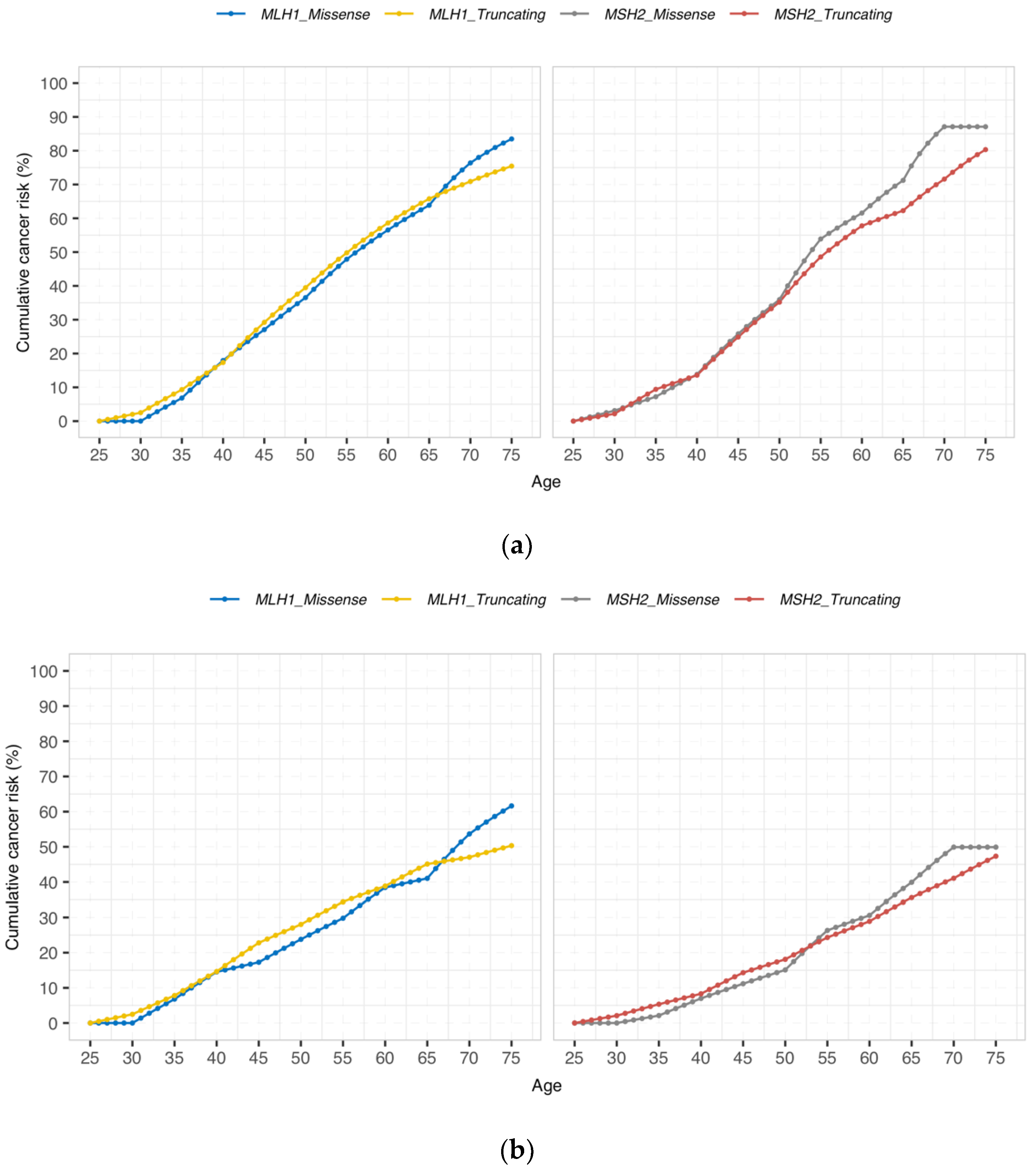
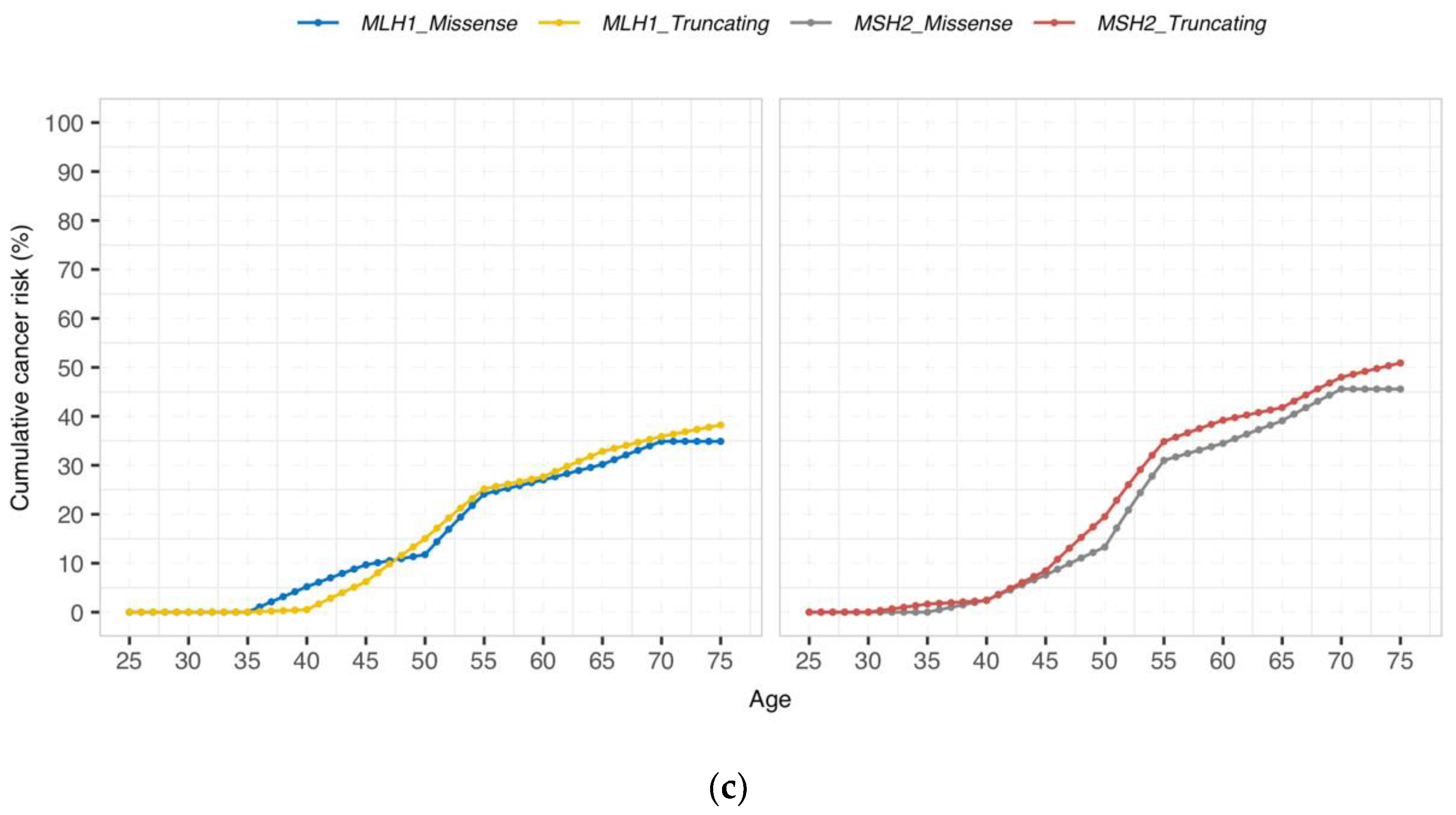
3.2. Cumulative Cancer Incidence by Gene and Type of Genetic Variant

The cumulative incidences by gene for any cancer, CRC, and endometrial cancer are detailed in Table 2 and illustrated in Figure 1. There were no significant differences between carriers with missense/aberrant splicing versus truncating variants at any age in any group. Moreover, no differences which could be considered non-significant trends were observed (p > 0.05 for all comparisons).
Cumulative incidences for any cancer at 50 years in path_MLH1 carriers with truncating or missense/aberrant splicing variants were 39.5% (95% CI (34.5–44.5)) and 36.5% (95% CI (26.6–46.5)), respectively, and in path_MSH2 carriers, 35.2% (95% CI (29.3–41.1)) and 36.0% (95% CI (23.9–48.0)), respectively. Corresponding cumulative incidences for CRC were 28.0% (95% CI (23.3–32.7)) versus 23.8% (95% CI (14.6–33.0)) for path_MLH1 carriers and 18.1% (95% CI (13.3–22.9)) versus 15.1% (95% CI (6.4–23.8)) for path_MSH2 carriers with truncating or missense/aberrant splicing variants, respectively. Corresponding cumulative incidences for endometrial cancer were 15.0% (95% CI (10.1–19.9)) versus 11.8% (95% CI (3.5–20.0)) for path_MLH1 carriers and 19.5% (95% CI (12.9–26.1)) versus 13.3% (95% CI (2.4–24.2)) for path_MSH2 carriers with truncating or missense/aberrant splicing variants, respectively.
Cumulative incidences for any cancer at 75 years in path_MLH1 carriers with truncating or missense/aberrant splicing variants were 75.4% (95% CI (69.1–81.8)) versus 83.5% (95% CI (71.4–95.6)), respectively, and in path_MSH2 carriers 80.3% (95% CI (73.3–87.4)) versus 87.1% (95% CI (75.6–98.6)), respectively. Corresponding cumulative incidences for CRC were 50.3% (95% CI (43.8–56.8)) versus 61.6% (95% CI (45.9–77.4)) for path_MLH1 carriers and 47.3% (95% CI (39.6–55.1)) versus 49.9% (95% CI (36.4–63.4)) for path_MSH2 carriers with truncating or missense/aberrant splicing variants, respectively. Corresponding cumulative incidences for endometrial cancer were 38.2% (95% CI (29.0–47.4)) versus 34.9% (95% CI (19.2–50.6)) for path_MLH1 carriers and 50.9% (95% CI (39.5–62.3)) versus 45.6% (95% CI (25.6–65.6)) for path_MSH2 carriers with truncating or missense/aberrant splicing variants, respectively.

4. Discussion and Conclusions

In contrast to expectations for the hypothesis we tested, carriers of truncating variants of either path_MLH1 and path_MSH2 had similar average cumulative incidences of cancers to carriers of missense or aberrant splicing variants affecting the corresponding gene. On average, carriers of both categories of pathogenic variants had the same high cumulative incidences of any cancer for both genes. The numbers of carriers in each of the groups were large enough to detect any major differences. Our findings will be of clinical interest when interpreting the results of genetic testing, and in planning preventive health care interventions in carriers. As reported previously [1,4], path_MSH2 carriers have higher incidence of other cancers than in the colorectum and endometrium, which is also reflected in the current results for carriers of both truncating and missense path_MSH2 variants. The cumulative cancer incidences for missense path_MSH2 carriers in the two highest age groups showed variation that was considered likely to be stochastic, reflecting the limited number of observation years.
We have previously reported that pathogenic variants in each of the MMR genes result in different risks for cancers in organs, including the colorectum, endometrium, ovaries, stomach, small bowel, bile duct, pancreas, and upper urinary tract [1]. Previously, only one study with a very limited number of cases attempted to address the issue of whether the type of pathogenic variant also resulted in different cancer risks but was inconclusive [13].
Some MMR gene variants may be associated with partial but compromised function. The POLYPHEN and SIFT algorithms [26,27] attribute distinct degrees of malfunctioning to different missense variants, and there are examples of aberrant splicing and missense variants in the BRCA-genes that are associated with intermediate cancer incidences [22,28,29]. We cannot rule out the possibility that the criteria applied by the InSiGHT database to classify variants lack the sensitivity to identify low-risk variants in MLH1 and MSH2. Indeed, a functional study showed that the MLH1 variant p.K618T that was classified benign by InSIGHT had an intermediate repair capacity of ~35% to 50% [30]. Carriers of such variants may be at moderately increased risk for cancer, but may not be offered appropriate health care. Less penetrant path_MMR variants may also present clinically as the autosomal recessive constitutional mismatch repair deficiency syndrome (CCMRD), but path_PMS2 and path_MSH6 variants account for the majority of such cases. Path_PMS2 variants associated with a milder heterozygous phenotype may be overrepresented, since it was shown that heterozygous relatives of CMMRD patients had a lower cumulative colon cancer risk (8.7%) than reported for path_PMS2 as a whole by the PLSD and others [1,31]. A difference in age at CRC diagnosis was found for path_PMS2 carriers when stratifying variants into those that lead to loss of RNA expression compared to those for which expression was preserved [32,33], but a similar relationship was not observed in a CMMRD family cohort. Host immune factors may also be involved in determining cancer incidence in LS. Carriers of path_MLH1 and path_MSH2 variants develop thousands of mismatch repair-deficient and potentially precancerous gastrointestinal crypts [34,35]. The frequency at which they progress to infiltrating cancers may be largely determined by the host immune system, rather than the nature of the inherited path_MMR variant [34,36]. Genetic modifiers may also contribute to the variation in cancer risk and phenotypic variability in path_MMR carriers, leading those with such genetic modifiers to be at increased risk of having further cancers [9].
The strengths of our study include its large sample size and its prospective design, but a potential weakness is selection bias at contributing centres that may have failed to identify some low-penetrance variants. We are also aware that there are many other possible categorizations of path_MMR variants that could be investigated for differences in associated cancer incidences using the PLSD data, but we hesitate to do so until we have other plausible hypotheses to test. Similarly, we did not test for differences between class 4 and class 5 variants in relation to cancer incidence, as numbers were not large enough to make this comparison.
The penetrance of the pathogenic MMR variants has no bearing on the classification of their pathogenicity. Having recruited sufficient numbers of carriers into PLSD to reach robust conclusions, we examined the hypothesis that missense or aberrant splicing variants may have lower incidence of cancer than truncating path_MLH1 and path_MSH2 variants. In contrast to our hypothesis, we found no difference. The results are of practical interest when presenting preventive health care options to carriers of path_MLH1 and path_MSH2 variants.

Author Contributions

P.M., J.-P.P. and M.D.-V. designed the study. M.D.-V. is the PLSD curator, J.-P.P. is the InSiGHT database curator, and P.M. is the PI to the PLSD. J.-P.P. scored the variants as truncating or missense/aberrant splicing. S.N. implemented code for cumulative risk and performed Figure 1. M.D.-V. and P.M. calculated the results. M.D.-V., J.-P.P., J.R.S., T.T.S. and P.M. wrote the manuscript. All authors contributed data to the PLSD and reviewed and approved the manuscript. All authors have read and agreed to the final version of the manuscript.

Funding

We acknowledge funding from the Norwegian Cancer Society, contract 194751-2017. The Colon CFR is supported in part by the National Cancer Institute of the National Institutes of Health under Award Number UM1CA167551. The Finnish contribution: Cancer Foundation Finland, Jane and Aatos Erkko Foundation, Emil Aaltonen Foundation, Finnish Medical Foundation, Sigrid Juselius Foundation, Instrumentarium Science Foundation, iCAN Flagship of the Academy of Finland. D.G.E. and E.J.C. are supported by the Manchester National Institute for Health Research (NIHR) Biomedical Research Centre (IS-BRC-1215-20007). The contribution from Wales was supported by the Wales Gene Park award from Health and Care Research Wales. The German Consortium for Familial Intestinal Cancer was supported by grants from the German Cancer Aid. Work by G.C., M.N., J.B., and M.P. was funded by the Spanish Ministry of Science and Innovation and cofounded by FEDER M.D. Catalonia (grants 2017SGR1282). The funding body had no role in the design of the study and collection, analysis, and interpretation of data and in writing the manuscript.

Institutional Review Board Statement

The study adhered to the principles set out in the Declaration of Helsinki. It was approved by the Oslo University Hospital ethical committee approval code S-02030 and its data governance rules by the Norwegian Data Inspectorate ref. 2001/2988-2. Genetic testing was performed with informed consent according to local and national requirements and all reporting centers exported only de-identified data to PLSD.

Informed Consent Statement

Not applicable.

Data Availability Statement

The cancer risk algorithm is available at the PLSD website (www.plsd.eu, accessed on 12 February 2021) that is based upon the results presented in this report and enables interactive calculation of remaining lifetime risks for cancer in any LS patient by giving their age, gender, and gene variant.

Conflicts of Interest

Seven authors (S.A., C.P., R.H., E.H.-F., V.S.-L., G.C., J.B.) of this publication are members of the European Reference Network on Genetic Tumor Risk Syndromes (ERN GENTURIS)-Project ID No. 739547. R.B. has received honoraria for lectures and advisory boards from AbbVie, Amgen, AstraZeneca, Bayer, BMS, Boehringer-Ingelheim, Illumina, Lilly, Merck-Serono, MSD, Novartis, Qiagen, Pfizer, Roche, and Targos MP Inc. R.B. is a Co-Founder and Scientific Advisor for Targos Mol. Pathology Inc. Kassel/Germany. T.T.S. is the CEO and co-owner of Healthfund Finland Oy and reports an interview honorarium from Boehringer Ingelheim Finland. J.B. has received honoraria for advisory boards from Astra Zeneca and MSD. G.C. is founder and co-owner of VCN Biosciences, Spain.

References

  1. Dominguez-Valentin, M.; Sampson, J.R.; Seppälä, T.T.; Broeke, S.W.T.; Be, J.-P.P.; Nakken, S.; Engel, C.; Aretz, S.; Jenkins, M.; Sunde, L.; et al. Cancer risks by gene, age, and gender in 6350 carriers of pathogenic mismatch repair variants: Findings from the Prospective Lynch Syndrome Database. Genet. Med. 2020, 22, 15–25. [Google Scholar] [CrossRef] [Green Version]
  2. Møller, P.; Seppälä, T.; Bernstein, I.; Holinski-Feder, E.; Sala, P.; Evans, D.G.; Lindblom, A.; Macrae, F.; Blanco, I.; Sijmons, R.; et al. Cancer incidence and survival in Lynch syndrome patients receiving colonoscopic and gynaecological surveillance: First report from the prospective Lynch syndrome database. Gut 2017, 66, 464–472. [Google Scholar] [CrossRef]
  3. Møller, P.; Seppälä, T.; Bernstein, I.; Holinski-Feder, E.; Sala, P.; Evans, D.G.; Lindblom, A.; Macrae, F.; Blanco, I.; Sijmons, R.; et al. Incidence of and survival after subsequent cancers in carriers of pathogenic MMR variants with previous cancer: A report from the prospective Lynch syndrome database. Gut 2017, 66, 1657–1664. [Google Scholar] [CrossRef]
  4. Møller, P.; Seppälä, T.; Bernstein, I.; Holinski-Feder, E.; Sala, P.; Evans, D.G.; Lindblom, A.; Macrae, F.; Blanco, I.; Sijmons, R.H.; et al. Cancer risk and survival in path_MMR carriers by gene and gender up to 75 years of age: A report from the Prospective Lynch Syndrome Database. Gut 2018, 67, 1306–1316. [Google Scholar] [CrossRef] [Green Version]
  5. Lynch, H.T.; Snyder, C.L.; Shaw, T.G.; Heinen, C.D.; Hitchins, M.P. Milestones of Lynch syndrome: 1895–2015. Nat. Rev. Cancer 2015, 15, 181–194. [Google Scholar] [CrossRef] [PubMed]
  6. Ryan, N.; Morris, J.; Green, K.; Lalloo, F.; Woodward, E.R.; Hill, J.; Crosbie, E.J.; Evans, D.G. Association of Mismatch Repair Mutation with Age at Cancer Onset in Lynch Syndrome. JAMA Oncol. 2017, 3, 1702–1706. [Google Scholar] [CrossRef]
  7. Seppälä, T.T.; Dominguez-Valentin, M.; Sampson, J.R.; Møller, P. Prospective observational data informs understanding and future management of Lynch syndrome: Insights from the Prospective Lynch Syndrome Database (PLSD). Fam. Cancer 2021, 20, 35–39. [Google Scholar] [CrossRef] [PubMed]
  8. Wijnen, J.T.; Brohet, R.M.; van Eijk, R.; Jagmohan–Changur, S.; Middeldorp, A.; Tops, C.M.; Van Puijenbroek, M.; Ausems, M.G.E.M.; García, E.G.; Hes, F.J.; et al. Chromosome 8q23.3 and 11q23.1 Variants Modify Colorectal Cancer Risk in Lynch Syndrome. Gastroenterology 2009, 136, 131–137. [Google Scholar] [CrossRef]
  9. Talseth-Palmer, B.A.; Wijnen, J.T.; Brenne, I.S.; Jagmohan-Changur, S.; Barker, D.; Ashton, K.A.; Tops, C.M.; Evans, T.-J.; McPhillips, M.; Groombridge, C.; et al. Combined analysis of three lynch syndrome cohorts confirms the modifying effects of 8q23.3 and 11q23.1 in MLH1 mutation carriers. Int. J. Cancer 2012, 132, 1556–1564. [Google Scholar] [CrossRef] [PubMed]
  10. Ten Broeke, S.W.; Van Der Klift, H.M.; Tops, C.M.; Aretz, S.; Bernstein, I.; Buchanan, D.D.; De La Chapelle, A.; Capella, G.; Clendenning, M.; Engel, C.; et al. Cancer Risks for PMS2-Associated Lynch Syndrome. J. Clin. Oncol. 2018, 36, 2961–2968. [Google Scholar] [CrossRef] [Green Version]
  11. Peltomäki, P.; Olkinuora, A.; Nieminen, T.T. Updates in the field of hereditary nonpolyposis colorectal cancer. Expert Rev. Gastroenterol. Hepatol. 2020, 14, 707–720. [Google Scholar] [CrossRef]
  12. Peltomäki, P. Update on Lynch syndrome genomics. Fam. Cancer 2016, 15, 385–393. [Google Scholar] [CrossRef] [Green Version]
  13. Cravo, M.; Afonso, A.; Lage, P.; Albuquerque, C.; Maia, L.; Lacerda, C.; Fidalgo, P.; Chaves, P.; Cruz, C.; Nobre-Leitão, C. Pathogenicity of missense and splice site mutations in hMSH2 and hMLH1 mismatch repair genes: Implications for genetic testing. Gut 2002, 50, 405–412. [Google Scholar] [CrossRef]
  14. Liu, B.; Parsons, R.E.; Hamilton, S.R.; Petersen, G.M.; Lynch, H.T.; Watson, P.; Markowitz, S.; Willson, J.K.V.; Green, J.; de la Chapelle, A.; et al. hMSH2 mutations in hereditary nonpolyposis colorectal cancer kindreds. Cancer Res. 1994, 54, 4590–4594. [Google Scholar] [PubMed]
  15. Papadopoulos, N.; Nicolaides, N.C.; Wei, Y.F.; Ruben, S.M.; Carter, K.C.; Rosen, C.A.; Haseltine, W.A.; Fleischmann, R.D.; Fraser, C.; Adams, M.D.; et al. Mutation of a mutL homolog in hereditary colon cancer. Science 1994, 263, 1625–1629. [Google Scholar] [CrossRef] [PubMed]
  16. Peltomaki, P.; Vasen, H. Mutations predisposing to hereditary nonpolyposis colorectal cancer: Database and results of a collaborative study. The International Collaborative Group on Hereditary Nonpolyposis Colorectal Cancer. Gastroenterology 1997, 113, 1146–1158. [Google Scholar] [CrossRef]
  17. Li, L.; Hamel, N.; Baker, K.; McGuffin, M.J.; Couillard, M.; Gologan, A.; Marcus, V.A.; Chodirker, B.; Chudley, A.; Stefanovici, C.; et al. A homozygousPMS2founder mutation with an attenuated constitutional mismatch repair deficiency phenotype. J. Med. Genet. 2015, 52, 348–352. [Google Scholar] [CrossRef] [PubMed]
  18. Auclair, J.; Busine, M.P.; Navarro, C.; Ruano, E.; Montmain, G.; Desseigne, F.; Saurin, J.C.; Lasset, C.; Bonadona, V.; Giraud, S.; et al. Systematic mRNA analysis for the effect ofMLH1 andMSH2 missense and silent mutations on aberrant splicing. Hum. Mutat. 2006, 27, 145–154. [Google Scholar] [CrossRef]
  19. Soukarieh, O.; Gaildrat, P.; Hamieh, M.; Drouet, A.; Baert-Desurmont, S.; Frébourg, T.; Tosi, M.; Martins, A. Exonic Splicing Mutations Are More Prevalent than Currently Estimated and Can Be Predicted by Using In Silico Tools. PLoS Genet. 2016, 12, e1005756. [Google Scholar] [CrossRef] [Green Version]
  20. Thompson, B.A.; Walters, R.; Parsons, M.T.; Dumenil, T.; Drost, M.; Tiersma, Y.; Lindor, N.M.; Tavtigian, S.V.; De Wind, N.; Spurdle, A.B.; et al. Contribution of mRNA Splicing to Mismatch Repair Gene Sequence Variant Interpretation. Front. Genet. 2020, 11, 798. [Google Scholar] [CrossRef]
  21. Maccaroni, E.; Bracci, R.; Giampieri, R.; Bianchi, F.; Belvederesi, L.; Brugiati, C.; Pagliaretta, S.; Del Prete, M.; Scartozzi, M.; Cascinu, S. Prognostic impact of mismatch repair genes germline defects in colorectal cancer patients: Are all mutations equal? Oncotarget 2015, 6, 38737–38748. [Google Scholar] [CrossRef] [Green Version]
  22. Møller, P. The Prospective Lynch Syndrome Database reports enable evidence-based personal precision health care. Hered. Cancer Clin. Pract. 2020, 18, 6–7. [Google Scholar] [CrossRef]
  23. Thompson, B.A.; Spurdle, A.B.; Plazzer, J.-P.; Greenblatt, M.S.; Akagi, K.; Al-Mulla, F.; Bapat, B.; Bernstein, I.; Capellá, G.; den Dunnen, J.T.; et al. Application of a 5-tiered scheme for standardized classification of 2360 unique mismatch repair gene variants in the InSiGHT locus-specific database. Nat. Genet. 2014, 46, 107–115. [Google Scholar] [CrossRef] [PubMed] [Green Version]
  24. Seppälä, T.; Pylvänäinen, K.; Evans, D.G.; Järvinen, H.; Renkonen-Sinisalo, L.; Bernstein, I.; Holinski-Feder, E.; Sala, P.; Lindblom, A.; Macrae, F.; et al. Colorectal cancer incidence in path_MLH1 carriers subjected to different follow-up protocols: A Prospective Lynch Syndrome Database report. Hered. Cancer Clin. Pract. 2017, 15, 18. [Google Scholar] [CrossRef] [Green Version]
  25. Seppälä, T.T.; Ahadova, A.; Dominguez-Valentin, M.; Macrae, F.; Evans, D.G.; Therkildsen, C.; Sampson, J.; Scott, R.; Burn, J.; Möslein, G.; et al. Lack of association between screening interval and cancer stage in Lynch syndrome may be accounted for by over-diagnosis; a prospective Lynch syndrome database report. Hered. Cancer Clin. Pract. 2019, 17, 8. [Google Scholar] [CrossRef]
  26. Adzhubei, I.; Jordan, D.; Sunyaev, S.R. Predicting Functional Effect of Human Missense Mutations Using PolyPhen-2. Curr. Protoc. Hum. Genet. 2013, 76, 7.20.1–7.20.41. [Google Scholar] [CrossRef] [Green Version]
  27. Ng, P.C.; Henikoff, S. SIFT: Predicting amino acid changes that affect protein function. Nucleic Acids Res. 2003, 31, 3812–3814. [Google Scholar] [CrossRef] [PubMed] [Green Version]
  28. Moghadasi, S.; Meeks, H.D.; Vreeswijk, M.P.; Janssen, L.A.; Borg, Å.; Ehrencrona, H.; Paulsson-Karlsson, Y.; Wappenschmidt, B.; Engel, C.; Gehrig, A.; et al. The BRCA1 c. 5096G>A p.Arg1699Gln (R1699Q) intermediate risk variant: Breast and ovarian cancer risk estimation and recommendations for clinical management from the ENIGMA consortium. J. Med. Genet. 2017, 55, 15–20. [Google Scholar] [CrossRef]
  29. Spurdle, A.B.; Whiley, P.J.; Thompson, B.; Feng, B.; Healey, S.; Brown, M.; Pettigrew, C.; Van Asperen, C.J.; Ausems, M.G.E.M.; Kattentidt-Mouravieva, A.A.; et al. BRCA1 R1699Q variant displaying ambiguous functional abrogation confers intermediate breast and ovarian cancer risk. J. Med. Genet. 2012, 49, 525–532. [Google Scholar] [CrossRef] [PubMed]
  30. Drost, M.; Ms, Y.T.; Thompson, B.A.; Frederiksen, J.H.; Keijzers, G.; Glubb, D.; Kathe, S.; Osinga, J.; Westers, H.; Pappas, L.; et al. A functional assay–based procedure to classify mismatch repair gene variants in Lynch syndrome. Genet. Med. 2019, 21, 1486–1496. [Google Scholar] [CrossRef]
  31. Ten Broeke, S.W.; Brohet, R.M.; Tops, C.M.; van der Klift, H.M.; Velthuizen, M.E.; Bernstein, I.; Munar, G.C.; Garcia, E.G.; Hoogerbrugge, N.; Letteboer, T.G.W.; et al. Lynch syndrome caused by germline PMS2 mutations: Delineating the cancer risk. J. Clin. Oncol. 2015, 33, 319–325. [Google Scholar] [CrossRef] [PubMed]
  32. Suerink, M.; Rodríguez-Girondo, M.; Van Der Klift, H.M.; Colas, C.; Brugieres, L.; Lavoine, N.; Jongmans, M.; Munar, G.C.; Evans, D.G.; Farrell, M.P.; et al. An alternative approach to establishing unbiased colorectal cancer risk estimation in Lynch syndrome. Genet. Med. 2019, 21, 2706–2712. [Google Scholar] [CrossRef] [PubMed]
  33. Suerink, M.; Van Der Klift, H.M.; Broeke, S.W.T.; Dekkers, O.M.; Bernstein, I.; Munar, G.C.; Garcia, E.G.; Hoogerbrugge, N.; Letteboer, T.G.W.; Menko, F.H.; et al. The effect of genotypes and parent of origin on cancer risk and age of cancer development in PMS2 mutation carriers. Genet. Med. 2015, 18, 405–409. [Google Scholar] [CrossRef]
  34. Ballhausen, A.; Przybilla, M.J.; Jendrusch, M.; Haupt, S.; Pfaffendorf, E.; Seidler, F.; Witt, J.; Sanchez, A.H.; Urban, K.; Draxlbauer, M.; et al. The shared frameshift mutation landscape of microsatellite-unstable cancers suggests immunoediting during tumor evolution. Nat. Commun. 2020, 11, 1–13. [Google Scholar] [CrossRef] [PubMed]
  35. Ahadova, A.; Doeberitz, M.V.K.; Bläker, H.; Kloor, M. CTNNB1-mutant colorectal carcinomas with immediate invasive growth: A model of interval cancers in Lynch syndrome. Fam. Cancer 2016, 15, 579–586. [Google Scholar] [CrossRef] [PubMed]
  36. Ahadova, A.; Seppälä, T.; Engel, C.; Gallon, R.; Burn, J.; Holinski-Feder, E.; Steinke-Lange, V.; Möslein, G.; Nielsen, M.; Broeke, S.T.; et al. The “unnatural” history of colorectal cancer in Lynch syndrome: Lessons from colonoscopy surveillance. Int. J. Cancer 2021, 148, 800–811. [Google Scholar] [CrossRef]
Figure 1. Cumulative incidence of (a) any cancer, (b) colorectal, and (c) endometrial cancer by gene and type of variant. There were no significant differences between carriers with missense/aberrant splicing versus truncating variants at any age in any groups (p > 0.05 for all comparisons).
Jcm 10 02856 g001aJcm 10 02856 g001b
Table 1. Categorization of the path_MLH1 and path_MSH2 carriers having inherited variants causing truncating or missense/aberrant splicing clinically actionable variants as defined in the InSiGHT database.
Categorization GroupVariant TypeGeneNumber of CarriersSum of the FUP YearsMean of the FUP Years95% CI
Missense or aberrant splicingAberrant SplicingMLH123318297.8(7.1–8.5)
Aberrant SplicingMSH235027787.9(7.4–8.4)
MissenseMLH134527157.9(7.4–8.4)
MissenseMSH21178837.5(6.7–8.3)
Total 104582057.9(7.6–8.2)
TruncatingCanonical SplicingMLH150147099.4(8.9–9.9)
Canonical SplicingMSH218516358.8(8.0–9.6)
Exon DeletionMLH1688764311.1(10.6–11.6)
Exon DeletionMSH257942077.3(6.9–7.7)
NonsenseMLH132428808.9(8.3–9.5)
NonsenseMSH260849298.1(7.7–8.5)
FrameshiftMLH148237227.7(7.3–8.1)
FrameshiftMSH256344167.8(7.4–8.2)
Total 393034,1418.7(8.5–8.9)
OthersExon DuplicationMLH1111(1.0–1.0)
Exon DuplicationMSH216714.4(2.7–6.1)
Inframe IndelMLH1857909.3(8.3–10.3)
Inframe IndelMSH2938118.7(7.7–9.7)
Initiation CodonMLH18364.5(1.5–7.5)
IntronicMSH23258.3(2.1–14.5)
UndefinedMLH11824913.8(10.7–16.9)
Total 2241983
FUP, follow-up years.
Table 2. Cumulative cancer incidences stratified by age, gene, variant, and organ.
Cumulative Incidences (95% CI)
AgeMLH1 Missense/Aberrant SplicingMLH1 TruncatingMSH2 Missense/Aberrant SplicingMSH2 Truncating
Any cancer300 (0–0)2.5 (0.3–4.7)3.1 (0–9.1)2.2 (0–4.6)
4017.9 (9.3–26.5)17.3 (13.1–21.6)13.8 (4.1–23.5)13.6 (8.9–18.2)
5036.5 (26.6–46.5)39.5 (34.5–44.5)36.0 (23.9–48.0)35.2 (29.3–41.1)
6056.6 (44.6–68.4)58.6 (53.4–63.9)61.6 (49.5–73.6)57.8 (51.4–64.1)
7076.4 (63.6–89.2)71.0 (65.1–76.7)87.1 (75.6–98.6)71.6 (64.4–78.8)
7583.5 (71.4–95.6)75.4 (69.1–81.8)87.1 (75.6–98.6)80.3 (73.3–87.4)
Colorectal cancer300 (0–0)2.5 (0.3–4.6)0 (0–0)2.1 (0–4.4)
4014.5 (6.5–22.6)14.6 (10.6–18.6)7.0 (0.3–13.6)8.3 (4.5–12.1)
5023.8 (14.6–33.0)28.0 (23.3–32.7)15.1 (6.4–23.8)18.1 (13.3–22.9)
6038.4 (26.5–50.4)38.9 (33.7–44.0)30.6 (19.7–41.5)28.9 (23.3–34.5)
7053.7 (39.0–68.3)47.0 (41.2–52.8)49.9 (36.4–63.4)41.1 (34.2–48.0)
7561.6 (45.9–77.4)50.3 (43.8–56.8)49.9 (36.4–63.4)47.3 (39.6–55.1)
Endometrial cancer300 (0–0)0 (0–0)0 (0–0)0 (0–0)
405.2 (0–10.9)0.5 (0–1.5)2.5 (0–7.2)2.4 (0–5.0)
5011.8 (3.5–20.0)15.0 (10.1–19.9)13.3 (2.4–24.2)19.5 (12.9–26.1)
6027.0 (13.9–40.1)27.7 (21.0–34.3)34.5 (17.6–51.3)39.2 (30.3–48.1)
7034.9 (19.2–50.6)35.9 (27.6–44.2)45.6 (25.6–65.6)48.0 (37.4–58.5)
7534.9 (19.2–50.6)38.2 (29.0–47.4)45.6 (25.6–65.6)50.9 (39.5–62.3)
CI, confidence interval.
Publisher’s Note: MDPI stays neutral with regard to jurisdictional claims in published maps and institutional affiliations.

