Article

Erythema Increase Predicts Psoriasis Improvement after Phototherapy

by
Trinidad Montero-Vilchez
1,2,
Antonio Martinez-Lopez
1,2,*,
Alvaro Sierra-Sanchez
2,
Miguel Soler-Gongora
3,
Eladio Jimenez-Mejias
4,
Alejandro Molina-Leyva
1,2,
Agustin Buendia-Eisman
3 and
Salvador Arias-Santiago
1,2,3
1
Dermatology Department, Hospital Universitario Virgen de las Nieves, Avenida de Madrid, 15, 18012 Granada, Spain
2
Instituto de Investigación Biosanitaria GRANADA, 18012 Granada, Spain
3
Dermatology Department, Faculty of Medicine, University of Granada, 18001 Granada, Spain
4
Epidemiology and Public Health Department, Faculty of Medicine, University of Granada, 18012 Granada, Spain
*
Author to whom correspondence should be addressed.
J. Clin. Med. 2021, 10(17), 3897; https://doi.org/10.3390/jcm10173897
Submission received: 16 July 2021 / Revised: 1 August 2021 / Accepted: 26 August 2021 / Published: 30 August 2021
(This article belongs to the Section Dermatology)

Abstract

:
Psoriasis is a major global health problem. There is a need to develop techniques to help physicians select the most appropriate cost-effective therapy for each patient. The main objectives of this study are (1) to evaluate changes in epidermal barrier function and skin homeostasis after phototherapy and (2) to explore potentially predictive values in epidermal barrier function and skin homeostasis to assess clinical improvement after fifteen sessions of phototherapy. A total of 76 subjects, 38 patients with plaque-type psoriasis and 38 gender- and age-matched healthy volunteers, were included in the study. Erythema, transepidermal water loss (TEWL), temperature, stratum corneum hydration (SCH), pH, sebum, and antioxidant capacity were measured before and after the first and fifteenth phototherapy session. Erythema (401.09 vs. 291.12 vs. 284.52 AU, p < 0.001) and TEWL (18.23 vs. 11.44 vs. 11.41 g·m−2·h−1, p < 0.001) were significantly higher at psoriatic plaques than in uninvolved psoriatic skin and healthy volunteers, respectively, while SCH was lower (9.71 vs. 44.64 vs. 40.00 AU, p < 0.001). After fifteen phototherapy sessions, TEWL (–5.19 g·m−2·h−1, p = 0.016) decreased while SCH (+7.01 AU, p = 0.013) and erythema (+30.82 AU, p = 0.083) increased at psoriatic plaques. An erythema increase exceeding 53.23 AU after the first phototherapy session, with a sensitivity of 71.4% and specificity of 84.2%, indicates that a patient may improve Psoriasis Area and Severity Index (PASI) by ≥3 points after fifteen phototherapy sessions. In conclusion, phototherapy improves epidermal barrier function in psoriatic patients and the erythema increase after one phototherapy session could help doctors select psoriasis patients who are more likely to respond to phototherapy.

1. Introduction

Psoriasis is a chronic, recurrent, multisystemic inflammatory disease [1] caused by a combination of immunological imbalances, genetic associations, and environmental factors [2]. Its prevalence around the word has been estimated at between 0.51% and 11.43% [3]. Psoriasis is considered a major global health problem [4]. Although the skin manifestations are often the only recognized symptoms of psoriasis [5], this disease is associated with multiple comorbidities [6,7,8,9] and impacts the patient’s quality of life [5,10]. Moreover, the economic burden of psoriasis is high, as in Europe the annual total cost per patient is EUR 6000–12,000 [11].
Multiple treatments are effective for psoriasis, including topical medicines, oral systemic prescriptions, phototherapy, and biologics [12]. Nevertheless, it is not known which type of patient would respond best to each treatment [13]. Moreover, tools to assess disease severity and treatment effectiveness are subjective [14]. Thus, there is a need to develop techniques to help physicians select the most appropriate cost-effective therapy for each patient [15].
Phototherapy is an effective, safe, and low-cost therapy for mild–moderate psoriasis, although many medical appointments are needed to see an improvement [16]. Several types of light and lasers have been developed to treat psoriasis, the narrowband ultraviolet light B (NB-UVB) being the most frequently used. NB-UVB wavelengths ranges from 311 to 313 nm. The starting dose is based on skin phototype or minimal erythema dose (MED), and two or three sessions per week are recommended [17]. Selecting the right patient profile for this treatment and accurately assessing disease severity would improve patient satisfaction and healthcare spending [13]. It would also be interesting to predict the response to assess home phototherapy effectiveness [18]. As the development of psoriasis plaques results from the deregulation of epidermal keratinocytes and immunity cells [19] and the phototherapy’s beneficial effect on psoriasis lesions is explained by it blocking epidermal hyperproliferation and an immunomodulatory effect [20], objective changes in the epidermal barrier function may help to select the right psoriasis patients for phototherapy treatment and to assess disease improvement. Epidermal barrier dysfunction in psoriasis patients has previously been reported, assessed by an increase in transepidermal water loss (TEWL) and a decrease in stratum corneum hydration (SCH) [21,22]. To date, only three studies have evaluated the variations in epidermal barrier function following phototherapy, displaying an improvement in TEWL and SCH [23,24,25].
Thus, the aims of this study are (1) to compare epidermal barrier function and skin homeostasis of healthy volunteers, uninvolved psoriatic skin, and psoriatic plaques, (2) to assess changes in epidermal barrier function and skin homeostasis after one session of phototherapy, (3) to assess changes in epidermal barrier function and skin homeostasis after fifteen phototherapy sessions, and (4) to explore potentially predictive values in epidermal barrier function and skin homeostasis to assess clinical improvement after fifteen phototherapy sessions.

2. Materials and Methods

2.1. Design

A cross-sectional study was conducted to evaluate epidermal barrier function and skin homeostasis disparities between healthy skin, uninvolved psoriatic skin, and psoriatic plaques.
A prospective observational study was carried out on patients with psoriasis to assess epidermal barrier function and skin homeostasis following fifteen phototherapy sessions. Psoriatic patients were exposed to fifteen phototherapy sessions, while healthy volunteers were only reviewed after this period of time without being exposed to phototherapy.

2.2. Setting

This study was conducted between September 2019 and March 2020 in the Dermatology Department of the Hospital Universitario Virgen de las Nieves in Granada, Spain.

2.3. Study Population

Inclusion Criteria:
  • Patients with established clinical diagnosis of active moderate-to-severe plaque-type psoriasis (minimum Psoriasis Area and Severity Index (PASI) score of 4) [1] selected by clinical criteria to attend phototherapy treatment with UVB narrowband (NB-UVB) [16].
  • Controls were healthy volunteers, gender- and age-matched (±3 years) with psoriasis patients. These volunteers were people who attended the Dermatology Department for trivial conditions such as melanocytic nevi or seborrheic keratoses. The same criteria were used to select the non-exposed group in the prospective study.
Exclusion Criteria:
  • For psoriasis patients, currently having non-plaque forms of psoriasis.
  • For healthy volunteers, having previous personal or family history of any inflammatory skin disease.
  • Clinical infection on the treatment area.
  • History of cancer or an immunocompromised disease.
  • Not signing the informed consent form.

2.4. Follow-Up and Exposure

Exposed subjects were evaluated before and after receiving the first phototherapy session and before and after the 15th phototherapy session. The starting dose for NB-UVB therapy and the dosage schedule were based on skin phototype following the current guidelines [17]. The frequency was two or three times a week depending on the patient’s availability. Non-exposed subjects were evaluated twice, on the same days as their exposed pair.

2.5. Variables

  • Clinical and sociodemographic variables. Gender, age, smoking and alcohol habit, psoriasis family history, and use of emollients were gathered by means of clinical interview. Psoriasis severity was assessed by the PASI and the body surface area (BSA). Every study patient was also evaluated with the Dermatology Life Quality Index (DLQI). Information about disease duration, previous treatment, the previous number of phototherapy sessions, session dose, and total cumulative dose was also collected.
  • Epidermal barrier function variables. Homeostasis parameters related to epidermal barrier function and skin homeostasis were measured. SCH (in arbitrary units, using Corneometer®CM825), TEWL (in g·m−2·h−1, using Tewameter®TM300), pH (using Skin-pH-Meter®PH905), erythema index (in arbitrary units, using Mexameter®MX18), sebum (in arbitrary units, usingSebumeter®SM815), and skin temperature (in °C, using Skin-ThermometerST500) were measured by a Multi Probe Adapter (MPA, Courage + Khazaka electronic GmbH, Köln, Germany). Total antioxidant capacity (TAC) was measured using eBQC® electrochemical method (Bioquochem S.L. (BQCkit), Asturias, Spain), and expressed in microcoulombs. TAC is divided into two sections: fast antioxidants (Q1), which have a lower oxidation potential, and slow antioxidants (Q2) [26]. All variables were measured at a psoriatic plaque on the elbow and at an uninvolved skin area near the elbow in psoriatic patients, while healthy subjects were measured at a skin area on their elbows. All parameters were measured ten times for each area, using their average for analysis. The measurements were taken in the same room. The average ambient air temperature at the time of the study was 22 ± 1°C, and the average ambient air humidity was 45% ± 3%.

2.6. Statistical Analysis

Descriptive statistics were used to present the sample characteristics. Continuous data were expressed as the mean ± standard deviation. The absolute and relative frequency distributions were estimated for qualitative variables. The Shapiro–Wilk test was used to check the normality of data distribution, and Levene’s test was used to check the homogeneity of variance. Linear regression models were constructed to compare continuous data between healthy skin and psoriatic patients. To predict PASI improvement after fifteen phototherapy sessions, cut-off points were generated using ROC curves for the changes of erythema and SCH after the first phototherapy session. To produce these ROC curves, the sensitivities and specificities for changes of erythema and SCH values after the first phototherapy that predict an improvement in PASI of ≥3 after the fifteenth phototherapy session were tabulated and the graphical ROC curve was generated by plotting true positive rate (sensitivity) on the y-axis against false positive rate (1-specificity) on the x-axis for the various values tabulated. To select the optimal cut-off point, the point nearest to the top-left-most corner of the ROC curve was chosen, giving equal weight to the importance of sensitivity and specificity. A p-value of <0.05 was considered statistically significant. Statistical Analyses were performed using the SPSS package (SPSS for Windows, Version 24.0 Chicago, IL, USA: SPSS Inc.).

2.7. Ethics

This study was authorized by the ethics committee of Hospital Universitario Virgen de las Nieves. The nature of the study was explained to all participants, who agreed to participate through verbal and written consent. The measurements taken were noninvasive, and patient data were kept confidential. All experiments were done in accordance with relevant guidelines and regulations.

3. Results

3.1. Skin Homeostasis Parameters between Healthy Participants and Psoriatic Patients

The study included 76 participants, consisting of 38 psoriatic patients and 38 healthy participants, Supplementary Table S1.
Differences in skin homeostasis parameters between healthy skin, uninvolved, and involved psoriatic skin before phototherapy were found, Table 1. Lower TEWL values were found in healthy skin compared with uninvolved psoriatic skin and psoriatic plaques (11.41 vs. 11.44 vs. 18.23 g·m−2·h−1, p < 0.001). Higher SCH values were observed in healthy skin compared with uninvolved psoriatic skin and psoriatic plaques (40.00 vs. 44.64 vs. 9.71 AU, p < 0.001). Lower temperature values were detected in uninvolved psoriatic skin than at psoriatic plaques (30.40 vs. 31.25 °C, p < 0.001). Lower erythema index was found in healthy skin than in uninvolved psoriatic skin and psoriatic plaques (284.52 vs. 291.12 vs. 401.09 AU, p < 0.001). Higher total antioxidant capacity was observed in uninvolved psoriatic skin than at psoriatic plaques (6.33 vs. 5.54 uC, p = 0.014). No differences were found in pH or sebum.

3.2. Differences in Skin Homeostasis Parameters after One Phototherapy Session

Skin homeostasis parameters were modified after one phototherapy session, Table 2. TEWL did not change at psoriatic plaques or in uninvolved skin after one phototherapy session. The effect of phototherapy on SCH values was different depending on the skin involvement. It was observed that SCH increased by 2.45 ± 0.72 AU (p = 0.002) at psoriatic plaques (but SCH was not modified in uninvolved skin (p = 0.126).
Temperature increased by 0.24 ± 0.10 °C at psoriatic plaques (p = 0.016). The erythema index increased by 31.42 ± 8.30 AU (p < 0.001) at psoriatic plaques, but no changes were observed in uninvolved skin.
Total antioxidant capacity was not modified at psoriatic plaques or in uninvolved skin after one phototherapy session. No differences in pH or sebum were observed.

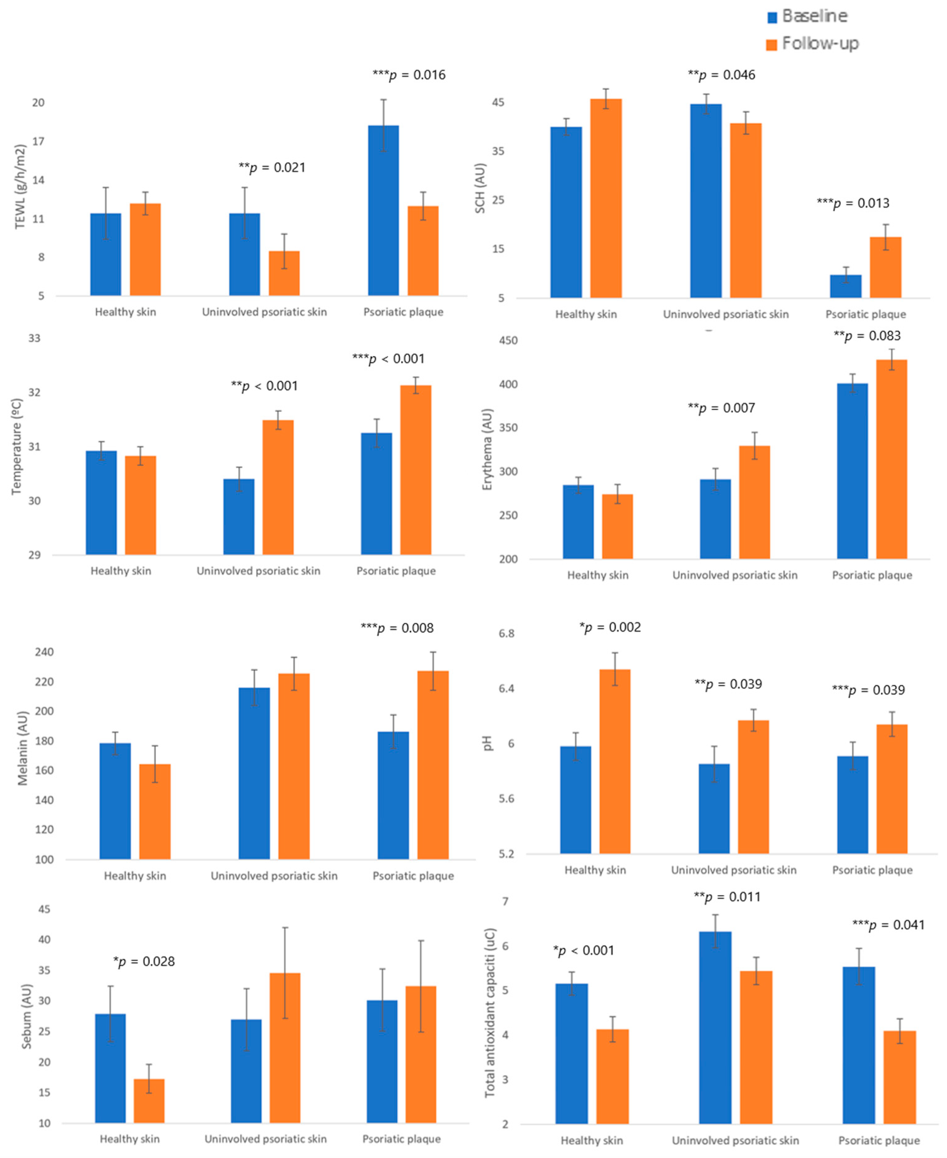
3.3. Skin Homeostasis Changes after Follow-Up

The prospective study included 76 subjects, where 52 (68.42%) met the requirements (26 psoriatic patients and 26 healthy participants). The mean session dose at baseline was 0.46 (0.31) J. Homeostasis parameters changed after follow-up, Table 3. TEWL decreased by 3.50 ± 1.41 g·m−2·h−1 in uninvolved skin (p = 0.021) and by 5.19 ± 2.00 g·m−2·h−1 at psoriatic plaques (p = 0.016). No effect was observed in healthy non-exposed skin. SCH increased by 7.01 ± 2.63 AU at psoriatic plaque (p = 0.013), while no changes were observed in healthy skin.
Temperature increased after phototherapy by 1.5 ± 0.26 °C in uninvolved skin (p < 0.001) and by 1.42 ± 0.28 °C at psoriatic plaques (p < 0.001), while it did not change in healthy non-exposed skin. Erythema increased by 31.83 ± 17.06 AU in uninvolved skin (p = 0.007), and an almost significant increase of 30.82 ± 17.06 AU was also observed at psoriatic plaques (p = 0.087), Figure 1.

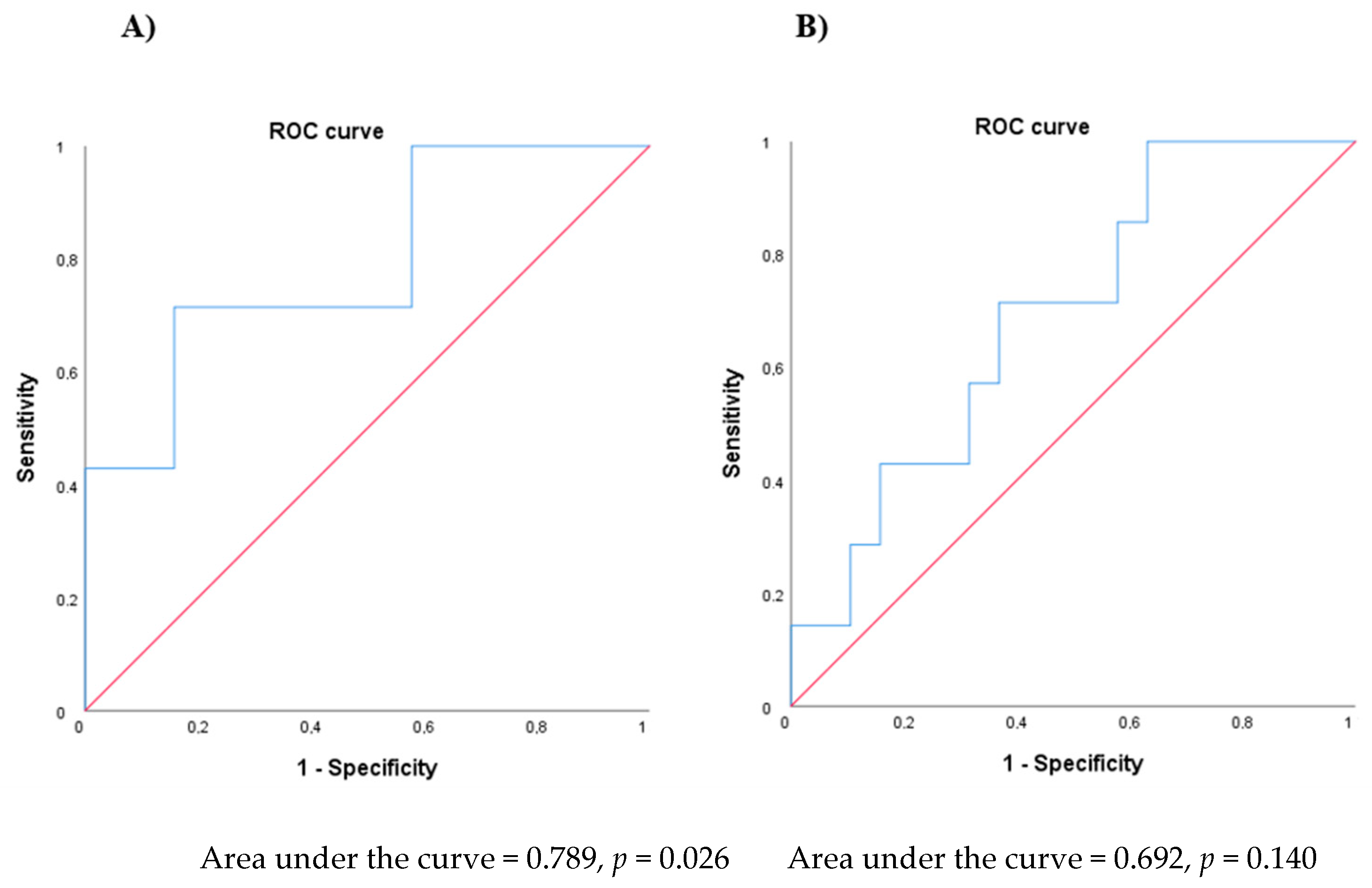
3.4. Skin Homeostasis Predicts PASI Improvement

After follow-up, PASI decreased by 3.13 ± 3.13 points, so patients were placed in two groups: PASI reduction < 3 and PASI reduction ≥ 3. Of the patients, 73.1% (19/26) were included in the first group and 26.9% (7/26) in the second. After the first phototherapy session, patients with a PASI improvement ≥ 3 showed a higher erythema increase (71.08 vs. 11.54 AU, p = 0.011), and an almost significant higher SCH increase (4.69 vs. 1.40; p = 0.141) and higher TEWL decrease (−4.97 vs. 0.86 g·m−2·h−1, p = 0.199).
A ROC curve was generated to determine an optimum cut-off value for erythema increases after one phototherapy session, which allowed clinical improvement after 15 phototherapy sessions to be predicted (area under the curve = 0.789, p = 0.026) (Figure 2A). A value for erythema increases exceeding 53.23 AU after the first phototherapy session, with a sensitivity of 71.4% and specificity of 84.2%, indicates that a patient may improve PASI by ≥3 points after fifteen phototherapy sessions.
SCH increases were also higher in patients with PASI improvement ≥ 3. An ROC curve was generated to determine an optimum cut-off value for SCH increase after one phototherapy session, which allowed clinical improvement after 15 phototherapy sessions to be predicted (area under the curve = 0.692, p = 0.1402) (Figure 2B). A value for SCH increases exceeding 1.06 AU after the first phototherapy session, with a sensitivity of 71.4% and specificity of 63.8%, indicates that a patient may improve PASI by ≥3 points after fifteen phototherapy sessions.
After calculating the different cut-off levels, we evaluated whether combined values may also predict clinical improvement. Patients with erythema increase > 53.23 AU and SCH increase > 1.06 AU after the first phototherapy session may improve PASI by ≥3 after 15 phototherapy sessions, with a sensitivity of 57.1% and a specificity of 94.7% (Supplementary Table S2).

4. Discussion

Differences in skin homeostasis parameters between healthy skin, uninvolved psoriatic skin, and psoriatic plaques have been observed. After one phototherapy session, temperature, erythema, and SCH increased at psoriatic plaques. Moreover, after fifteen phototherapy sessions, decreased TEWL and increased SCH and temperature levels at psoriatic plaques were observed. Phototherapy could improve epidermal barrier function and skin homeostasis in psoriatic patients, and erythema increases after one phototherapy session could help clinicians select psoriasis patients with more probability of responding to phototherapy.
In agreement with previous reports, it has been observed that the whole epidermal barrier is affected in psoriatic patients, not only at psoriatic plaques [27]. Other research also found higher TEWL at psoriatic plaques than in uninvolved psoriatic skin and healthy controls [21,22,27] and lower SCH values at psoriatic plaques than in uninvolved psoriatic skin and healthy controls [21,27,28]. The differences in TEWL and SCH values between psoriatic plaques and uninvolved psoriatic skin may be explained by a low AQP3 expression in plaques [29]. Temperature and erythema were also higher at psoriatic skin, probably due to its inflammatory pathogenesis [30]. Moreover, TEWL and temperature at psoriatic plaques were noted as useful tools for evaluating psoriasis severity [27].
The role of phototherapy on epidermal barrier function and skin homeostasis is not well known. Our results found an improvement in epidermal barrier function and skin homeostasis after phototherapy. Recently, it has been observed that SCH decreased, and TEWL, erythema, and temperature increased at psoriatic plaques after only one phototherapy session [25]. Moreover, it was shown that phototherapy increased SCH and decreased TEWL after fourteen [24] and twenty-four [23] phototherapy sessions, without information regarding other skin homeostasis parameters. Our study found increased SCH at psoriatic plaques following only one phototherapy session and increased SCH and decreased TEWL at psoriatic plaques after fifteen phototherapy sessions. Moreover, in contrast with previous studies, we also included a non-exposed group with follow-up to prove that changes in SCH are not because of time. Changes in SCH might be due to the inhibition of epidermal hyperproliferation caused by phototherapy [20,31]. SCH and TEWL changes were greater at psoriatic plaques than in uninvolved psoriatic skin, which might underline a local effect on psoriasis plaques [32,33]. Temperature and erythema index rose after the phototherapy session, in agreement with previous reports [25,34,35,36]. Assessment of temperature and erythema increase may help clinicians optimize phototherapy to treat patients with an effective dosage without adverse events. The pH increased in healthy skin, uninvolved psoriatic skin, and psoriatic plaques, suggesting that time may have an effect on pH changes. Antioxidant capacity also decreased in healthy skin, uninvolved psoriatic skin, and psoriatic plaque. This fact might mean that the time have also an impact in antioxidant capacity or that the sticks used might lose their capacity to measure the antioxidant capacity along the time. There is little information regarding the effect of phototherapy on antioxidant capacity. Oxidative stress has been evaluated by measuring different parameters of a blood sample, with controversial results. Darlenski et al. found a slight decrease in the detoxifying activity of catalase without significant differences after phototherapy [24]. On the other hand, Pektas et al. observed total oxidant status and oxidative stress index increased after phototherapy [37]. Our results showed total antioxidant capacity decreases after phototherapy, in agreement with this research by Pektas.
Brazzelli et al. suggested that SCH improvement at psoriatic plaques might precede clinical improvement [23]. As far as we know, it is not known which parameters might predict clinical improvement in psoriatic patients treated with phototherapy. We observed that SCH changes after one phototherapy session might predict PASI improvement after fifteen phototherapy sessions. Moreover, a value for erythema increases exceeding 53.23 AU after the first phototherapy session, with a sensitivity of 71.4% and specificity of 84.2%, indicates that a patient may improve PASI by ≥3 points after fifteen phototherapy sessions. This research could help clinicians select psoriatic patients for phototherapy treatment. Therefore, patients who do not reach this value of erythema after the first session can be treated with another therapeutic alternative. Moreover, this research would also be interesting for selecting candidates for home phototherapy, as patients who have an erythema increase exceeding 53.23 AU after the first phototherapy session may improve during treatment.
This study has some limitations. (1) The variation of the homeostasis parameters depending on external conditions. Nevertheless, to improve outcome reliability, all participants were measured by the same researcher in the same room and the ambient conditions were measured. (2) The loss of patients observed during follow-up as COVID-19 broke out during the follow-up period and the activity of dermatology practices was greatly reduced.

5. Conclusions

As far as we know, this is the first study to propose a cut-off point in erythema increases after one phototherapy session to select psoriasis patients with more likelihood of responding to fifteen phototherapy sessions. This could increase the treatment’s cost-effectiveness and reduce indirect costs and hospital visits for patients with probable low response.

Supplementary Materials

The following are available online at www.mdpi.com/article/10.3390/jcm10173897/s1, Table S1. Characteristics of the participants included in the study. Table S2. Sensitivity and specificity in the prediction of clinical improvement after 15 phototherapy sessions based on the skin homeostasis changes after one phototherapy session.

Author Contributions

Conceptualization, T.M.-V. and S.A.-S.; methodology, T.M.-V. and S.A.-S.; software, A.M.-L. (Alejandro Molina-Leyva), A.B.-E.; validation, T.M.-V., A.M.-L. (Antonio Martinez-Lopez) and S.A.-S.; formal analysis, T.M.-V. and S.A.-S.; investigation, T.M.-V. and S.A.-S.; resources, S.A.-S.; data curation, T.M.-V., A.S.-S., and M.S.-G.; writing—original draft preparation, T.M.-V. and S.A.-S.; writing—review and editing, T.M.-V., E.J.-M. and S.A.-S.; visualization, T.M.-V., A.M.-L. (Alejandro Molina-Leyva), A.M.-L. (Antonio Martinez-Lopez) and S.A.-S.; project administration, S.A.-S.; funding acquisition, S.A.-S. All authors have read and agreed to the published version of the manuscript.

Funding

This research received no external funding.

Institutional Review Board Statement

The study was conducted according to the guidelines of the Declaration of Helsinki, and approved by the Ethics Committee of Hospital Universitario Virgen de las Nieves, Granada, Spain (protocol code V01 and date of approval 19/05/2019).

Informed Consent Statement

Informed consent was obtained from all subjects involved in the study.

Data Availability Statement

The data presented in this study are available on request from the corresponding author.

Acknowledgments

We would like to thank all the individuals who generously shared their time to participate in this research. The results of this study are part of the PhD work of Trinidad Montero-Vilchez.

Conflicts of Interest

The authors declare no conflict of interest.

References

  1. Boehncke, W.H.; Schon, M.P. Psoriasis. Lancet 2015, 386, 983–994. [Google Scholar] [CrossRef]
  2. Frischknecht, L.; Vecellio, M.; Selmi, C. The role of epigenetics and immunological imbalance in the etiopathogenesis of psoriasis and psoriatic arthritis. Ther. Adv. Musculoskelet Dis. 2019, 11, 1759720X19886505. [Google Scholar] [CrossRef]
  3. Michalek, I.M.; Loring, B.; John, S.M. A systematic review of worldwide epidemiology of psoriasis. J. Eur. Acad. Dermatol. Venereol. 2017, 31, 205–212. [Google Scholar] [CrossRef] [PubMed]
  4. WHO. World Health Organization. EXECUTIVE BOARD. EB133/5. 133rd Session. 5 April 2013. Provisional Agenda Item 6.2. Psoriasis. 2013. Available online: https://apps.who.int/gb/ebwha/pdf_files/EB133/B133_5-en.pdf (accessed on 13 May 2020).
  5. Mehrmal, S.; Uppal, P.; Nedley, N.; Giesey, R.L.; Delost, G.R. The global, regional, and national burden of psoriasis in 195 countries and territories, 1990 to 2017: A systematic analysis from the Global Burden of Disease Study 2017. J. Am. Acad. Dermatol. 2021, 84, 46–52. [Google Scholar] [CrossRef] [PubMed]
  6. Arias-Santiago, S.; Orgaz-Molina, J.; Castellote-Caballero, L.; Arrabal-Polo, M.A.; Garcia-Rodriguez, S.; Perandres-Lopez, R. Atheroma plaque, metabolic syndrome and inflammation in patients with psoriasis. Eur. J. Dermatol. 2012, 22, 337–344. [Google Scholar] [CrossRef] [Green Version]
  7. Martinez-Lopez, A.; Blasco-Morente, G.; Giron-Prieto, M.S.; Arrabal-Polo, M.A.; Luque-Valenzuela, M.; Luna-Del Castillo, J.D. Linking of psoriasis with osteopenia and osteoporosis: A cross-sectional study. Indian J. Dermatol. Venereol. Leprol. 2019, 85, 153–159. [Google Scholar] [CrossRef] [PubMed]
  8. Oliveira Mde, F.; Rocha Bde, O.; Duarte, G.V. Psoriasis: Classical and emerging comorbidities. An. Bras. Dermatol. 2015, 90, 9–20. [Google Scholar] [CrossRef] [Green Version]
  9. Orgaz-Molina, J.; Buendia-Eisman, A.; Arrabal-Polo, M.A.; Ruiz, J.C.; Arias-Santiago, S. Deficiency of serum concentration of 25-hydroxyvitamin D in psoriatic patients: A case-control study. J. Am. Acad. Dermatol. 2012, 67, 931–938. [Google Scholar] [CrossRef]
  10. Martinez-Garcia, E.; Arias-Santiago, S.; Valenzuela-Salas, I.; Garrido-Colmenero, C.; Garcia-Mellado, V.; Buendia-Eisman, A. Quality of life in persons living with psoriasis patients. J. Am. Acad. Dermatol. 2014, 71, 302–307. [Google Scholar] [CrossRef]
  11. Feldman, S.R.; Burudpakdee, C.; Gala, S.; Nanavaty, M.; Mallya, U.G. The economic burden of psoriasis: A systematic literature review. Expert Rev. Pharmacoecon. Outcomes Res. 2014, 14, 685–705. [Google Scholar] [CrossRef]
  12. Stiff, K.M.; Glines, K.R.; Porter, C.L.; Cline, A.; Feldman, S.R. Current pharmacological treatment guidelines for psoriasis and psoriatic arthritis. Expert Rev. Clin. Pharmacol. 2018, 11, 1209–1218. [Google Scholar] [CrossRef] [PubMed]
  13. Florek, A.G.; Wang, C.J.; Armstrong, A.W. Treatment preferences and treatment satisfaction among psoriasis patients: A systematic review. Arch. Dermatol. Res. 2018, 310, 271–319. [Google Scholar] [CrossRef] [PubMed]
  14. Bozek, A.; Reich, A. The reliability of three psoriasis assessment tools: Psoriasis area and severity index, body surface area and physician global assessment. Adv. Clin. Exp. Med. 2017, 26, 851–856. [Google Scholar] [CrossRef] [PubMed] [Green Version]
  15. Albaghdadi, A. Current and Under Development Treatment Modalities of Psoriasis: A Review. Endocr. Metab. Immune Disord. Drug Targets 2017, 17, 189–199. [Google Scholar] [CrossRef] [PubMed]
  16. Yanovsky, R.L.; Huang, K.P.; Buzney, E.A. Optimizing Narrowband UVB Phototherapy Regimens for Psoriasis. Dermatol. Clin. 2020, 38, 1–10. [Google Scholar] [CrossRef] [PubMed]
  17. Elmets, C.A.; Lim, H.W.; Stoff, B.; Connor, C.; Cordoro, K.M.; Lebwohl, M. Joint American Academy of Dermatology-National Psoriasis Foundation guidelines of care for the management and treatment of psoriasis with phototherapy. J. Am. Acad. Dermatol. 2019, 81, 775–804. [Google Scholar] [CrossRef] [Green Version]
  18. Hung, R.; Ungureanu, S.; Edwards, C.; Gambles, B.; Anstey, A.V. Home phototherapy for psoriasis: A review and update. Clin. Exp. Dermatol. 2015, 40, 827–833, quiz 32-3. [Google Scholar] [CrossRef]
  19. Nestle, F.O.; Kaplan, D.H.; Barker, J. Psoriasis. N. Engl. J. Med. 2009, 361, 496–509. [Google Scholar] [CrossRef]
  20. Yu, Z.; Wolf, P. How It Works: The Immunology Underlying Phototherapy. Dermatol. Clin. 2020, 38, 37–53. [Google Scholar] [CrossRef]
  21. Takahashi, H.; Tsuji, H.; Minami-Hori, M.; Miyauchi, Y.; Iizuka, H. Defective barrier function accompanied by structural changes of psoriatic stratum corneum. J. Dermatol. 2014, 41, 144–148. [Google Scholar] [CrossRef]
  22. Nikam, V.N.; Monteiro, R.C.; Dandakeri, S.; Bhat, R.M. Transepidermal Water Loss in Psoriasis: A Case-control Study. Indian Dermatol. Online J. 2019, 10, 267–271. [Google Scholar]
  23. Brazzelli, V.; Barbagallo, T.; Prestinari, F.; Rona, C.; De Silvestri, A.; Trevisan, V. Non-invasive evaluation of tacalcitol plus puva versus tacalcitol plus UVB-NB in the treatment of psoriasis: “Right-left intra-individual pre/post comparison design”. Int. J. Immunopathol. Pharmacol. 2005, 18, 755–760. [Google Scholar] [CrossRef] [Green Version]
  24. Darlenski, R.; Hristakieva, E.; Aydin, U.; Gancheva, D.; Gancheva, T.; Zheleva, A. Epidermal barrier and oxidative stress parameters improve during in 311nm narrow band UVB phototherapy of plaque type psoriasis. J. Dermatol. Sci. 2018, 91, 28–34. [Google Scholar] [CrossRef]
  25. Montero-Vilchez, T.; Soler-Gongora, M.; Martinez-Lopez, A.; Ana, F.G.; Buendia-Eisman, A.; Molina-Leyva, A. Epidermal barrier changes in patients with psoriasis: The role of phototherapy. Photodermatol. Photoimmunol. Photomed. 2021, 37, 285–292. [Google Scholar] [CrossRef] [PubMed]
  26. Rey, S.; Gómez, E.; Muñoz-Cimadevilla, H.; Hevia, D. Fast and Accurate Electrochemical Measurement of Total Antioxidant Capacity as an Alternative to Spectrophotometrical Methods. Biomed. J. Sci. Tech. Res. 2018, 11, 8376. [Google Scholar]
  27. Montero-Vilchez, T.; Segura-Fernandez-Nogueras, M.V.; Perez-Rodriguez, I.; Soler-Gongora, M.; Martinez-Lopez, A.; Fernandez-Gonzalez, A. Skin Barrier Function in Psoriasis and Atopic Dermatitis: Transepidermal Water Loss and Temperature as Useful Tools to Assess Disease Severity. J. Clin. Med. 2021, 10, 359. [Google Scholar] [CrossRef]
  28. Cannavo, S.P.; Guarneri, F.; Giuffrida, R.; Aragona, E.; Guarneri, C. Evaluation of cutaneous surface parameters in psoriatic patients. Skin Res. Technol. 2017, 23, 41–47. [Google Scholar] [CrossRef] [PubMed]
  29. Lee, Y.; Je, Y.J.; Lee, S.S.; Li, Z.J.; Choi, D.K.; Kwon, Y.B. Changes in transepidermal water loss and skin hydration according to expression of aquaporin-3 in psoriasis. Ann. Dermatol. 2012, 24, 168–174. [Google Scholar] [CrossRef]
  30. Gran, F.; Kerstan, A.; Serfling, E.; Goebeler, M.; Muhammad, K. Current Developments in the Immunology of Psoriasis. Yale J. Biol. Med. 2020, 93, 97–110. [Google Scholar]
  31. Mehta, D.; Lim, H.W. Ultraviolet B Phototherapy for Psoriasis: Review of Practical Guidelines. Am. J. Clin. Dermatol. 2016, 17, 125–133. [Google Scholar] [CrossRef]
  32. Ozawa, M.; Ferenczi, K.; Kikuchi, T.; Cardinale, I.; Austin, L.M.; Coven, T.R. 312-nanometer ultraviolet B light (narrow-band UVB) induces apoptosis of T cells within psoriatic lesions. J. Exp. Med. 1999, 189, 711–718. [Google Scholar] [CrossRef]
  33. Van de Kerkhof, P.C.M.; de Gruijl, F.R. Phototherapy in the perspective of the chronicity of psoriasis. J. Eur. Acad. Dermatol. Venereol. 2020, 34, 926–931. [Google Scholar] [CrossRef]
  34. Chen, H.K.; Waite, G.N.; Miller, P.L.; Hughes, E.F.; Waite, L.R. Monitoring temperature and light exposure of biosamples exposed to ultraviolet and low energy radiation. Biomed. Sci. Instrum. 2007, 43, 312–317. [Google Scholar]
  35. Kwon, I.H.; Kwon, H.H.; Na, S.J.; Youn, J.I. Could colorimetric method replace the individual minimal erythemal dose (MED) measurements in determining the initial dose of narrow-band UVB treatment for psoriasis patients with skin phototype III-V? J. Eur. Acad. Dermatol. Venereol. 2013, 27, 494–498. [Google Scholar] [CrossRef] [PubMed]
  36. Kwon, I.H.; Woo, S.M.; Choi, J.W.; Kwon, H.H.; Youn, J.I. Recovery from tanning induced by narrow-band UVB phototherapy in brown-skinned individuals with psoriasis: Twelve-month follow-up. J. Dermatol. Sci. 2010, 57, 12–18. [Google Scholar] [CrossRef] [PubMed]
  37. Pektas, S.D.; Akoglu, G.; Metin, A.; Neselioglu, S.; Erel, O. Evaluation of systemic oxidant/antioxidant status and paraoxonase 1 enzyme activities in psoriatic patients treated by narrow band ultraviolet B phototherapy. Redox Rep. 2013, 18, 200–204. [Google Scholar] [CrossRef] [PubMed]
Figure 1. Homeostasis skin parameters in healthy skin, uninvolved psoriatic skin, and psoriatic plaques before and after follow-up. AU, arbitrary units; SCH, stratum corneum hydration; TEWL, transepidermal water loss; uC, microcoulombs. * p-value after using a linear regression model adjusted by emollient use to compare homeostasis parameters between control and uninvolved psoriatic skin before phototherapy. ** p-value after using a linear regression model adjusted by emollient use to compare homeostasis parameters between control and psoriatic plaques before phototherapy. *** p-value after using Student’s t-test for paired samples to compare homeostasis parameters between uninvolved psoriatic skin and psoriatic plaques before phototherapy. Only p-values of <0.05 are shown.
Jcm 10 03897 g001
Figure 2. Receiver operating characteristic (ROC) curve for the values of erythema increases after one phototherapy session. (A) A receiver operating characteristic (ROC) curve was created to determine the optimal cut-off value of erythema increases after one phototherapy session to predict PASI improvement in patients with psoriasis after fifteen phototherapy sessions (area under curve = 0.789, p = 0.026). An erythema increase exceeding 53.23 AU after the first phototherapy session had high probability of improving PASI by ≥3 points after fifteen phototherapy sessions (sensitivity = 71.4%; specificity = 84.2%). (B) A receiver operating characteristic (ROC) curve was created to determine the optimal cut-off value of stratum corneum hydration (SCH) increases after one phototherapy session to predict PASI improvement in patients with psoriasis after fifteen phototherapy sessions (area under curve = 0.692, p = 0.140). An SCH increase exceeding 1.06 AU after the first phototherapy session had high probability of improving PASI by ≥3 points after fifteen phototherapy sessions (sensitivity = 71.4%; specificity = 63.8%).
Jcm 10 03897 g002
Table 1. Homeostasis parameters in healthy skin, uninvolved psoriatic skin, and involved psoriatic skin at baseline.
Healthy Skin at Baseline (n = 38)Uninvolved Psoriatic Skin
at Baseline
(n = 38)
Psoriatic Plaques
at Baseline
(n = 38)
p *p **p ***
TEWL
(g·m−2h−1)
11.41 (6.63)11.44 (8.11)18.23 (9.46)0.792<0.001 **<0.001 ***
SCH (AU)40.00 (10.50)44.64 (12.49)9.71 (9.81)0.073<0.001 **<0.001 ***
Temperature (°C)30.92 (1.04)30.40 (1.34)31.25 (1.59)0.0800.280<0.001 ***
Erythema (AU)284.52 (55.54)291.12 (75.43)401.09 (64.51)0.574<0.001 **<0.001 ***
pH5.98 (0.63)5.86 (0.64)5.91 (0.47)0.3210.3010.728
Sebum (AU)27.91 (26.95)26.97 (30.50)30.14 (30.38)0.9570.0560.386
Q1 (uC)0.86 (0.2)1.15 (0.46)0.96 (0.45)0.001 *0.1760.001 ***
Q2 (uC)4.30 (1.37)5.20 (1.85)4.57 (2.16)0.028 *0.5650.026 ***
QT (uC)5.16 (1.53)6.33 (2.26)5.54 (2.53)0.015 *0.4740.014 ***
AU, arbitrary units; Q1, fast antioxidant capacity; Q2, slow antioxidant capacity; QT, total antioxidant capacity; SCH, stratum corneum hydration; TEWL, transepidermal water loss; uC, microcoulombs. The data are expressed as means (standard deviation). * p-value after using a linear regression model adjusted by emollient use to compare homeostasis parameters between healthy skin and uninvolved psoriatic skin at baseline. ** p-value after using a linear regression model adjusted by emollient use to compare homeostasis parameters between healthy skin and psoriatic plaques at baseline. *** p-value after using Student’s t-test for paired samples to compare homeostasis parameters between uninvolved psoriatic skin and psoriatic plaques at baseline.
Table 2. Homeostasis parameters for uninvolved psoriatic skin and psoriatic plaques after one phototherapy session.
Uninvolved Psoriatic Skin
after One Phototherapy Session
(n = 38)
Psoriatic Plaques
after One Phototherapy Session
(n = 38)
Mean Difference in Uninvolved Skin after vs. before PhototherapyMean Difference at Psoriatic Plaques after vs. before Phototherapyp *p **p ***
TEWL
(g·m−2h−1)
10.78 (8.84)17.72 (8.46)−0.66 (0.87)−0.52 (0.94)<0.001 *0.450.568
SCH (AU)42.78 (11.26)12.16 (10.77)−1.86 (1.19)2.45 (0.72)<0.001 *0.1260.002 ***
Temperature (°C)30.54 (1.54)31.49 (1.42)0.14 (0.13)0.24 (0.1)<0.001 *0.2970.016 ***
Erythema (AU)294.11 (78.14)432.51 (81.91)2.98 (6.19)31.42 (8.30)<0.001 *0.6330.001 ***
pH5.84 (0.54)6.04 (0.51)−0.03 (0.11)0.13 (0.18)0.0810.8150.1
Sebum (AU)30.21 (27.40)27.71 (17.19)0.97 (3.65)−3.41 (3.84)0.5710.7920.381
Q1 (uC)1.09 (0.32)0.93 (0.39)−0.05 (0.07)−0.03 (0.06)0.010 *0.420.66
Q2 (uC)5.00 (1.31)4.51 (1.57)−0.18 (0.26)−0.09 (0.24)0.026 *0.4940.744
QT (uC)6.09 (1.55)5.44 (1.91)−0.21 (0.32)−0.11 (0.28)0.013 *0.5050.703
AU, arbitrary units; Q1, fast antioxidant capacity; Q2, slow antioxidant capacity; QT, total antioxidant capacity; SCH, stratum corneum hydration; TEWL, transepidermal water loss; uC, microcoulombs. The data is expressed are means (standard deviation). * p-value after using Student’s t-test for paired samples to compare homeostasis parameters between uninvolved psoriatic skin and psoriatic plaques after one phototherapy session. ** p-value after using Student’s t-test for paired samples to compare homeostasis parameters in uninvolved psoriatic skin before and after one phototherapy session. *** p-value after using Student’s t-test for paired samples to compare homeostasis parameters at psoriatic plaques before and after one phototherapy session.
Table 3. Homeostasis parameters in healthy skin, uninvolved psoriatic skin, and psoriatic plaques after fifteen phototherapy sessions.
Healthy Skin after Follow-Up
(n = 26)
Uninvolved Psoriatic Skin
after Phototherapy
(n = 26)
Psoriatic Plaques
after Phototherapy
(n = 26)
Mean Difference in Healthy Skin after Follow-UpMean Difference in Uninvolved Skin after vs. before PhototherapyMean Difference at Psoriatic Plaques after vs. before Phototherapyp *p **p ***
TEWL
(g·m−2h−1)
12.18 (4.5)8.48 (6.77)11.98 (5.45)0.30 (1.11)−3.50 (1.41)−5.19 (2.00)0.7860.021 **0.016 ***
SCH (AU)45.73 (10.13)40.78 (11.70)17.45 (13.41)4.18 (1.96)−6.10 (2.91)7.01 (2.63)0.530.046 **0.013 ***
Temperature
(°C)
30.93 (1.39)31.49 (0.88)32.13 (0.75)−0.01 (0.25)1.5 (0.26)1.42 (0.28)0.537<0.00 1 **<0.001 ***
Erythema (AU)274.13 (55.65)329.57 (79.44)428.15 (61.82)−13.50 (6.78)31.83 (17.06)30.82 (17.06)0.680.007 *0.083
pH6.54 (0.59)6.20 (0.28)6.26 (0.36)0.65 (0.18)0.37 (0.16)0.37 (0.16)0.002 *0.039 **0.039 ***
Sebum (AU)17.27 (11.90)35.00 (39.18)32.26 (38.78)−10.38 (4.45)12.83 (9.51)7.17 (9.80)0.028 *0.1900.472
Q1 (uC)0.72 (0.27)0.87 (0.25)0.85 (0.30)−0.17 (0.04)−0.39 (0.09)−0.22 (0.11)<0.001 *<0.001 **0.059
Q2 (uC)3.39 (1.20)4.32 (1.37)4.07 (1.20)−1.03 (0.26)−1.24 (0.43)−1.01 (0.49)0.001 *0.009 *0.049 *
QT (uC)4.13 (1.42)5.44 (1.57)4.90 (1.43)−1.18 (0.27)−1.36 (0.49)−1.23 (0.57)<0.001 *0.011 *0.041 *
AU, arbitrary units; Q1, fast antioxidant capacity; Q2, slow antioxidant capacity; QT, total antioxidant capacity; SCH, stratum corneum hydration; TEWL, transepidermal water loss; uC, microcoulombs. The data are expressed as means (standard deviation). * p-value after using Student’s t-test for paired samples to compare homeostasis parameters in healthy skin before and after the follow-up. ** p-value after using Student’s t-test for paired samples to compare homeostasis parameters in uninvolved psoriatic skin before and after fifteen phototherapy sessions. *** p-value after using Student’s t-test for paired samples to compare homeostasis parameters at psoriatic plaques before and after fifteen phototherapy sessions.
Publisher’s Note: MDPI stays neutral with regard to jurisdictional claims in published maps and institutional affiliations.

