
New Candidates for Biomarkers and Drug Targets of Ischemic Stroke—A First Dynamic LC-MS Human Serum Proteomic Study

 , , and
Abstract
:1. Introduction
2. Materials and Methods
2.1. Bioethics Statement, Protocol Approvals and Patient Consents
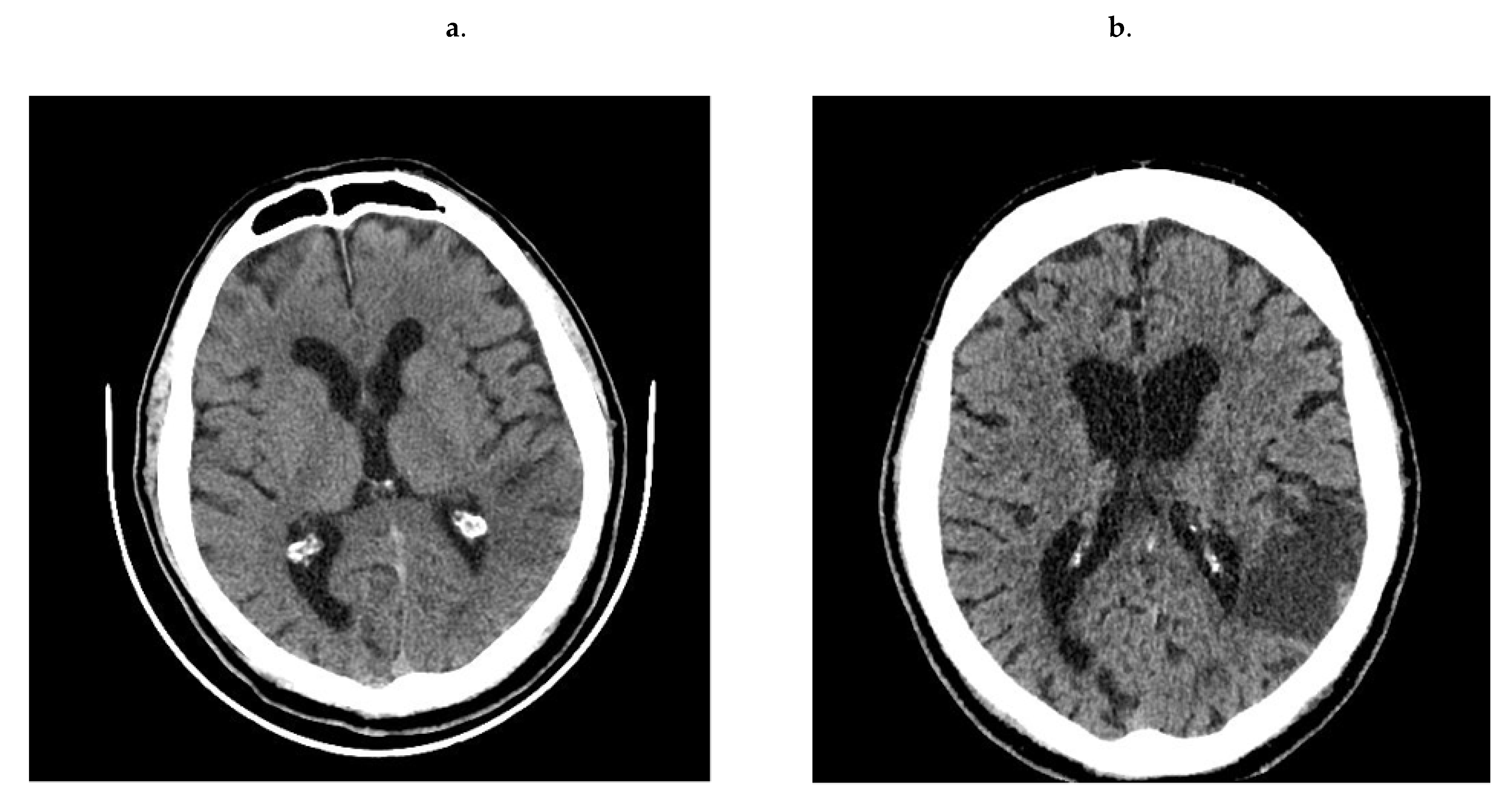
2.2. Recruitment of Patients, Study Design and Groups Description
2.3. Blood Collection
2.4. Preparation of Plasma for Proteomic Analysis
2.5. Quantitative and Qualitative Determination of Proteins-Proteomic Analysis by LC-MS
2.6. Statistical Analyses
3. Results
3.1. Baseline Characteristics of Patients
3.2. LC-MS Results
4. Discussion
4.1. Basic Biochemical Tests
4.2. Proteomics Analysis
5. Conclusions
Author Contributions
Funding
Institutional Review Board Statement
Informed Consent Statement
Data Availability Statement
Conflicts of Interest
References
- Mozaffarian, D.; Benjamin, E.J.; Go, A.S.; Arnett, D.K.; Blaha, M.J.; Cushman, M.; de Ferranti, S.; Després, J.P.; Fullerton, H.J.; Howard, V.J.; et al. American Heart Association Statistics Committee and Stroke Statistics Subcommittee. Heart disease and stroke statistics-2015 update: A report from the American Heart Association. Circulation 2015, 131, e29–e322. [Google Scholar] [CrossRef] [PubMed] [Green Version]
- Qiu, R.; Gao, Y.; Hou, D.; Wang, Y.; Yu, C.; Wang, W.; Liu, S.; Gao, C.; Tong, X.; Wu, J. Association between hs–CRP Levels and the Outcomes of Patients with Small–Artery Occlusion. Front. Aging Neurosci. 2016, 8, 191. [Google Scholar] [CrossRef] [PubMed] [Green Version]
- Belayev, L.; Busto, R.; Zhao, W.; Ginsberg, M.D. Quantitative evaluation of blood–brain barrier permeability following middle cerebral artery occlusion in rats. Brain Res. 1996, 739, 88–96. [Google Scholar] [CrossRef]
- Merten, C.L.; Knitelius, H.O.; Assheuer, J.; Bergmann–Kurz, B.; Hedde, J.P.; Bewermeyer, H. MRI of acute cerebral infarcts.; increased contrast enhancement with continuous infusion of gadolinium. Neuroradiology 1999, 41, 242–248. [Google Scholar] [CrossRef] [PubMed]
- Reynolds, M.A.; Kirchick, H.J.; Dahlen, J.R.; Anderberg, J.M.; McPherson, P.H.; Nakamura, K.K.; Laskowitz, D.T.; Valkirs, G.E.; Buechler, K.F. Early biomarkers of stroke. Clin. Chem. 2003, 49, 1733–1739. [Google Scholar] [CrossRef] [PubMed]
- Kim, B.; Winstein, C. Can Neurological Biomarkers of Brain Impairment Be Used to Predict Poststroke Motor Recovery? A Systematic Review. Neurorehabil. Neural. Repair. 2017, 31, 3–24. [Google Scholar] [CrossRef] [PubMed]
- Miao, Y.; Liao, J.K. Potential serum biomarkers in the pathophysiological processes of stroke. Expert. Rev. Neurother. 2014, 14, 173–185. [Google Scholar] [CrossRef] [PubMed] [Green Version]
- Fontes, J.D.; Yamamoto, J.F.; Larson, M.G.; Wang, N.; Dallmeier, D.; Rienstra, M.; Schnabel, R.B.; Vasan, R.S.; Keaney, J.F., Jr.; Benjamin, E.J. Clinical correlates of change in inflammatory biomarkers: The Framingham Heart Study. Atherosclerosis 2013, 228, 217–223. [Google Scholar] [CrossRef] [PubMed] [Green Version]
- Huang, P.; Lo, L.H.; Chen, Y.C.; Lin, R.T.; Shiea, J.; Liu, C.K. Serum free hemoglobin as a novel potential biomarker for acute ischemic stroke. J. Neurol. 2009, 256, 625–631. [Google Scholar] [CrossRef]
- Dassan, P.; Keir, G.; Brown, M.M. Criteria for a clinically informative serum biomarker in acute ischaemic stroke: A review of S100B. Cerebrovasc. Dis. 2009, 27, 295–302. [Google Scholar] [CrossRef] [PubMed]
- Nigro, N.; Wildi, K.; Mueller, C.; Schuetz, P.; Mueller, B.; Fluri, F.; Christ-Crain, M.; Katan, M. BNP but Not s-cTnln is associated with cardioembolic aetiology and predicts short- and long-term prognosis after cerebrovascular events. PLoS ONE 2014, 9, e102704. [Google Scholar] [CrossRef] [Green Version]
- Whiteley, W.; Tseng, M.C.; Sandercock, P. Blood biomarkers in the diagnosis of ischemic stroke: A systematic review. Stroke 2008, 39, 2902–2909. [Google Scholar] [CrossRef] [PubMed]
- Thompson, A.; Gao, P.; Orfei, L.; Watson, S.; Di Angelantonio, E.; Kaptoge, S.; Ballantyne, C.; Cannon, C.P.; Criqui, M.; et al.; Lp-PLA(2) Studies Collaboration Lipoprotein-associated phospholipase A(2) and risk of coronary disease, stroke, and mortality: Collaborative analysis of 32 prospective studies. Lancet 2010, 375, 1536–1544. [Google Scholar] [CrossRef] [Green Version]
- del Zoppo, G.J.; Levy, D.E.; Wasiewski, W.W.; Pancioli, A.M.; Demchuk, A.M.; Trammel, J.; Demaerschalk, B.M.; Kaste, M.; Albers, G.W.; Ringelstein, E.B. Hyperfibrinogenemia and functional outcome from acute ischemic stroke. Stroke 2009, 40, 1687–1691. [Google Scholar] [CrossRef] [Green Version]
- Wańkowicz, P.; Staszewski, J.; Dębiec, A.; Nowakowska-Kotas, M.; Szylińska, A.; Turoń-Skrzypińska, A.; Rotter, I. Pre-Stroke Statin Therapy Improves In-Hospital Prognosis Following Acute Ischemic Stroke Associated with Well-Controlled Nonvalvular Atrial Fibrillation. J. Clin. Med. 2021, 10, 3036. [Google Scholar] [CrossRef] [PubMed]
- Vasan, R.S. Biomarkers of cardiovascular disease: Molecular basis and practical considerations. Circulation 2006, 16, 2335–2362. [Google Scholar] [CrossRef] [PubMed]
- Chamorro, A.; Vila, N.; Ascaso, C.; Saiz, A.; Montalvo, J.; Alonso, P.; Tolosa, E. Early prediction of stroke severity. Role of the erythrocyte sedimentation rate. Stroke 1995, 26, 573–576. [Google Scholar] [CrossRef]
- Malinowska, A.; Kistowski, M.; Bakun, M. Diffprot—Software for non–parametric statistical analysis of differential proteomics data. J. Proteomics 2012, 75, 4062–4073. [Google Scholar] [CrossRef] [PubMed]
- Reina, S.A.; Llabre, M.M.; Allison, M.A.; Wilkins, J.T.; Mendez, A.J.; Arnan, M.K.; Schneiderman, N.; Sacco, R.L.; Carnethon, M.; Delaney, J.A. HDL cholesterol and stroke risk: The Multi-Ethnic Study of Atherosclerosis. Atherosclerosis 2015, 243, 314–319. [Google Scholar] [CrossRef] [PubMed] [Green Version]
- Tanne, D.; Yaari, S.; Goldbourt, U. High-density lipoprotein cholesterol and risk of ischemic stroke mortality. A 21-year follow-up of 8586 men from the Israeli Ischemic Heart Disease Study. Stroke 1997, 28, 83–87. [Google Scholar] [CrossRef]
- Lindenstrom, E.; Boysen, G.; Nyboe, J. Influence of total cholesterol, high density lipoprotein cholesterol, and triglycerides on risk of cerebrovascular disease: The Copenhagen City Heart Study. BMJ 1994, 309, 11–15. [Google Scholar] [CrossRef] [Green Version]
- Milton, M.; Smith, P.D. It’s All about Timing: The Involvement of Kir4.1 Channel Regulation in Acute Ischemic Stroke Pathology. Front. Cell Neurosci. 2018, 12, 36. [Google Scholar] [CrossRef] [Green Version]
- Gariballa, S.E.; Robinson, T.G.; Fotherby, M.D. Hypokalemia and potassium excretion in stroke patients. J. Am. Geriatr. Soc. 1997, 45, 1454–1458. [Google Scholar] [CrossRef] [PubMed]
- Adebamowo, S.N.; Spiegelman, D.; Flint, A.J.; Willett, W.C.; Rexrode, K.M. Intakes of magnesium, potassium, and calcium and the risk of stroke among men. Int. J. Stroke 2015, 10, 1093–1100. [Google Scholar] [CrossRef]
- Vinceti, M.; Filippini, T.; Crippa, A.; de Sesmaisons, A.; Wise, L.A.; Orsini, N. Meta-Analysis of Potassium Intake and the Risk of Stroke. J. Am. Heart Assoc. 2016, 5, e004210. [Google Scholar] [CrossRef]
- Hunt, B.D.; Cappuccio, F.P. Potassium intake and stroke risk: A review of the evidence and practical considerations for achieving a minimum target. Stroke 2014, 45, 1519–1522. [Google Scholar] [CrossRef] [PubMed] [Green Version]
- Gijsbers, L.; Dower, J.I.; Schalkwijk, C.G.; Kusters, Y.H.; Bakker, S.J.; Hollman, P.C.; Geleijnse, J.M. Effects of sodium and potassium supplementation on endothelial function: A fully controlled dietary intervention study. Br. J. Nutr. 2015, 114, 1419–1426. [Google Scholar] [CrossRef] [PubMed] [Green Version]
- Palmer, B.F.; Clegg, D.J. Achieving the benefits of a high-potassium, paleolithic diet, without the toxicity. Mayo Clin. Proc. 2016, 91, 496–508. [Google Scholar] [CrossRef] [PubMed] [Green Version]
- Ghahremanfard, F.; Asghari, N.; Ghorbani, R.; Samaei, A.; Ghomi, H.; Tamadon, M. The relationship between mean platelet volume and severity of acute ischemic brain stroke. Neurosciences 2013, 18, 147–151. [Google Scholar] [PubMed]
- Ciancarelli, I.; De Amicis, D.; Di Massimo, C.; Pistarini, C.; Ciancarelli, M.G. Mean Platelet Volume During Ischemic Stroke is a Potential Pro–inflammatory Biomarker in the Acute Phase and During Neurorehabilitation Not Directly Linked to Clinical Outcome. Curr. Neurovasc. Res. 2016, 13, 177–183. [Google Scholar] [CrossRef] [PubMed]
- Lee, C.D.; Folsom, A.R.; Nieto, F.J.; Chambless, L.E.; Shahar, E.; Wolfe, D.A. White blood cell count and incidence of coronary heart disease and ischemic stroke and mortality from cardiovascular disease in African–American and White men and women: Atherosclerosis risk in communities study. Am. J. Epidemiol. 2001, 154, 758–764. [Google Scholar] [CrossRef] [PubMed]
- Elkind, M.S.; Sciacca, R.R.; Boden–Albala, B.; Rundek, T.; Paik, M.C.; Sacco, R.L. Relative elevation in baseline leukocyte count predicts first cerebral infarction. Neurology 2005, 64, 2121–2125. [Google Scholar] [CrossRef] [PubMed]
- Miyazono, K.; Okabe, T.; Urabe, A.; Takaku, F.; Heldin, C.H. Purification and properties of an endothelial cell growth factor from human platelets. J. Biol. Chem. 1987, 262, 4098–4103. [Google Scholar] [CrossRef]
- Schwartz, M. Thymidine phosphorylase from Escherichia coli. Properties and kinetics. Eur. J. Biochem. 1971, 21, 191–198. [Google Scholar] [CrossRef]
- Miyadera, K.; Sumizawa, T.; Haraguchi, M.; Yoshida, H.; Konstanty, W.; Yamada, Y.; Akiyama, S. Role of thymidine phosphorylase activity in the angiogenic effect of platelet derived endothelial cell growth factor/thymidine phosphorylase. Cancer Res. 1995, 55, 1687–1690. [Google Scholar] [PubMed]
- Ikeda, R.; Furukawa, T.; Kitazono, M.; Ishitsuka, K.; Okumura, H.; Tani, A.; Sumizawa, T.; Haraguchi, M.; Komatsu, M.; Uchimiya, H.; et al. Molecular basis for the inhibition of hypoxia–induced apoptosis by 2–deoxy–D–ribose. Biochem. Biophys. Res. Commun. 2002, 291, 806–812. [Google Scholar] [CrossRef]
- Ishikawa, F.; Miyazono, K.; Hellman, U.; Drexler, H.; Wernstedt, C.; Hagiwara, K.; Usuki, K.; Takaku, F.; Risau, W.; Heldin, C.H. Identification of angiogenic activity and the cloning and expression of platelet–derived endothelial cell growth factor. Nature 1989, 338, 557–562. [Google Scholar] [CrossRef]
- Li, W.; Gigante, A.; Perez-Perez, M.J.; Yue, H.; Hirano, M.; McIntyre, T.M.; Silverstein, R.L. Thymidine Phosphorylase Participates in Platelet Signaling and Promotes Thrombosis. Circ. Res. 2014, 115, 997–1006. [Google Scholar] [CrossRef] [Green Version]
- Chapouly, C.; Tadesse, A.A.; Horng, S.; Castro, K.; Zhang, J.; Asp, L.; Loo, H.; Laitman, B.M.; Mariani, J.N.; Straus Farber, R.; et al. Astrocytic TYMP and VEGFA drive blood–brain barrier opening in inflammatory central nervous system lesions. Brain 2015, 138, 1548–1567. [Google Scholar] [CrossRef] [PubMed] [Green Version]
- Asai, K.; Nakanishi, K.; Isobe, I.; Eksioglu, Y.Z.; Hirano, A.; Hama, K.; Miyamoto, T.; Kato, T. Neurotrophic action of gliostatin on cortical neurons. Identity of gliostatin and platelet–derived endothelial cell growth factor. J. Biol. Chem. 1992, 267, 20311–20316. [Google Scholar] [CrossRef]
- Jessen, K.R.; Mirsky, R. Glial cells in the enteric nervous system contain glial fibrillary acidic protein. Nature 1980, 286, 736–737. [Google Scholar] [CrossRef]
- Hayashi, T.; Wang, X.Q.; Zhang, H.Z.; Deguchi, K.; Nagotani, S.; Sehara, Y.; Tsuchiya, A.; Nagai, M.; Shoji, M.; Abe, K. Induction of platelet derived–endothelial cell growth factor in the brain after ischemia. Neurol. Res. 2007, 29, 463–468. [Google Scholar] [CrossRef] [PubMed]
- Belcher, A.; Zulfiker, A.H.M.; Li, O.Q.; Yue, H.; Gupta, A.S.; Li, W. Targeting Thymidine Phosphorylase with Tipiracil Hydrochloride Attenuates Thrombosis Without Increasing Risk of Bleeding in Mice. Arterioscler. Thromb. Vasc. Biol. 2021, 41, 668–682. [Google Scholar] [CrossRef]
- Cho, G.W.; Koh, S.H.; Kim, M.H.; Yoo, A.R.; Noh, M.Y.; Oh, S.; Kim, S.H. The neuroprotective effect of erythropoietin–transduced human mesenchymal stromal cells in an animal model of ischemic stroke. Brain Res. 2010, 1353, 1–13. [Google Scholar] [CrossRef]
- Li, W.; Yue, H. Thymidine phosphorylase: A potential new target for treating cardiovascular disease. Trends Cardiovasc. Med. 2018, 28, 157–171. [Google Scholar] [CrossRef]
- Hollander, Z.; Dai, D.L.; Putko, B.N.; Yogasundaram, H.; Wilson–McManus, J.E.; Thompson, R.B.; Khan, A.; West, M.L.; McManus, B.M.; Oudit, G.Y. Gender–specific plasma proteomic biomarkers in patients with Anderson–Fabry disease. Eur. J. Heart Fail. 2015, 17, 291–300. [Google Scholar] [CrossRef] [Green Version]
- Udby, L.; Sørensen, O.E.; Pass, J.; Johnsen, A.H.; Behrendt, N.; Borregaard, N.; Kjeldsen, L. Cysteine–rich secretory protein 3 is a ligand of alpha1B–glycoprotein in human plasma. Biochemistry 2004, 43, 12877–12886. [Google Scholar] [CrossRef] [PubMed]
- Helgeland, E.; Breivik, L.E.; Vaudel, M.; Svendsen, Ø.S.; Garberg, H.; Nordrehaug, J.E.; Berven, F.S.; Jonassen, A.K. Exploring the human plasma proteome for humoral mediators of remote ischemic preconditioning–a word of caution. PLoS ONE 2014, 9, e109279. [Google Scholar] [CrossRef] [Green Version]
- Kinouchi, H.; Sharp, F.R.; Hill, M.P.; Koistinaho, J.; Sagar, S.M.; Chan, P.H. Induction of 70–kDa heat shock protein and hsp70 mRNA following transient focal cerebral ischemia in the rat. J. Cereb. Blood Flow Metab. 1993, 13, 105–115. [Google Scholar] [CrossRef] [Green Version]
- Liu, Y.; Jiang, S.; Yang, P.Y.; Zhang, Y.F.; Li, T.J.; Rui, Y.C. EF1A1/HSC70 Cooperatively Suppress Brain Endothelial Cell Apoptosis via Regulating JNK Activity. CNS Neurosci. Ther. 2016, 22, 836–844. [Google Scholar] [CrossRef] [PubMed]
- Chen, A.; Liao, W.P.; Lu, Q.; Wong, W.S.; Wong, P.T. Upregulation of dihydropyrimidinase–related protein 2, spectrin alpha II chain.; heat shock cognate protein 70 pseudogene 1 and tropomodulin 2 after focal cerebral ischemia in rats–a proteomics approach. Neurochem. Int. 2007, 50, 1078–1086. [Google Scholar] [CrossRef]
- Calabrese, V.; Scapagnini, G.; Ravagna, A.; Colombrita, C.; Spadaro, F.; Butterfield, D.A.; Giuffrida Stella, A.M. Increased expression of heat shock proteins in rat brain during aging: Relationship with mitochondrial function and glutathione redox state. Mech. Ageing Dev. 2004, 125, 325–335. [Google Scholar] [CrossRef]
- Stricher, F.; Macri, C.; Ruff, M.; Muller, S. HSPA8/HSC70 chaperone protein: Structure.; function.; and chemical targeting. Autophagy 2013, 9, 1937–1954. [Google Scholar] [CrossRef] [Green Version]
- Daugaard, M.; Rohde, M.; Jäättelä, M. The heat shock protein 70 family: Highly homologous proteins with overlapping and distinct functions. FEBS Lett. 2007, 581, 3702–3710. [Google Scholar] [CrossRef] [Green Version]
- Habryka, A.; Gogler-Pigłowska, A.; Sojka, D.; Kryj, M.; Krawczyk, Z.; Scieglinska, D. Cell type–dependent modulation of the gene encoding heat shock protein HSPA2 by hypoxia–inducible factor HIF–1: Down–regulation in keratinocytes and up–regulation in HeLa cells. Biochim. Biophys. Acta 2015, 1849, 1155–1169. [Google Scholar] [CrossRef] [PubMed]
- Alawieh, A.; Elvington, A.; Tomlinson, S. Complement in the Homeostatic and Ischemic Brain. Front. Immunol. 2015, 6, 417. [Google Scholar] [CrossRef] [PubMed] [Green Version]
- Gorsuch, W.B.; Chrysanthou, E.; Schwaeble, W.J.; Stahl, G.L. The complement system in ischemia-reperfusion injuries. Immunobiology 2012, 217, 1026–1033. [Google Scholar] [CrossRef] [Green Version]
- Arumugam, T.V.; Woodruff, T.M.; Lathia, J.D.; Selvaraj, P.K.; Mattson, M.P.; Taylor, S.M. Neuroprotection in stroke by complement inhibition and immunoglobulin therapy. Neuroscience 2009, 158, 1074–1789. [Google Scholar] [CrossRef] [PubMed] [Green Version]
- Pedersen, E.D.; Waje-Andreassen, U.; Vedeler, C.A.; Aamodt, G.; Mollnes, T.E. Systemic complement activation following human acute ischaemic stroke. Clin. Exp. Immunol. 2004, 137, 117–122. [Google Scholar] [CrossRef]
- Alawieh, A.; Elvington, A.; Zhu, H.; Yu, J.; Kindy, M.S.; Atkinson, C.; Tomlinson, S. Modulation of post-stroke degenerative and regenerative processes and subacute protection by site-targeted inhibition of the alternative pathway of complement. J. Neuroinflamm. 2015, 12, 247. [Google Scholar] [CrossRef] [Green Version]
- Macrez, R.; Ali, C.; Toutirais, O.; Le Mauff, B.; Defer, G.; Dirnagl, U.; Vivien, D. Stroke and the immune system: From pathophysiology to new therapeutic strategies. Lancet Neurol. 2011, 10, 471–480. [Google Scholar] [CrossRef]
- Orsini, F.; De Blasio, D.; Zangari, R.; Zanier, E.R.; De Simoni, M.-G. Versatility of the complement system in neuroinflammation, neurodegeneration and brain homeostasis. Front. Cell Neurosci. 2014, 8, 380. [Google Scholar] [CrossRef] [Green Version]
- Bhatia, M.; Howard, S.C.; Clark, T.G.; Neale, R.; Qizilbash, N.; Murphy, M.F.; Rothwell, P.M. Apolipoproteins as predictors of ischaemic stroke in patients with a previous transient ischaemic attack. Cerebrovasc. Dis. 2006, 21, 323–328. [Google Scholar] [CrossRef]
- Kargman, D.E.; Tuck, C.; Berglund, L.; Lin, I.F.; Mukherjee, R.S.; Thompson, E.V.; Jones, J.; Boden–Albala, B.; Paik, M.C.; Sacco, R.L. Lipid and lipoprotein levels remain stable in acute ischemic stroke: The Northern Manhattan Stroke Study. Atherosclerosis 1998, 139, 391–399. [Google Scholar] [CrossRef]
- Park, J.H.; Hong, K.S.; Lee, E.J.; Lee, J.; Kim, D.E. High levels of apolipoprotein B/AI ratio are associated with intracranial atherosclerotic stenosis. Stroke 2011, 42, 3040–3046. [Google Scholar] [CrossRef] [Green Version]
- Vuilleumier, N.; Rossier, M.F.; Pagano, S.; Python, M.; Charbonney, E.; Nkoulou, R.; James, R.; Reber, G.; Mach, F.; Roux–Lombard, P. Anti–apolipoprotein A–1 IgG as an independent cardiovascular prognostic marker affecting basal heart rate in myocardial infarction. Eur. Heart J. 2010, 31, 815–823. [Google Scholar] [CrossRef] [PubMed] [Green Version]
- Rodríguez–Yáñez, M.; Sobrino, T.; Arias, S.; Vázquez–Herrero, F.; Brea, D.; Blanco, M.; Leira, R.; Castellanos, M.; Serena, J.; Vivancos, J.; et al. Early biomarkers of clinical–diffusion mismatch in acute ischemic stroke. Stroke 2011, 42, 2813–2818. [Google Scholar] [CrossRef] [Green Version]
- Wang, Y.; Reheman, A.; Spring, C.M.; Kalantari, J.; Marshall, A.H.; Wolberg, A.S.; Gross, P.L.; Weitz, J.I.; Rand, M.L.; Mosher, D.F.; et al. Plasma fibronectin supports hemostasis and regulates thrombosis. J. Clin. Investig. 2014, 124, 4281–4293. [Google Scholar] [CrossRef] [Green Version]
- Ning, M.; Sarracino, D.A.; Kho, A.T.; Guo, S.; Lee, S.R.; Krastins, B.; Buonanno, F.S.; Vizcaíno, J.A.; Orchard, S.; McMullin, D.; et al. Proteomic temporal profile of human brain endothelium after oxidative stress. Stroke 2011, 42, 37–43. [Google Scholar] [CrossRef] [PubMed] [Green Version]
- Silva, Y.; Leira, R.; Tejada, J.; Lainez, J.M.; Castillo, J.; Dávalos, A. Stroke Project. Cerebrovascular Diseases Group of the Spanish Neurological Society. Molecular signatures of vascular injury are associated with early growth of intracerebral hemorrhage. Stroke 2005, 36, 86–91. [Google Scholar] [CrossRef]
- Castellanos, M.; Sobrino, T.; Millán, M.; García, M.; Arenillas, J.; Nombela, F.; Brea, D.; Perez de la Ossa, N.; Serena, J.; Vivancos, J.; et al. Serum cellular fibronectin and matrix metalloproteinase–9 as screening biomarkers for the prediction of parenchymal hematoma after thrombolytic therapy in acute ischemic stroke: A multicenter confirmatory study. Stroke 2007, 38, 1855–1859. [Google Scholar] [CrossRef] [PubMed] [Green Version]
- Wang, H.; Li, W.; Zhu, S.; Li, J.; D’Amore, J.; Ward, M.F.; Yang, H.; Wu, R.; Jahnen–Dechent, W.; Tracey, K.J.; et al. Peripheral administration of fetuin–A attenuates early cerebral ischemic injury in rats. J. Cereb. Blood Flow. Metab. 2010, 30, 493–504. [Google Scholar] [CrossRef] [PubMed] [Green Version]
- Sezer, S.; Uçar, F.; Ulusoy, E.K.; Erdogan, S.; Bilen, S.; Züngün, C.; Uysal, S.; Erdemli, H.K. Serum amyloid A, fetuin–A, and pentraxin–3 levels in patients with ischemic stroke: Novel prognostic biomarkers? Turkish J. Med. Sci. 2014, 44, 16–23. [Google Scholar] [CrossRef]
- Paley, E.L.; Paley, D.E.; Merkulova-Rainon, T.; Subbarayan, P.R. Hypoxia signature of splice forms of tryptophanyl–tRNAsynthetase marks pancreatic cancer cells with distinct metastatic abilities. Pancreas 2011, 40, 1043–1056. [Google Scholar] [CrossRef] [PubMed]
- Zeng, R.; Chen, Y.C.; Zeng, Z.; Liu, W.Q.; Jiang, X.F.; Liu, R.; Qiang, O.; Li, X. Effect of mini–tyrosyl–tRNAsynthetase/mini–tryptophanyl–tRNAsynthetase on ischemic angiogenesis in rats: Proliferation and migration of endothelial cells. Heart Vessel. 2011, 26, 69–80. [Google Scholar] [CrossRef]
- Zeng, R.; Wang, M.; You, G.Y.; Yue, R.Z.; Chen, Y.C.; Zeng, Z.; Liu, R.; Qiang, O.; Zhang, L. Effect of Mini–Tyrosyl–tRNASynthetase/Mini–Tryptophanyl–tRNA Synthetase on Angiogenesis in Rhesus Monkeys after Acute Myocardial Infarction. Cardiovasc. Ther. 2016, 34, 4–12. [Google Scholar] [CrossRef] [Green Version]
- Messmer-Blust, A.; An, X.; Li, J. Hypoxia–regulated angiogenic inhibitors. Trends Cardiovasc. Med. 2009, 19, 252–256. [Google Scholar] [CrossRef] [PubMed] [Green Version]
- Zangari, R.; Zanier, E.R.; Torgano, G.; Bersano, A.; Beretta, S.; Beghi, E.; Casolla, B.; Checcarelli, N.; Lanfranconi, S.; Maino, A.; et al. Early ficolin–1 is a sensitive prognostic marker for functional outcome in ischemic stroke. J. Neuroinflamm. 2016, 13, 16. [Google Scholar] [CrossRef] [Green Version]
- Füst, G.; Munthe–Fog, L.; Illes, Z.; Széplaki, G.; Molnar, T.; Pusch, G.; Hirschberg, K.; Szegedi, R.; Széplaki, Z.; Prohászka, Z.; et al. Low ficolin–3 levels in early follow–up serum samples are associated with the severity and unfavorable outcome of acute ischemic stroke. J. Neuroinflamm. 2011, 8, 185. [Google Scholar] [CrossRef] [Green Version]


| Stroke (Mean ± SEM) n = 31 | Control (Mean± SEM) n = 28 | p | ||||||
|---|---|---|---|---|---|---|---|---|
| Women | 13 (42%) | 14 (50%) | 0.38 | |||||
| Age | [y] | 62.68 | ± | 9.35 | 62.00 | ± | 11.40 | 0.80 |
| Hemoglobin | [g/dL] | 14.39 | ± | 1.63 | 13.78 | ± | 1.63 | 0.16 |
| Hematocrit | [%] | 42.65 | ± | 4.45 | 40.93 | ± | 4.81 | 0.16 |
| RBC | [m/µL] | 4.79 | ± | 0.59 | 4.68 | ± | 0.58 | 0.50 |
| WBC | [k/µL] | 9.16 | ± | 3.48 | 7.05 | ± | 2.55 | 0.01 * |
| PLT | [k/µL] | 254 | ± | 195 | 244 | ± | 46 | 0.14 |
| MPV | [fl] | 9.38 | ± | 1.26 | 10.90 | ± | 0.83 | 0.00 * |
| hsCRP | [mg/L] | 5.55 | ± | 6.03 | 5.52 | ± | 5.96 | 0.99 |
| Potassium | [mmol/L] | 3.85 | ± | 0.34 | 4.15 | ± | 0.42 | 0.00 * |
| Sodium | [mmol/L] | 139 | ± | 2.50 | 139.38 | ± | 5.23 | 0.06 |
| Glucose | [mg/dL] | 130 | ± | 50 | 108 | ± | 51 | 0.12 |
| Urea | [mg/dL] | 33.7 | ± | 12.7 | 29.5 | ± | 13.1 | 0.22 |
| Creatinine | [mg/dL] | 0.93 | ± | 0.27 | 0.89 | ± | 0.27 | 0.57 |
| AST | [IU/L] | 19.00 | ± | 7.06 | 23.50 | ± | 12.00 | 0.22 |
| ALT | [IU/L] | 21.63 | ± | 9.79 | 26.65 | ± | 15.75 | 0.31 |
| Total bilirubin | [mg/dL] | 0.54 | ± | 0.07 | 0.72 | ± | 0.29 | 0.30 |
| TCh | [mg/dL] | 184.27 | ± | 48.20 | 210.95 | ± | 52.34 | 0.06 |
| HDL | [mg/dL] | 49.20 | ± | 13.60 | 58.36 | ± | 15.87 | 0.03 * |
| LDL | [mg/dL] | 108.83 | ± | 41.47 | 126.22 | ± | 47.50 | 0.16 |
| TG | [mg/dL] | 137.23 | ± | 92.44 | 144.48 | ± | 84.09 | 0.77 |
| TSH | [µIU/L] | 3.03 | ± | 3.56 | 1.47 | ± | 0.85 | 0.06 |
| APTT | [s] | 27.66 | ± | 3.58 | 28.61 | ± | 5.22 | 0.45 |
| PT/INR | 0.98 | ± | 0.10 | 0.99 | ± | 0.05 | 0.18 | |
| Protein Name | Comparison | q Value | Ratio | Fold Change | Peptides | |
|---|---|---|---|---|---|---|
| Complement factor B | A vs. C | p1 | 0.13460 | 0.51 | 1.97 | 69 |
| p2 | 0.37151 | 0.49 | 2.04 | 69 | ||
| B vs. C | p1 | 0.00032 * | 0.52 | 1.92 | 70 | |
| p2 | 0.00015 * | 0.53 | 1.87 | 70 | ||
| A + B vs. C | p1 | 0.03326 * | 0.56 | 1.80 | 70 | |
| p2 | 0.15461 | 0.59 | 1.69 | 69 | ||
| Apolipoprotein A-I | A vs. C | p1 | 1.00000 | 0.69 | 1.45 | 405 |
| p2 | 1.00000 | 0.75 | 1.34 | 405 | ||
| B vs. C | p1 | 0.00021 * | 0.63 | 1.59 | 405 | |
| p2 | 0.00034 * | 0.65 | 1.53 | 405 | ||
| A + B vs. C | p1 | 1.00000 | 0.64 | 1.57 | 407 | |
| p2 | 1.00000 | 0.62 | 1.61 | 405 | ||
| Fibronectin | A vs. C | p1 | 0.78238 | 1.57 | 1.57 | 12 |
| p2 | 0.68263 | 1.43 | 1.43 | 12 | ||
| B vs. C | p1 | 0.08008 | 1.95 | 1.95 | 12 | |
| p2 | 0.03980 * | 2.15 | 2.15 | 12 | ||
| A + B vs. C | p1 | 0.24051 | 1.43 | 1.43 | 12 | |
| p2 | 0.77102 | 1.40 | 1.40 | 12 | ||
| Alpha-2-HS-glycoprotein | A vs. C | p1 | 0.69703 | 1.84 | 1.84 | 126 |
| p2 | 0.72959 | 1.66 | 1.66 | 126 | ||
| B vs. C | p1 | 0.01408 * | 1.62 | 1.62 | 126 | |
| p2 | 0.00015 * | 1.63 | 1.63 | 126 | ||
| A + B vs. C | p1 | 0.29914 | 1.39 | 1.39 | 126 | |
| p2 | 0.87082 | 1.53 | 1.53 | 126 | ||
| Alpha-1B-glycoprotein | A vs. C | p1 | 0.00290 * | 0.54 | 1.87 | 55 |
| p2 | 0.04753 * | 0.52 | 1.91 | 54 | ||
| B vs. C | p1 | 1.00000 | 0.56 | 1.78 | 55 | |
| p2 | 0.90000 | 0.57 | 1.74 | 53 | ||
| A + B vs. C | p1 | 0.05477 | 0.46 | 2.19 | 55 | |
| p2 | 0.00146 * | 0.40 | 2.53 | 54 | ||
| Heat shock protein Hsp70 family † | A vs. C | p1 | 0.00663 * | 2.19 | 2.19 | 1 |
| B vs. C | p1 | 1.00000 | 2.11 | 2.11 | 1 | |
| A + B vs. C | p1 | 1.00000 | 1.51 | 1.51 | 1 | |
| Thymidine phosphorylase | A vs. C | p1 | 0.77044 | 0.23 | 4.35 | 1 |
| B vs. C | p1 | 0.57787 | 0.13 | 7.55 | 1 | |
| A + B vs. C | p1 | 0.00425 * | 0.12 | 8.05 | 1 | |
| Tryptophan--tRNAligase, cytoplasmic | A vs. C | p1 | 0.85112 | 0.44 | 2.29 | 1 |
| B vs. C | p1 | 0.00163 * | 0.18 | 5.55 | 1 | |
| A + B vs. C | p1 | 0.00230 * | 0.25 | 3.96 | 1 | |
| Ficolin-2 | A vs. C | p1 | 0.67919 | 2.77 | 2.77 | 5 |
| p2 | 0.68670 | 2.11 | 2.11 | 5 | ||
| B vs. C | p1 | 0.01077 * | 6.53 | 6.53 | 5 | |
| p2 | 0.04484 * | 5.18 | 5.18 | 5 | ||
| A + B vs. C | p1 | 0.29495 | 1.67 | 1.67 | 5 | |
| p2 | 0.34767 | 2.55 | 2.55 | 5 | ||
| Beta-Ala-His dipeptidase | A vs. C | p1 | 0.14225 | 6.47 | 6.47 | 7 |
| p2 | 0.35263 | 6.10 | 6.10 | 7 | ||
| B vs. C | p1 | 0.08966 | 2.58 | 2.58 | 7 | |
| p1 | 0.06369 | 3.13 | 3.13 | 7 | ||
| A + B vs. C | p1 | 0.04009 * | 1.49 | 1.49 | 7 | |
| p2 | 0.13244 | 1.16 | 1.16 | 7 |
Publisher’s Note: MDPI stays neutral with regard to jurisdictional claims in published maps and institutional affiliations. |
© 2022 by the authors. Licensee MDPI, Basel, Switzerland. This article is an open access article distributed under the terms and conditions of the Creative Commons Attribution (CC BY) license (https://creativecommons.org/licenses/by/4.0/).
Share and Cite
Turek-Jakubowska, A.; Dębski, J.; Jakubowski, M.; Szahidewicz-Krupska, E.; Gawryś, J.; Gawryś, K.; Janus, A.; Trocha, M.; Doroszko, A. New Candidates for Biomarkers and Drug Targets of Ischemic Stroke—A First Dynamic LC-MS Human Serum Proteomic Study. J. Clin. Med. 2022, 11, 339. https://doi.org/10.3390/jcm11020339
Turek-Jakubowska A, Dębski J, Jakubowski M, Szahidewicz-Krupska E, Gawryś J, Gawryś K, Janus A, Trocha M, Doroszko A. New Candidates for Biomarkers and Drug Targets of Ischemic Stroke—A First Dynamic LC-MS Human Serum Proteomic Study. Journal of Clinical Medicine. 2022; 11(2):339. https://doi.org/10.3390/jcm11020339
Chicago/Turabian StyleTurek-Jakubowska, Aleksandra, Janusz Dębski, Maciej Jakubowski, Ewa Szahidewicz-Krupska, Jakub Gawryś, Karolina Gawryś, Agnieszka Janus, Małgorzata Trocha, and Adrian Doroszko. 2022. "New Candidates for Biomarkers and Drug Targets of Ischemic Stroke—A First Dynamic LC-MS Human Serum Proteomic Study" Journal of Clinical Medicine 11, no. 2: 339. https://doi.org/10.3390/jcm11020339