

Li-Doped Bioactive Ceramics: Promising Biomaterials for Tissue Engineering and Regenerative Medicine
 ,
,  and
and
Abstract
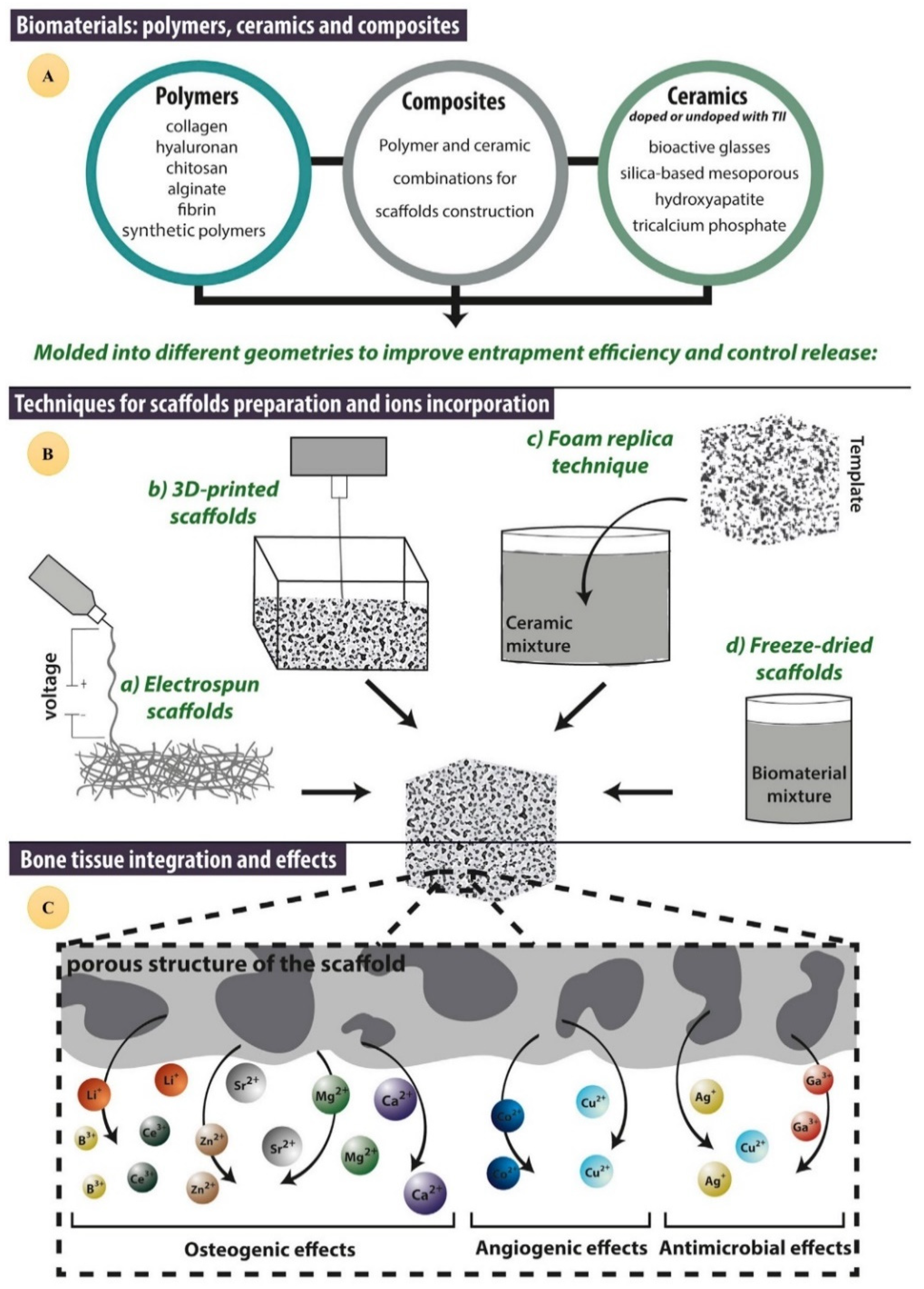
:1. Introduction
2. Lithium and Its Biological Effects
2.1. Lithium and Stem Cell Fate
2.2. Lithium and Osteogenesis
2.3. Lithium and Bone and Cartilage Regeneration
2.4. Lithium and Wound Healing
2.5. Lithium and Nerve Regeneration
2.6. Lithium and Antibacterial and Antiviral Activities
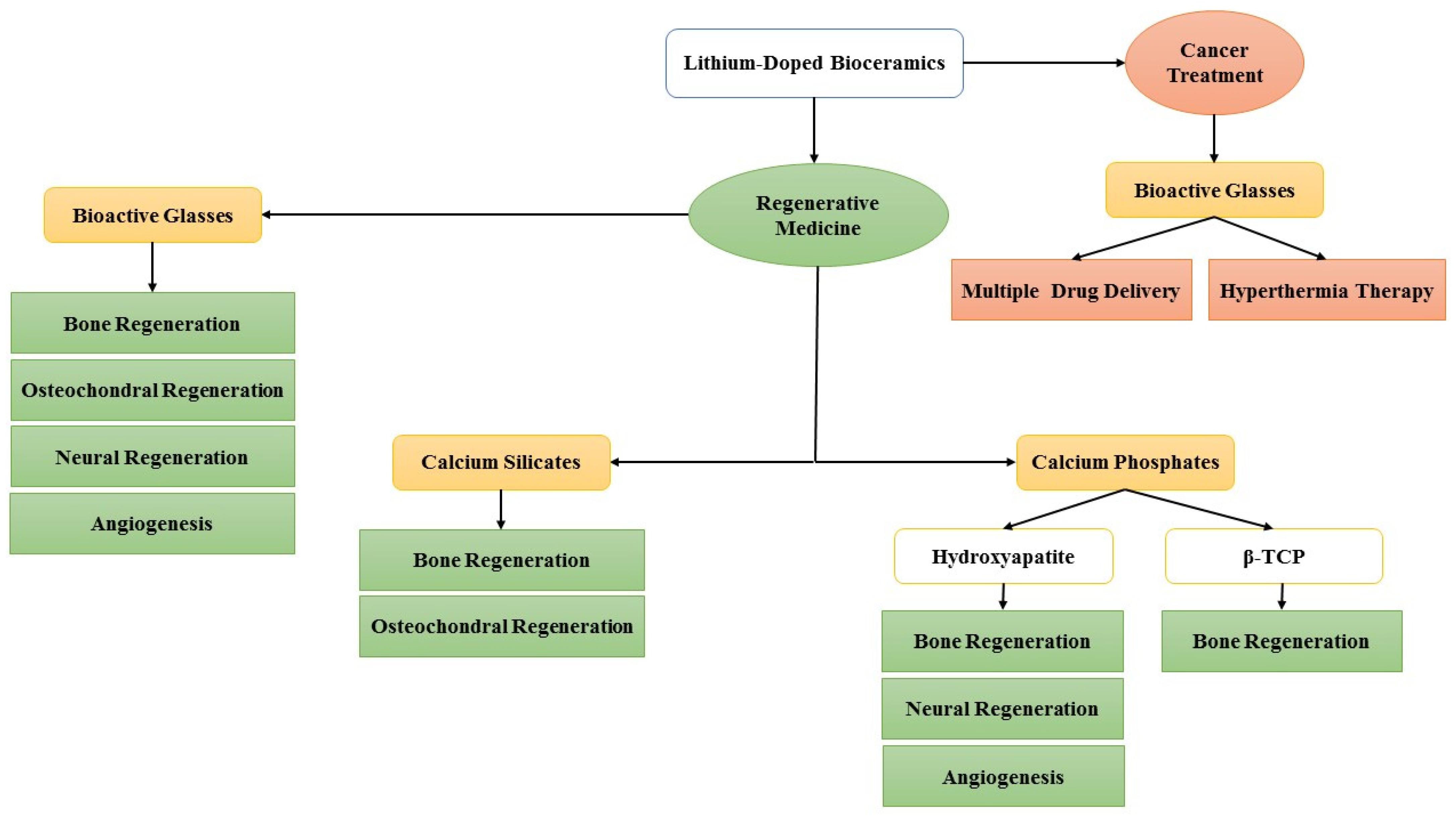
3. Lithium-Doped Bioceramics in Regenerative Medicine
3.1. Lithium-Doped Bioactive Glasses (Li-BGs)
3.2. Lithium-Doped Calcium Phosphates
3.3. Other Lithium-Doped Bioceramics
3.4. Lithium-Doped Bioceramics for Anticancer Applications
4. Conclusions
Author Contributions
Funding
Data Availability Statement
Conflicts of Interest
References
- Farmani, A.R.; Nekoofar, M.H.; Barough, S.E.; Azami, M.; Rezaei, N.; Najafipour, S.; Ai, J. Application of Platelet Rich Fibrin in Tissue Engineering: Focus on Bone Regeneration. Platelets 2021, 32, 183–188. [Google Scholar] [CrossRef] [PubMed]
- Khademi, F.; Soleimani, M.; Verdi, J.; Tavangar, S.M.; Sadroddiny, E.; Massumi, M.; Ai, J. Human endometrial stem cells differentiation into functional hepatocyte-like cells. Cell Biol. Int. 2014, 38, 825–834. [Google Scholar] [CrossRef] [PubMed]
- Asadpour, S.; Kargozar, S.; Moradi, L.; Ai, A.; Nosrati, H.; Ai, J. Natural biomacromolecule based composite scaffolds from silk fibroin, gelatin and chitosan toward tissue engineering applications. Int. J. Biol. Macromol. 2020, 154, 1285–1294. [Google Scholar] [CrossRef] [PubMed]
- Rezapour-Lactoee, A.; Yeganeh, H.; Ostad, S.N.; Gharibi, R.; Mazaheri, Z.; Ai, J. Thermoresponsive polyurethane/siloxane membrane for wound dressing and cell sheet transplantation: In-vitro and in-vivo studies. Mater. Sci. Eng. C 2016, 69, 804–814. [Google Scholar] [CrossRef]
- Asadpour, S.; Yeganeh, H.; Ai, J.; Kargozar, S.; Rashtbar, M.; Seifalian, A.; Ghanbari, H. Polyurethane-Polycaprolactone Blend Patches: Scaffold Characterization and Cardiomyoblast Adhesion, Proliferation, and Function. ACS Biomater. Sci. Eng. 2018, 4, 4299–4310. [Google Scholar] [CrossRef]
- Hasanzadeh, E.; Ebrahimi-Barough, S.; Mirzaei, E.; Azami, M.; Tavangar, S.M.; Mahmoodi, N.; Basiri, A.; Ai, J. Preparation of fibrin gel scaffolds containing MWCNT/PU nanofibers for neural tissue engineering. J. Biomed. Mater. Res. Part A 2019, 107, 802–814. [Google Scholar] [CrossRef]
- Noory, P.; Navid, S.; Zanganeh, B.M.; Talebi, A.; Borhani-Haghighi, M.; Gholami, K.; Manshadi, M.D.; Abbasi, M. Human menstrual blood stem cell-derived granulosa cells participate in ovarian follicle formation in a rat model of premature ovarian failure in vivo. Cell. Reprogramming 2019, 21, 249–259. [Google Scholar] [CrossRef]
- Jabari, A.; Gilani, M.A.S.; Koruji, M.; Gholami, K.; Mohsenzadeh, M.; Khadivi, F.; Gashti, N.G.; Nikmahzar, A.; Mojaverrostami, S.; Talebi, A.; et al. Three-dimensional co-culture of human spermatogonial stem cells with Sertoli cells in soft agar culture system supplemented by growth factors and Laminin. Acta Histochem. 2020, 122, 151572. [Google Scholar] [CrossRef]
- Gholami, K.; Vermeulen, M.; Del Vento, F.; de Michele, F.; Giudice, M.G.; Wyns, C. The air-liquid interface culture of the mechanically isolated seminiferous tubules embedded in agarose or alginate improves in vitro spermatogenesis at the expense of attenuating their integrity. In Vitro Cell. Dev. Biol.-Anim. 2020, 56, 261–270. [Google Scholar] [CrossRef]
- Ashouri Movassagh, S.; Ashouri Movassagh, S.; Dehkordi, M.B.; Pourmand, G.; Gholami, K.; Talebi, A.; Esfandyari, S.; Jabari, A.; Samadian, A.; Abbasi, M. Isolation, identification and differentiation of human spermatogonial cells on three-dimensional decellularized sheep testis. Acta Histochem. 2020, 122, 151623. [Google Scholar] [CrossRef]
- Bakhshandeh, B.; Zarrintaj, P.; Oftadeh, M.O.; Keramati, F.; Fouladiha, H.; Sohrabi-Jahromi, S.; Ziraksaz, Z. Tissue engineering; strategies, tissues, and biomaterials. Biotechnol. Genet. Eng. Rev. 2017, 33, 144–172. [Google Scholar] [CrossRef] [PubMed]
- Han, F.; Wang, J.; Ding, L.; Hu, Y.; Li, W.; Yuan, Z.; Guo, Q.; Zhu, C.; Yu, L.; Wang, H.; et al. Tissue Engineering and Regenerative Medicine: Achievements, Future, and Sustainability in Asia. Front. Bioeng. Biotechnol. 2020, 8, 83. [Google Scholar] [CrossRef] [PubMed]
- Khademhosseini, A.; Langer, R. A decade of progress in tissue engineering. Nat. Protoc. 2016, 11, 1775–1781. [Google Scholar] [CrossRef] [PubMed]
- Pina, S.; Ribeiro, V.P.; Marques, C.F.; Maia, F.R.; Silva, T.H.; Reis, R.L.; Oliveira, J.M. Scaffolding strategies for tissue engineering and regenerative medicine applications. Materials 2019, 12, 1824. [Google Scholar] [CrossRef]
- Kwon, S.G.; Kwon, Y.W.; Lee, T.W.; Park, G.T.; Kim, J.H. Recent advances in stem cell therapeutics and tissue engineering strategies. Biomater. Res. 2018, 22, 36. [Google Scholar] [CrossRef]
- Willerth, S.M.; Sakiyama-Elbert, S.E. Combining stem cells and biomaterial scaffolds for constructing tissues and cell delivery. StemJournal 2019, 1, 1–25. [Google Scholar] [CrossRef]
- Hayat, H.; Hayat, H.; Dwan, B.F.; Gudi, M.; Bishop, J.O.; Wang, P. A Concise Review: The Role of Stem Cells in Cancer Progression and Therapy. OncoTargets Ther. 2021, 14, 2761. [Google Scholar] [CrossRef]
- Pérez, L.M.; de Lucas, B.; Gálvez, B.G. Unhealthy stem cells: When health conditions upset stem cell properties. Cell. Physiol. Biochem. 2018, 46, 1999–2016. [Google Scholar] [CrossRef]
- Dhesi, A.S.; Morelli, S.S. Endometriosis: A role for stem cells. Women’s Health 2015, 11, 35–49. [Google Scholar] [CrossRef]
- Mahdavinezhad, F.; Gharaei, R.; Farmani, A.R.; Hashemi, F.; Kouhestani, M.; Amidi, F. The Potential Relationship Between Different Human Female Reproductive Disorders and Sperm Quality in Female Genital Tract. Reprod. Sci. 2021, 29, 695–710. [Google Scholar] [CrossRef]
- Lee, E.J.; Kasper, F.K.; Mikos, A.G. Biomaterials for tissue engineering. Ann. Biomed. Eng. 2014, 42, 323–337. [Google Scholar] [CrossRef] [PubMed]
- Sharma, K.; Mujawar, M.A.; Kaushik, A. State-of-Art Functional Biomaterials for Tissue Engineering. Front. Mater. 2019, 6, 172. [Google Scholar] [CrossRef]
- Kohane, D.S.; Langer, R. Polymeric Biomaterials in Tissue Engineering. Pediatric Res. 2008, 63, 487–491. [Google Scholar] [CrossRef]
- Baino, F.; Novajra, G.; Vitale-Brovarone, C. Bioceramics and Scaffolds: A Winning Combination for Tissue Engineering. Front. Bioeng. Biotechnol. 2015, 3, 202. [Google Scholar] [CrossRef]
- Zhao, H.; Liu, M.; Zhang, Y.; Yin, J.; Pei, R. Nanocomposite hydrogels for tissue engineering applications. Nanoscale 2020, 12, 14976–14995. [Google Scholar] [CrossRef]
- Prasad, K.; Bazaka, O.; Chua, M.; Rochford, M.; Fedrick, L.; Spoor, J.; Symes, R.; Tieppo, M.; Collins, C.; Cao, A.; et al. Metallic Biomaterials: Current Challenges and Opportunities. Materials 2017, 10, 884. [Google Scholar] [CrossRef]
- Prakasam, M.; Locs, J.; Salma-Ancane, K.; Loca, D.; Largeteau, A.; Berzina-Cimdina, L. Biodegradable Materials and Metallic Implants—A Review. J. Funct. Biomater. 2017, 8, 44. [Google Scholar] [CrossRef]
- Yusop, A.H.; Bakir, A.A.; Shaharom, N.A.; Abdul Kadir, M.R.; Hermawan, H. Porous Biodegradable Metals for Hard Tissue Scaffolds: A Review. Int. J. Biomater. 2012, 2012, 641430. [Google Scholar] [CrossRef]
- Bazaka, O. Metallic Implants for Biomedical Applications. In Metallic Implants for Biomedical Applications; Royal Society of Chemistry: London, UK, 2021. [Google Scholar]
- Chowdhury, S.K.; Nagarjuna, V.; Bhaskar, B. Metallic Biomaterials in Tissue Engineering: Retrospect and Prospects. In Biomaterials in Tissue Engineering and Regenerative Medicine: From Basic Concepts to State of the Art Approaches; Bhaskar, B., Rao, P.S., Kasoju, N., Nagarjuna, V., Baadhe, R.R., Eds.; Springer: Singapore, 2021; pp. 19–60. [Google Scholar]
- Jodati, H.; Yılmaz, B.; Evis, Z. A review of bioceramic porous scaffolds for hard tissue applications: Effects of structural features. Ceram. Int. 2020, 46 Part. B, 15725–15739. [Google Scholar] [CrossRef]
- Zhou, Y.; Wu, C.; Chang, J. Bioceramics to regulate stem cells and their microenvironment for tissue regeneration. Mater. Today 2019, 24, 41–56. [Google Scholar] [CrossRef]
- Mazzoni, E.; Iaquinta, M.R.; Lanzillotti, C.; Mazziotta, C.; Maritati, M.; Montesi, M.; Sprio, S.; Tampieri, A.; Tognon, M.; Martini, F. Bioactive Materials for Soft Tissue Repair. Front. Bioeng. Biotechnol. 2021, 9, 613787. [Google Scholar] [CrossRef]
- Wang, X.; Xue, J.; Ma, B.; Wu, J.; Chang, J.; Gelinsky, M.; Wu, C. Black Bioceramics: Combining Regeneration with Therapy. Adv. Mater. 2020, 32, 2005140. [Google Scholar] [CrossRef]
- Yu, Q.; Chang, J.; Wu, C. Silicate bioceramics: From soft tissue regeneration to tumor therapy. J. Mater. Chem. B 2019, 7, 5449–5460. [Google Scholar] [CrossRef]
- Hench, L.L. Bioceramics: From Concept to Clinic. J. Am. Ceram. Soc. 1991, 74, 1487–1510. [Google Scholar] [CrossRef]
- Gerhardt, L.-C.; Boccaccini, A.R. Bioactive Glass and Glass-Ceramic Scaffolds for Bone Tissue Engineering. Materials 2010, 3, 3867–3910. [Google Scholar] [CrossRef]
- Eliaz, N.; Metoki, N. Calcium Phosphate Bioceramics: A Review of Their History, Structure, Properties, Coating Technologies and Biomedical Applications. Materials 2017, 10, 334. [Google Scholar] [CrossRef]
- Ben-Nissan, B.; Pezzotti, G. Bioceramics processing routes and mechanical evaluation. J. Ceram. Soc. Jpn. 2002, 110, 601–608. [Google Scholar] [CrossRef]
- Swain, M.V.; He, L.H. 4—Mechanical properties of bioceramics. In Bioceramics and their Clinical Applications; Kokubo, T., Ed.; Woodhead Publishing: Sawston, UK, 2008; pp. 78–105. [Google Scholar]
- Ginebra, M.P.; Espanol, M.; Maazouz, Y.; Bergez, V.; Pastorino, D. Bioceramics and bone healing. EFORT Open Rev. 2018, 3, 173–183. [Google Scholar] [CrossRef]
- Kokubo, T. Bioceramics and Their Clinical Applications; Elsevier: Amsterdam, The Netherlands, 2008. [Google Scholar]
- Glasses, B. An Introduction to Bioceramics; Structure; World Scientific: Singapore, 2019. [Google Scholar]
- Hench, L.L. An Introduction to Bioceramics; World Scientific: Singapore, 1993. [Google Scholar]
- Vallet-Regí, M.; Izquierdo-Barba, I.; Colilla, M. Structure and functionalization of mesoporous bioceramics for bone tissue regeneration and local drug delivery. Philos. Trans. R. Soc. A Math. Phys. Eng. Sci. 2012, 370, 1400–1421. [Google Scholar] [CrossRef]
- El-Ghannam, A. Bioceramic Drug Delivery System for Cancer Treatment and Regenerative Medicine. Key Eng. Mater. 2016, 696, 245–249. [Google Scholar] [CrossRef]
- Zhuang, H.; Lin, R.; Liu, Y.; Zhang, M.; Zhai, D.; Huan, Z.; Wu, C. Three-Dimensional-Printed Bioceramic Scaffolds with Osteogenic Activity for Simultaneous Photo/Magnetothermal Therapy of Bone Tumors. ACS Biomater. Sci. Eng. 2019, 5, 6725–6734. [Google Scholar] [CrossRef]
- Zhang, K.; Zhou, Y.; Xiao, C.; Zhao, W.; Wu, H.; Tang, J.; Li, Z.; Yu, S.; Li, X.; Min, L.; et al. Application of hydroxyapatite nanoparticles in tumor-associated bone segmental defect. Sci. Adv. 2019, 5, eaax6946. [Google Scholar] [CrossRef] [PubMed]
- Sedighi, O.; Alaghmandfard, A.; Montazerian, M.; Baino, F. A critical review of bioceramics for magnetic hyperthermia. J. Am. Ceram. Soc. 2021, 105, 1723–1747. [Google Scholar] [CrossRef]
- Mouriño, V.; Cattalini, J.P.; Boccaccini, A.R. Metallic ions as therapeutic agents in tissue engineering scaffolds: An overview of their biological applications and strategies for new developments. J. R. Soc. Interface 2012, 9, 401–419. [Google Scholar] [CrossRef]
- Schatkoski, V.M.; do Amaral Montanheiro, T.L.; de Menezes, B.R.C.; Pereira, R.M.; Rodrigues, K.F.; Ribas, R.G.; da Silva, D.M.; Thim, G.P. Current advances concerning the most cited metal ions doped bioceramics and silicate-based bioactive glasses for bone tissue engineering. Ceram. Int. 2021, 47, 2999–3012. [Google Scholar] [CrossRef]
- Sprio, S.; Dapporto, M.; Preti, L.; Mazzoni, E.; Iaquinta, M.R.; Martini, F.; Tognon, M.; Pugno, N.M.; Restivo, E.; Visai, L.; et al. Enhancement of the Biological and Mechanical Performances of Sintered Hydroxyapatite by Multiple Ions Doping. Front. Mater. 2020, 7, 224. [Google Scholar] [CrossRef]
- Mouriño, V.; Vidotto, R.; Cattalini, J.P.; Boccaccini, A.R. Enhancing biological activity of bioactive glass scaffolds by inorganic ion delivery for bone tissue engineering. Curr. Opin. Biomed. Eng. 2019, 10, 23–34. [Google Scholar] [CrossRef]
- Jakobsson, E.; Argüello-Miranda, O.; Chiu, S.W.; Fazal, Z.; Kruczek, J.; Nunez-Corrales, S.; Pandit, S.; Pritchet, L. Towards a unified understanding of lithium action in basic biology and its significance for applied biology. J. Membr. Biol. 2017, 250, 587–604. [Google Scholar] [CrossRef]
- Oruch, R.; Elderbi, M.A.; Khattab, H.A.; Pryme, I.F.; Lund, A. Lithium: A review of pharmacology, clinical uses, and toxicity. Eur. J. Pharmacol. 2014, 740, 464–473. [Google Scholar] [CrossRef]
- Lodders, K. Solar system abundances and condensation temperatures of the elements. Astrophys. J. 2003, 591, 1220. [Google Scholar] [CrossRef]
- Coppen, A. Lithium in unipolar depression and the prevention of suicide. J. Clin. Psychiatry 2000, 61, 52–56. [Google Scholar] [PubMed]
- Shorter, E. The history of lithium therapy. Bipolar Disord. 2009, 11 (Suppl. S2), 4–9. [Google Scholar] [CrossRef] [PubMed]
- Ruffalo, M.L. A brief history of lithium treatment in psychiatry. Prim. Care Companion CNS Disord. 2017, 19, 27325. [Google Scholar] [CrossRef]
- Hashimoto, Y.; Kotake, K.; Watanabe, N.; Fujiwara, T.; Sakamoto, S. Lamotrigine in the maintenance treatment of bipolar disorder. Cochrane Database Syst. Rev. 2021, 9, CD013575. [Google Scholar] [CrossRef] [PubMed]
- MAEDA, Y. Influence of ionic conditions on cell differentiation and morphogenesis of the cellular slime molds. Dev. Growth Differ. 1970, 12, 217–227. [Google Scholar] [CrossRef] [PubMed]
- Klein, P.S.; Melton, D.A. A molecular mechanism for the effect of lithium on development. Proc. Natl. Acad. Sci. USA 1996, 93, 8455–8459. [Google Scholar] [CrossRef]
- Corbella, B.; Vieta, E. Molecular targets of lithium action. Acta Neuropsychiatr. 2003, 15, 316–340. [Google Scholar] [CrossRef]
- Ward, M.E.; Musa, M.N.; Bailey, L. Clinical pharmacokinetics of lithium. J. Clin. Pharmacol. 1994, 34, 280–285. [Google Scholar] [CrossRef]
- Ishii, N.; Terao, T.; Hirakawa, H. The Present State of Lithium for the Prevention of Dementia Related to Alzheimer’s Dementia in Clinical and Epidemiological Studies: A Critical Review. Int. J. Environ. Res. Public Health 2021, 18, 7756. [Google Scholar] [CrossRef]
- Shine, B.; McKnight, R.F.; Leaver, L.; Geddes, J.R. Long-term effects of lithium on renal, thyroid, and parathyroid function: A retrospective analysis of laboratory data. Lancet 2015, 386, 461–468. [Google Scholar] [CrossRef] [Green Version]
- Gitlin, M. Lithium side effects and toxicity: Prevalence and management strategies. Int. J. Bipolar Disord. 2016, 4, 27. [Google Scholar] [CrossRef] [PubMed]
- Peng, Y.-Y. Reversible hand tremors, downbeat nystagmus, and an unsteady gait with nontoxic lithium level. Clin. Case Rep. 2019, 7, 599–600. [Google Scholar] [CrossRef] [PubMed]
- Lehman, A.F.; Lieberman, J.A.; Dixon, L.B.; McGlashan, T.H.; Miller, A.L.; Perkins, D.O.; Kreyenbuhl, J.; McIntyre, J.S.; Charles, S.C.; Altshuler, K.; et al. Practice guideline for the treatment of partients with schizophrenia. Am. J. Psychiatry. 2004, 161 (Suppl. S2), i–iv+ 1–56. [Google Scholar]
- Mitchell, P.B. Therapeutic drug monitoring of psychotropic medications. British J. Clinical Pharmacol. 2001, 52 (Suppl. S1), 45–54. [Google Scholar]
- Baek, J.; Kinrys, G.; Nierenberg, A. Lithium tremor revisited: Pathophysiology and treatment. Acta Psychiatr. Scand. 2014, 129, 17–23. [Google Scholar] [CrossRef]
- Lee, M.S.; Lessell, S. Lithium-induced periodic alternating nystagmus. Neurology 2003, 60, 344. [Google Scholar] [CrossRef]
- Özerdem, A.; Tunca, Z.; Çımrın, D.; Hıdıroğlu, C.; Ergör, G. Female vulnerability for thyroid function abnormality in bipolar disorder: Role of lithium treatment. Bipolar Disord. 2014, 16, 72–82. [Google Scholar] [CrossRef]
- Albert, U.; De Cori, D.; Blengino, G.; Bogetto, F.; Maina, G. Lithium treatment and potential long-term side effects: A systematic review of the literature. Riv. Psichiatr. 2014, 49, 12–21. [Google Scholar]
- Martinsson, L. Lithium treatment and cancer incidence in bipolar disorder. Bipolar Disord. 2016, 18, 33–40. [Google Scholar] [CrossRef]
- Ge, W.; Jakobsson, E. Systems biology understanding of the effects of lithium on cancer. Front. Oncol. 2019, 9, 296. [Google Scholar] [CrossRef]
- Ryves, W.J.; Harwood, A.J. Lithium Inhibits Glycogen Synthase Kinase-3 by Competition for Magnesium. Biochem. Biophys. Res. Commun. 2001, 280, 720–725. [Google Scholar] [CrossRef] [PubMed]
- Greenblatt, D.Y.; Ndiaye, M.; Chen, H.; Kunnimalaiyaan, M. Lithium inhibits carcinoid cell growth in vitro. Am. J. Transl. Res. 2010, 2, 248. [Google Scholar] [PubMed]
- Arena, A.; Capozza, A.B.; Orlando, M.E.; Curro, F.; Losi, E.; Chillemi, S.; Mesiti, M.; Merendino, R.A. In vitro effects of lithium chloride on TNFα and IL-6 production by monocytes from breast cancer patients. J. Chemother. 1997, 9, 219–226. [Google Scholar] [CrossRef] [PubMed]
- Suganthi, M.; Sangeetha, G.; Gayathri, G.; Ravi Sankar, B. Biphasic dose-dependent effect of lithium chloride on survival of human hormone-dependent breast cancer cells (MCF-7). Biol. Trace Elem. Res. 2012, 150, 477–486. [Google Scholar] [CrossRef]
- Rouhani, M.; Goliaei, B.; Khodagholi, F.; Nikoofar, A. Lithium increases radiosensitivity by abrogating DNA repair in breast cancer spheroid culture. Arch. Iran. Med. 2014, 17, 352–360. [Google Scholar]
- O’Donovan, T.R.; Rajendran, S.; O’Reilly, S.; O’Sullivan, G.C.; McKenna, S.L. Lithium modulates autophagy in esophageal and colorectal cancer cells and enhances the efficacy of therapeutic agents in vitro and in vivo. PLoS ONE 2015, 10, e0134676. [Google Scholar] [CrossRef]
- Vidal, F.; De Araujo, W.M.; Cruz, A.L.; Tanaka, M.N.; Viola, J.P.; Morgado-Díaz, J.A. Lithium reduces tumorigenic potential in response to EGF signaling in human colorectal cancer cells. Int. J. Oncol. 2011, 38, 1365–1373. [Google Scholar]
- Li, H.; Huang, K.; Liu, X.; Liu, J.; Lu, X.; Tao, K.; Wang, G.; Wang, J. Lithium chloride suppresses colorectal cancer cell survival and proliferation through ROS/GSK-3β/NF-κB signaling pathway. Oxidative Med. Cell. Longev. 2014, 2014, 241864. [Google Scholar] [CrossRef]
- de Araujo, W.M.; Robbs, B.K.; Bastos, L.G.; de Souza, W.F.; Vidal, F.C.; Viola, J.P.; Morgado Diaz, J.A. PTEN overexpression cooperates with lithium to reduce the malignancy and to increase cell death by apoptosis via PI3K/Akt suppression in colorectal cancer cells. J. Cell. Biochem. 2016, 117, 458–469. [Google Scholar] [CrossRef]
- Cammarota, F.; Conte, A.; Aversano, A.; Muto, P.; Ametrano, G.; Riccio, P.; Turano, M.; Valente, V.; Delrio, P.; Izzo, P.; et al. Lithium chloride increases sensitivity to photon irradiation treatment in primary mesenchymal colon cancer cells. Mol. Med. Rep. 2020, 21, 1501–1508. [Google Scholar] [CrossRef]
- Wang, J.S.; Wang, C.L.; Wen, J.F.; Wang, Y.J.; Hu, Y.B.; Ren, H.Z. Lithium inhibits proliferation of human esophageal cancer cell line Eca-109 by inducing a G2/M cell cycle arrest. World J. Gastroenterol. WJG 2008, 14, 3982. [Google Scholar] [CrossRef] [PubMed]
- Elmaci, I.; Altinoz, M.A. A metabolic inhibitory cocktail for grave cancers: Metformin, pioglitazone and lithium combination in treatment of pancreatic cancer and glioblastoma multiforme. Biochem. Genet. 2016, 54, 573–618. [Google Scholar] [CrossRef] [PubMed]
- Novetsky, A.P.; Thompson, D.M.; Zighelboim, I.; Thaker, P.H.; Powell, M.A.; Mutch, D.G.; Goodfellow, P.J. Lithium chloride and inhibition of glycogen synthase kinase 3β as a potential therapy for serous ovarian cancer. Int. J. Gynecol. Cancer 2013, 23, 361–366. [Google Scholar] [CrossRef]
- Wang, X.; Luo, C.; Cheng, X.; Lu, M. Lithium and an EPAC-specific inhibitor ESI-09 synergistically suppress pancreatic cancer cell proliferation and survival. Acta Biochim. Biophys. Sin. 2017, 49, 573–580. [Google Scholar] [CrossRef]
- Hossein, G.; Zavareh, V.A.; Fard, P.S. Combined treatment of androgen-independent prostate cancer cell line DU145 with chemotherapeutic agents and lithium chloride: Effect on growth arrest and/or apoptosis. Avicenna J. Med. Biotechnol. 2012, 4, 75. [Google Scholar] [PubMed]
- Erguven, M.; Oktem, G.; Kara, A.N.; Bilir, A. Lithium chloride has a biphasic effect on prostate cancer stem cells and a proportional effect on midkine levels. Oncol. Lett. 2016, 12, 2948–2955. [Google Scholar] [CrossRef]
- Sun, A.; Shanmugam, I.; Song, J.; Terranova, P.F.; Thrasher, J.B.; Li, B. Lithium suppresses cell proliferation by interrupting E2F–DNA interaction and subsequently reducing S–phase gene expression in prostate cancer. Prostate 2007, 67, 976–988. [Google Scholar] [CrossRef]
- Koong, S.S.; Reynolds, J.C.; Movius, E.G.; Keenan, A.M.; Ain, K.B.; Lakshmanan, M.C.; Robbins, J. Lithium as a potential adjuvant to 131I therapy of metastatic, well differentiated thyroid carcinoma. J. Clin. Endocrinol. Metab. 1999, 84, 912–916. [Google Scholar] [CrossRef]
- Adler, J.T.; Hottinger, D.G.; Kunnimalaiyaan, M.; Chen, H. Inhibition of growth in medullary thyroid cancer cells with histone deacetylase inhibitors and lithium chloride. J. Surg. Res. 2010, 159, 640–644. [Google Scholar] [CrossRef]
- Barbaro, D.; Grosso, M.; Boni, G.; Lapi, P.; Pasquini, C.; Orsini, P.; Turco, A.; Meucci, G.; Marzola, M.C.; Berti, P.; et al. Recombinant human TSH and ablation of post-surgical thyroid remnants in differentiated thyroid cancer: The effect of pre-treatment with furosemide and furosemide plus lithium. Eur. J. Nucl. Med. Mol. Imaging 2010, 37, 242–249. [Google Scholar] [CrossRef]
- Thakur, S.; Tobey, A.; Klubo-Gwiezdzinska, J. The Role of Lithium in Management of Endocrine Tumors—A Comprehensive Review. Front. Oncol. 2019, 9, 1092. [Google Scholar] [CrossRef] [PubMed]
- Lan, Y.; Liu, X.; Zhang, R.; Wang, K.; Wang, Y.; Hua, Z.C. Lithium enhances TRAIL-induced apoptosis in human lung carcinoma A549 cells. Biometals 2013, 26, 241–254. [Google Scholar] [CrossRef] [PubMed]
- Matsebatlela, T.; Gallicchio, V.; Becker, R. Lithium modulates cancer cell growth, apoptosis, gene expression and cytokine production in HL-60 promyelocytic leukaemia cells and their drug-resistant sub-clones. Biol. Trace Elem. Res. 2012, 149, 323–330. [Google Scholar] [CrossRef] [PubMed]
- Li, L.; Song, H.; Zhong, L.; Yang, R.; Yang, X.Q.; Jiang, K.L.; Liu, B.Z. Lithium chloride promotes apoptosis in human leukemia NB4 cells by inhibiting glycogen synthase kinase-3 beta. Int. J. Med. Sci. 2015, 12, 805. [Google Scholar] [CrossRef]
- Chen, H.; Wang, N.; Burmeister, M.; McInnis, M.G. MicroRNA expression changes in lymphoblastoid cell lines in response to lithium treatment. Int. J. Neuropsychopharmacol. 2009, 12, 975–981. [Google Scholar] [CrossRef] [Green Version]
- Maeng, Y.S.; Lee, R.; Lee, B.; Choi, S.I.; Kim, E.K. Lithium inhibits tumor lymphangiogenesis and metastasis through the inhibition of TGFBIp expression in cancer cells. Sci. Rep. 2016, 6, 20739. [Google Scholar] [CrossRef]
- Motoi, Y.; Shimada, K.; Ishiguro, K.; Hattori, N. Lithium and autophagy. ACS Chem. Neurosci. 2014, 5, 434–442. [Google Scholar] [CrossRef]
- Thorburn, A.; Thamm, D.H.; Gustafson, D.L. Autophagy and cancer therapy. Mol. Pharmacol. 2014, 85, 830–838. [Google Scholar] [CrossRef]
- Yun, C.W.; Lee, S.H. The roles of autophagy in cancer. Int. J. Mol. Sci. 2018, 19, 3466. [Google Scholar] [CrossRef]
- Pérez-Hernández, M.; Arias, A.; Martínez-García, D.; Pérez-Tomás, R.; Quesada, R.; Soto-Cerrato, V. Targeting autophagy for cancer treatment and tumor chemosensitization. Cancers 2019, 11, 1599. [Google Scholar] [CrossRef]
- De Sarno, P.; Axtell, R.C.; Raman, C.; Roth, K.A.; Alessi, D.R.; Jope, R.S. Lithium prevents and ameliorates experimental autoimmune encephalomyelitis. J. Immunol. 2008, 181, 338–345. [Google Scholar] [CrossRef] [PubMed]
- Wu, M.Y.; Wang, E.J.; Feng, D.; Li, M.; Richard, D.Y.; Lu, J.H. Pharmacological insights into autophagy modulation in autoimmune diseases. Acta Pharm. Sin. B 2021, 11, 3364–3378. [Google Scholar] [CrossRef] [PubMed]
- Gallicchio, V.S. Lithium effects on stem cells-advances in stem cell application in clinical medicine. Adv. Cell Sci. Tissue Cult. 2018, 2, 14–24. [Google Scholar] [CrossRef]
- Leucht, P.; Lee, S.; Yim, N. Wnt signaling and bone regeneration: Can’t have one without the other. Biomaterials 2019, 196, 46–50. [Google Scholar] [CrossRef] [PubMed]
- Zhu, Z.; Yin, J.; Guan, J.; Hu, B.; Niu, X.; Jin, D.; Wang, Y.; Zhang, C. Lithium stimulates human bone marrow derived mesenchymal stem cell proliferation through GSK-3β-dependent β-catenin/Wnt pathway activation. FEBS J. 2014, 281, 5371–5389. [Google Scholar] [CrossRef] [PubMed]
- Nassar, A.; Azab, A.N. Effects of lithium on inflammation. ACS Chem. Neurosci. 2014, 5, 451–458. [Google Scholar] [CrossRef] [PubMed]
- Albayrak, A.; Halici, Z.; Polat, B.; Karakus, E.; Cadirci, E.; Bayir, Y.; Kunak, S.; Karcioglu, S.S.; Yigit, S.; Unal, D.; et al. Protective effects of lithium: A new look at an old drug with potential antioxidative and anti-inflammatory effects in an animal model of sepsis. Int. Immunopharmacol. 2013, 16, 35–40. [Google Scholar] [CrossRef] [PubMed]
- Tan, Z.; Zhou, B.; Zheng, J.; Huang, Y.; Zeng, H.; Xue, L.; Wang, D. Lithium and Copper Induce the Osteogenesis-Angiogenesis Coupling of Bone Marrow Mesenchymal Stem Cells via Crosstalk between Canonical Wnt and HIF-1α Signaling Pathways. Stem Cells Int. 2021, 2021, 6662164. [Google Scholar] [CrossRef]
- Guo, S.; Arai, K.; Stins, M.F.; Chuang, D.M.; Lo, E.H. Lithium upregulates vascular endothelial growth factor in brain endothelial cells and astrocytes. Stroke 2009, 40, 652–655. [Google Scholar] [CrossRef] [PubMed]
- Zeilbeck, L.F.; Müller, B.; Knobloch, V.; Tamm, E.R.; Ohlmann, A. Differential angiogenic properties of lithium chloride in vitro and in vivo. PLoS ONE 2014, 9, e95546. [Google Scholar] [CrossRef] [PubMed]
- Ferensztajn-Rochowiak, E.; Rybakowski, J.K. The effect of lithium on hematopoietic, mesenchymal and neural stem cells. Pharmacol. Rep. 2016, 68, 224–230. [Google Scholar] [CrossRef] [PubMed]
- Tettamanti, G.; Carata, E.; Montali, A.; Dini, L.; Fimia, G.M. Autophagy in development and regeneration: Role in tissue remodelling and cell survival. Eur. Zool. J. 2019, 86, 113–131. [Google Scholar] [CrossRef]
- Perrotta, C.; Cattaneo, M.G.; Molteni, R.; De Palma, C. Autophagy in the Regulation of Tissue Differentiation and Homeostasis. Front. Cell Dev. Biol. 2020, 8, 1563. [Google Scholar] [CrossRef] [PubMed]
- Morgan, E.F.; Unnikrisnan, G.U.; Hussein, A.I. Bone Mechanical Properties in Healthy and Diseased States. Annu. Rev. Biomed. Eng. 2018, 20, 119–143. [Google Scholar] [CrossRef]
- Hart, N.H.; Nimphius, S.; Rantalainen, T.; Ireland, A.; Siafarikas, A.; Newton, R.U. Mechanical basis of bone strength: Influence of bone material, bone structure and muscle action. J. Musculoskelet. Neuronal Interact. 2017, 17, 114–139. [Google Scholar]
- Chao, L.; Jiao, C.; Liang, H.; Xie, D.; Shen, L.; Liu, Z. Analysis of Mechanical Properties and Permeability of Trabecular-Like Porous Scaffold by Additive Manufacturing. Front. Bioeng. Biotechnol. 2021, 9, 779854. [Google Scholar] [CrossRef]
- Ghassemi, T.; Shahroodi, A.; Ebrahimzadeh, M.H.; Mousavian, A.; Movaffagh, J.; Moradi, A. Current Concepts in Scaffolding for Bone Tissue Engineering. Arch. Bone Jt. Surg. 2018, 6, 90–99. [Google Scholar]
- Tang, G.H.; Xu, J.; Chen, R.J.; Qian, Y.F.; Shen, G. Lithium delivery enhances bone growth during midpalatal expansion. J. Dent. Res. 2011, 90, 336–340. [Google Scholar] [CrossRef]
- Arioka, M.; Takahashi-Yanaga, F.; Sasaki, M.; Yoshihara, T.; Morimoto, S.; Takashima, A.; Mori, Y.; Sasaguri, T. Acceleration of bone development and regeneration through the Wnt/β-catenin signaling pathway in mice heterozygously deficient for GSK-3β. Biochem. Biophys. Res. Commun. 2013, 440, 677–682. [Google Scholar] [CrossRef]
- Zeng, Y.T.; Fu, B.; Tang, G.H.; Zhang, L.; Qian, Y.F. Effects of lithium on extraction socket healing in rats assessed with micro-computed tomography. Acta Odontol. Scand. 2013, 71, 1335–1340. [Google Scholar] [CrossRef]
- Lewicki, M.; Paez, H.; Mandalunis, P.M. Effect of lithium carbonate on subchondral bone in sexually mature Wistar rats. Exp. Toxicol. Pathol. 2006, 58, 197–201. [Google Scholar] [CrossRef] [PubMed]
- Li, J.; Khavandgar, Z.; Lin, S.H.; Murshed, M. Lithium chloride attenuates BMP-2 signaling and inhibits osteogenic differentiation through a novel WNT/GSK3-independent mechanism. Bone 2011, 48, 321–331. [Google Scholar] [CrossRef] [PubMed]
- Baghaban Eslaminejad, M.; Talkhabi, M.; Zeynali, B. Effect of Lithium chloride on proliferation and bone differentiation of rat marrow-derived mesenchymal stem cells in culture. Iran. J. Basic Med. Sci. 2008, 11, 143–151. [Google Scholar]
- Bolek, D.; Pytlik, M. Effects of lithium on bone mechanical properties in the presence and deficiency of estrogens in rats. Pharmacol. Rep. 2010, 62, 74–75. [Google Scholar] [CrossRef]
- Jin, Y.; Xu, L.; Hu, X.; Liao, S.; Pathak, J.L.; Liu, J. Lithium chloride enhances bone regeneration and implant osseointegration in osteoporotic conditions. J. Bone Miner. Metab. 2017, 35, 497–503. [Google Scholar] [CrossRef]
- Duarte, P.M.; Miranda, T.S.; Marins, L.M.; Perez, E.G.; Copes, L.G.; Tonietto, C.B.; Montalli, V.A.; Malta, F.S.; Napimoga, M.H. Systemic Lithium Chloride Administration Improves Tooth Extraction Wound Healing in Estrogen-Deficient Rats. Braz. Dent. J. 2020, 31, 640–649. [Google Scholar] [CrossRef]
- Rattanawarawipa, P.; Pavasant, P.; Osathanon, T.; Sukarawan, W. Effect of lithium chloride on cell proliferation and osteogenic differentiation in stem cells from human exfoliated deciduous teeth. Tissue Cell 2016, 48, 425–431. [Google Scholar] [CrossRef]
- Tang, L.; Chen, Y.; Pei, F.; Zhang, H. Lithium chloride modulates adipogenesis and osteogenesis of human bone marrow-derived mesenchymal stem cells. Cell. Physiol. Biochem. 2015, 37, 143–152. [Google Scholar] [CrossRef]
- Satija, N.K.; Sharma, D.; Afrin, F.; Tripathi, R.P.; Gangenahalli, G. High throughput transcriptome profiling of lithium stimulated human mesenchymal stem cells reveals priming towards osteoblastic lineage. PLoS ONE 2013, 8, e55769. [Google Scholar] [CrossRef]
- Vachhani, K.; Pagotto, A.; Wang, Y.; Whyne, C.; Nam, D. Design of experiments confirms optimization of lithium administration parameters for enhanced fracture healing. J. Biomech. 2018, 66, 153–158. [Google Scholar] [CrossRef]
- Zamani, A.; Omrani, G.R.; Nasab, M.M. Lithium’s effect on bone mineral density. Bone 2009, 44, 331–334. [Google Scholar] [CrossRef] [PubMed]
- Chen, Z.; Klein, T.; Murray, R.Z.; Crawford, R.; Chang, J.; Wu, C.; Xiao, Y. Osteoimmunomodulation for the development of advanced bone biomaterials. Mater. Today 2016, 19, 304–321. [Google Scholar] [CrossRef]
- Lee, J.; Byun, H.; Madhurakkat Perikamana, S.K.; Lee, S.; Shin, H. Current advances in immunomodulatory biomaterials for bone regeneration. Adv. Healthc. Mater. 2019, 8, 1801106. [Google Scholar] [CrossRef] [PubMed]
- Raghavendra, P.B.; Lee, E.; Parameswaran, N. Regulation of macrophage biology by lithium: A new look at an old drug. J. Neuroimmune Pharmacol. 2014, 9, 277–284. [Google Scholar] [CrossRef]
- Batoon, L.; Millard, S.M.; Wullschleger, M.E.; Preda, C.; Wu, A.C.K.; Kaur, S.; Tseng, H.W.; Hume, D.A.; Levesque, J.P.; Raggatt, L.J.; et al. CD169+ macrophages are critical for osteoblast maintenance and promote intramembranous and endochondral ossification during bone repair. Biomaterials 2019, 196, 51–66. [Google Scholar] [CrossRef]
- Yang, C.; Wang, W.; Zhu, K.; Liu, W.; Luo, Y.; Yuan, X.; Wang, J.; Cheng, T.; Zhang, X. Lithium chloride with immunomodulatory function for regulating titanium nanoparticle-stimulated inflammatory response and accelerating osteogenesis through suppression of MAPK signaling pathway. Int. J. Nanomed. 2019, 14, 7475. [Google Scholar] [CrossRef]
- Geng, D.; Wu, J.; Shao, H.; Zhu, S.; Wang, Y.; Zhang, W.; Ping, Z.; Hu, X.; Zhu, X.; Xu, Y.; et al. Pharmaceutical inhibition of glycogen synthetase kinase 3 beta suppresses wear debris-induced osteolysis. Biomaterials 2015, 69, 12–21. [Google Scholar] [CrossRef]
- Arioka, M.; Takahashi-Yanaga, F.; Sasaki, M.; Yoshihara, T.; Morimoto, S.; Hirata, M.; Mori, Y.; Sasaguri, T. Acceleration of bone regeneration by local application of lithium: Wnt signal-mediated osteoblastogenesis and Wnt signal-independent suppression of osteoclastogenesis. Biochem. Pharmacol. 2014, 90, 397–405. [Google Scholar] [CrossRef]
- Glenske, K.; Donkiewicz, P.; Köwitsch, A.; Milosevic-Oljaca, N.; Rider, P.; Rofall, S.; Franke, J.; Jung, O.; Smeets, R.; Schnettler, R.; et al. Applications of metals for bone regeneration. Int. J. Mol. Sci. 2018, 19, 826. [Google Scholar] [CrossRef]
- Li, J.; Yao, Q.; Xu, Y.; Zhang, H.; Li, L.L.; Wang, L. Lithium chloride-releasing 3D printed scaffold for enhanced cartilage regeneration. Med. Sci. Monit. Int. Med. J. Exp. Clin. Res. 2019, 25, 4041. [Google Scholar] [CrossRef]
- Nemoto, E.; Sakisaka, Y.; Tsuchiya, M.; Tamura, M.; Nakamura, T.; Kanaya, S.; Shimonishi, M.; Shimauchi, H. Wnt3a signaling induces murine dental follicle cells to differentiate into cementoblastic/osteoblastic cells via an osterix-dependent pathway. J. Periodontal Res. 2016, 51, 164–174. [Google Scholar] [CrossRef] [PubMed]
- Rahman, S.U.; Oh, J.H.; Cho, Y.D.; Chung, S.H.; Lee, G.; Baek, J.H.; Ryoo, H.M.; Woo, K.M. Fibrous topography-potentiated canonical Wnt signaling directs the odontoblastic differentiation of dental pulp-derived stem cells. ACS Appl. Mater. Interfaces 2018, 10, 17526–17541. [Google Scholar] [CrossRef] [PubMed]
- Zhao, Y.; Yuan, X.; Bellido, T.; Helms, J.A. A correlation between Wnt/beta-catenin signaling and the rate of dentin secretion. J. Endod. 2019, 45, 1357–1364.e1. [Google Scholar] [CrossRef]
- Hara, M.; Horibe, K.; Mori, H.; Nakamura, H. The role of canonical Wnt signaling in dentin bridge formation. J. Oral Biosci. 2021, 63, 199–209. [Google Scholar] [CrossRef]
- Ali, M.; Okamoto, M.; Komichi, S.; Watanabe, M.; Huang, H.; Takahashi, Y.; Hayashi, M. Lithium-containing surface pre-reacted glass fillers enhance hDPSC functions and induce reparative dentin formation in a rat pulp capping model through activation of Wnt/β-catenin signaling. Acta Biomater. 2019, 96, 594–604. [Google Scholar] [CrossRef] [PubMed]
- Ishimoto, K.; Hayano, S.; Yanagita, T.; Kurosaka, H.; Kawanabe, N.; Itoh, S.; Ono, M.; Kuboki, T.; Kamioka, H.; Yamashiro, T. Topical application of lithium chloride on the pulp induces dentin regeneration. PLoS ONE 2015, 10, e0121938. [Google Scholar] [CrossRef]
- Eduardo, C.D.P.; Simões, A.; de Freitas, P.M.; Arana-Chavez, V.E.; Nicolau, J.; Gentil, V. Dentin decalcification during lithium treatment: Case report. Spec. Care Dent. 2013, 33, 91–95. [Google Scholar] [CrossRef]
- Gao, S.; Wang, Y.; Wang, X.; Lin, P.; Hu, M. Effect of lithium ions on cementoblasts in the presence of lipopolysaccharide in vitro. Exp. Ther. Med. 2015, 9, 1277–1282. [Google Scholar] [CrossRef] [Green Version]
- Wang, W.; Yan, X.; Lin, Y.; Ge, H.; Tan, Q. Wnt7a promotes wound healing by regulation of angiogenesis and inflammation: Issues on diabetes and obesity. J. Dermatol. Sci. 2018, 91, 124–133. [Google Scholar] [CrossRef]
- Fathke, C.; Wilson, L.; Shah, K.; Kim, B.; Hocking, A.; Moon, R.; Isik, F. Wnt signaling induces epithelial differentiation during cutaneous wound healing. BMC Cell Biol. 2006, 7, 4. [Google Scholar] [CrossRef]
- Cheon, S.S.; Wei, Q.; Gurung, A.; Youn, A.; Bright, T.; Poon, R.; Whetstone, H.; Guha, A.; Alman, B.A. Beta-catenin regulates wound size and mediates the effect of TGF-beta in cutaneous healing. FASEB J. 2006, 20, 692–701. [Google Scholar] [CrossRef] [PubMed]
- Houschyar, K.S.; Momeni, A.; Pyles, M.N.; Maan, Z.N.; Whittam, A.J.; Siemers, F. Wnt signaling induces epithelial differentiation during cutaneous wound healing. Organogenesis 2015, 11, 95–104. [Google Scholar] [CrossRef] [PubMed]
- Pandit, V.; Nesbitt, S.R.; Kim, D.Y.; Mixon, A.; Kotha, S.P. Combinatorial therapy using negative pressure and varying lithium dosage for accelerated wound healing. J. Mech. Behav. Biomed. Mater. 2015, 44, 173–178. [Google Scholar] [CrossRef] [PubMed]
- Yuan, J.; Hou, Q.; Chen, D.; Zhong, L.; Dai, X.; Zhu, Z.; Li, M.; Fu, X. Chitosan/LiCl composite scaffolds promote skin regeneration in full-thickness loss. Sci. China Life Sci. 2019, 63, 552–562. [Google Scholar] [CrossRef]
- Ma, Y.; Li, L.; Qian, J.; Qu, W.; Luo, R.; Wu, F.; Chen, R. Materials and structure engineering by magnetron sputtering for advanced lithium batteries. Energy Storage Mater. 2021, 39, 203–224. [Google Scholar] [CrossRef]
- Farber, P.L.; Isoldi, F.C.; Ferreira, L.M. Electric Factors in Wound Healing. Adv. Wound Care 2020, 10, 461–476. [Google Scholar] [CrossRef]
- Jeong, S.H.; Lee, Y.; Lee, M.G.; Song, W.J.; Park, J.U.; Sun, J.Y. Accelerated wound healing with an ionic patch assisted by a triboelectric nanogenerator. Nano Energy 2021, 79, 105463. [Google Scholar] [CrossRef]
- Rajendran, S.B.; Challen, K.; Wright, K.L.; Hardy, J.G. Electrical Stimulation to Enhance Wound Healing. J. Funct. Biomater. 2021, 12, 40. [Google Scholar] [CrossRef]
- Makoukji, J.; Belle, M.; Meffre, D.; Stassart, R.; Grenier, J.; Shackleford, G.G.; Fledrich, R.; Fonte, C.; Branchu, J.; Goulard, M.; et al. Lithium enhances remyelination of peripheral nerves. Proc. Natl. Acad. Sci. USA 2012, 109, 3973–3978. [Google Scholar] [CrossRef]
- Nouri, M.; Rasouli, M.R.; Rahimian, R.; Asadi-Amoli, F.; Dehpour, A.R. Lithium improves regeneration after sciatic nerve traumatic injury in rat. J. Reconstr. Microsurg. 2009, 25, 151. [Google Scholar] [CrossRef]
- Gu, X.K.; Li, X.R.; Lu, M.L.; Xu, H. Lithium promotes proliferation and suppresses migration of Schwann cells. Neural Regen. Res. 2020, 15, 1955. [Google Scholar] [PubMed]
- Zhang, D.; Wang, F.; Zhai, X.; Li, X.H.; He, X.J. Lithium promotes recovery of neurological function after spinal cord injury by inducing autophagy. Neural Regen. Res. 2018, 13, 2191. [Google Scholar] [CrossRef] [PubMed]
- Su, H.; Yuan, Q.; Qin, D.; Yang, X.; Wong, W.M.; So, K.F.; Wu, W. Lithium enhances axonal regeneration in peripheral nerve by inhibiting glycogen synthase kinase 3β activation. BioMed Res. Int. 2014, 2014, 658753. [Google Scholar] [CrossRef] [PubMed]
- Fu, R.; Tang, Y.; Ling, Z.M.; Li, Y.Q.; Cheng, X.; Song, F.H.; Zhou, L.H.; Wu, W. Lithium enhances survival and regrowth of spinal motoneurons after ventral root avulsion. BMC Neurosci. 2014, 15, 84. [Google Scholar] [CrossRef]
- Fang, X.Y.; Zhang, W.M.; Zhang, C.F.; Wong, W.M.; Li, W.; Wu, W.; Lin, J.H. Lithium accelerates functional motor recovery by improving remyelination of regenerating axons following ventral root avulsion and reimplantation. Neuroscience 2016, 329, 213–225. [Google Scholar] [CrossRef]
- Kocman, A.E.; Dag, I.; Sengel, T.; Soztutar, E.; Canbek, M. The effect of lithium and lithium-loaded hyaluronic acid hydrogel applications on nerve regeneration and recovery of motor functions in peripheral nerve injury. Rendiconti Lincei. Sci. Fis. Nat. 2020, 31, 889–904. [Google Scholar] [CrossRef]
- Hasan, J.; Crawford, R.J.; Ivanova, E.P. Antibacterial surfaces: The quest for a new generation of biomaterials. Trends Biotechnol. 2013, 31, 295–304. [Google Scholar] [CrossRef]
- Arnold, C.P.; Merryman, M.S.; Harris-Arnold, A.; McKinney, S.A.; Seidel, C.W.; Loethen, S.; Proctor, K.N.; Guo, L.; Alvarado, A.S. Pathogenic shifts in endogenous microbiota impede tissue regeneration via distinct activation of TAK1/MKK/p38. eLife 2016, 5, e16793. [Google Scholar] [CrossRef]
- Abnave, P.; Ghigo, E. Role of the immune system in regeneration and its dynamic interplay with adult stem cells. In Seminars in Cell & Developmental Biology; Elsevier: Amsterdam, The Netherlands, 2019. [Google Scholar]
- Huang, X.; Xu, W.; Li, M.; Zhang, P.; Zhang, Y.S.; Ding, J.; Chen, X. Antiviral biomaterials. Matter 2021, 4, 1892–1918. [Google Scholar] [CrossRef]
- Vandeven, N.; Nghiem, P. Pathogen-driven cancers and emerging immune therapeutic strategies. Cancer Immunol. Res. 2014, 2, 9–14. [Google Scholar] [CrossRef]
- Vogelmann, R.; Amieva, M.R. The role of bacterial pathogens in cancer. Curr. Opin. Microbiol. 2007, 10, 76–81. [Google Scholar] [CrossRef] [PubMed]
- Rajagopalan, D.; Jha, S. An epi(c)genetic war: Pathogens, cancer and human genome. Biochim. Biophys. Acta (BBA)-Rev. Cancer 2018, 1869, 333–345. [Google Scholar] [CrossRef] [PubMed]
- Morales-Sánchez, A.; Fuentes-Pananá, E.M. Human Viruses and Cancer. Viruses 2014, 6, 4047–4079. [Google Scholar] [CrossRef] [PubMed]
- Godoy-Gallardo, M.; Eckhard, U.; Delgado, L.M.; de Roo Puente, Y.J.; Hoyos-Nogués, M.; Gil, F.J.; Perez, R.A. Antibacterial approaches in tissue engineering using metal ions and nanoparticles: From mechanisms to applications. Bioact. Mater. 2021, 6, 4470–4490. [Google Scholar] [CrossRef]
- Lieb, J. Lithium and antidepressants: Stimulating immune function and preventing and reversing infection. Med. Hypotheses 2007, 69, 8–11. [Google Scholar] [CrossRef] [PubMed]
- Lieb, J. The immunostimulating and antimicrobial properties of lithium and antidepressants. J. Infect. 2004, 49, 88–93. [Google Scholar] [CrossRef] [PubMed]
- Spuch, C.; López-García, M.; Rivera-Baltanás, T.; Rodrígues-Amorím, D.; Olivares, J.M. Does lithium deserve a place in the treatment against COVID-19? A preliminary observational study in six patients, case report. Front. Pharmacol. 2020, 11, 1347. [Google Scholar] [CrossRef]
- Choi, Y.; Bowman, J.W.; Jung, J.U. Autophagy during viral infection—A double-edged sword. Nat. Rev. Microbiol. 2018, 16, 341–354. [Google Scholar] [CrossRef]
- Farmani, A.R.; Mahdavinezhad, F.; Moslemi, R.; Mehrabi, Z.; Noori, A.; Kouhestani, M.; Noroozi, Z.; Ai, J.; Rezaei, N. Anti-IgE monoclonal antibodies as potential treatment in COVID-19. Immunopharmacol. Immunotoxicol. 2021, 43, 259–264. [Google Scholar] [CrossRef]
- Nowak, J.K.; Walkowiak, J. Lithium and coronaviral infections. A scoping review. F1000Research 2020, 9, 93. [Google Scholar] [CrossRef]
- Murru, A.; Manchia, M.; Hajek, T.; Nielsen, R.E.; Rybakowski, J.K.; Sani, G.; Schulze, T.G.; Tondo, L.; Bauer, M. Lithium’s antiviral effects: A potential drug for COVID-19 disease? Int. J. Bipolar Disord. 2020, 8, 21. [Google Scholar] [CrossRef] [PubMed]
- Qaswal, A.B.; Suleiman, A.; Guzu, H.; Harb, T.A.; Atiyat, B. The potential role of lithium as an antiviral agent against SARS-CoV-2 via membrane depolarization: Review and hypothesis. Sci. Pharm. 2021, 89, 11. [Google Scholar] [CrossRef]
- Farmani, A.R.; Mahdavinezhad, F.; Scagnolari, C.; Kouhestani, M.; Mohammadi, S.; Ai, J.; Shoormeij, M.H.; Rezaei, N. An overview on tumor treating fields (TTFields) technology as a new potential subsidiary biophysical treatment for COVID-19. Drug Deliv. Transl. Res. 2021, 12, 1605–1615. [Google Scholar] [CrossRef]
- Mahdavinezhad, F.; Farmani, A.R.; Pakniat, H.; Taghavi, S.; Gharaei, R.; Valipour, J.; Amidi, F. COVID-19 and varicocele: The possible overlap factors and the common therapeutic approaches. Am. J. Reprod. Immunol. 2021, 87, e13518. [Google Scholar] [CrossRef] [PubMed]
- Farmani, A.R.; Swanson, R.J.; Mahdavinezhad, F.; Shoormeij, M.H.; Mohammadi, S.; Moeinzadeh, A.; Ghazipour, F.; Ai, J. Potential Application of Picosecond Pulsed Electric Field (PPEF): Advanced Bioelectrical Technology for Potential COVID-19 Treatment. J. New Mater. Electrochem. Syst. 2021, 24, 293–296. [Google Scholar] [CrossRef]
- Li, H.J.; Gao, D.S.; Li, Y.T.; Wang, Y.S.; Liu, H.Y.; Zhao, J. Antiviral effect of lithium chloride on porcine epidemic diarrhea virus in vitro. Res. Vet. Sci. 2018, 118, 288–294. [Google Scholar] [CrossRef]
- Sui, X.; Yin, J.; Ren, X. Antiviral effect of diammonium glycyrrhizinate and lithium chloride on cell infection by pseudorabies herpesvirus. Antivir. Res. 2010, 85, 346–353. [Google Scholar] [CrossRef]
- Chen, Y.; Kong, D.; Cai, G.; Jiang, Z.; Jiao, Y.; Shi, Y.; Li, H.; Wang, C. Novel antiviral effect of lithium chloride on mammalian orthoreoviruses in vitro. Microb. Pathog. 2016, 93, 152–157. [Google Scholar] [CrossRef]
- Zhao, Y.; Yan, K.; Wang, Y.; Cai, J.; Wei, L.; Li, S.; Xu, W.; Li, M. Lithium chloride confers protection against viral myocarditis via suppression of coxsackievirus B3 virus replication. Microb. Pathog. 2020, 144, 104169. [Google Scholar] [CrossRef]
- Liang, C.; Jiang, Q.; Yu, Y.; Xu, T.; Sun, H.; Deng, F.; Yu, X. Antibacterial Evaluation of Lithium-Loaded Nanofibrous Poly (L-Lactic Acid) Membranes Fabricated via an Electrospinning Strategy. Front. Bioeng. Biotechnol. 2021, 9, 334. [Google Scholar] [CrossRef]
- Al-Jubory, S.O.; Mohammedaliwadai, G.; Jheel, W. Antibacterial Activity by Lithiumnanoparticles has been Synthesis with Muse Spp. Peels. Ann. Rom. Soc. Cell Biol. 2021, 25, 2181–2189. [Google Scholar]
- Cruzetta, L.; Garcia, I.M.; de Souza Balbinot, G.; Motta, A.S.; Collares, F.M.; Sauro, S.; CB Leitune, V. Evaluation of the Physicochemical and Antibacterial Properties of Experimental Adhesives Doped with Lithium Niobate. Polymers 2020, 12, 1330. [Google Scholar] [CrossRef] [PubMed]
- Rao, Y.; Wang, W.; Tan, F.; Cai, Y.; Lu, J.; Qiao, X. Sol–gel preparation and antibacterial properties of Li-doped MgO nanoplates. Ceram. Int. 2014, 40, 14397–14403. [Google Scholar] [CrossRef]
- Padmanabhan, V.P.; TSN, S.N.; Sagadevan, S.; Hoque, M.E.; Kulandaivelu, R. Advanced lithium substituted hydroxyapatite nanoparticles for antimicrobial and hemolytic studies. New J. Chem. 2019, 43, 18484–18494. [Google Scholar] [CrossRef]
- Cordero, H.P.; Cid, R.C.; Dosque, M.D.; Ibacache, R.C.; Fluxá, P.P. Li-doped bioglass® 45S5 for potential treatment of prevalent oral diseases. J. Dent. 2021, 105, 103575. [Google Scholar] [CrossRef]
- Jones, J.R. Review of bioactive glass: From Hench to hybrids. Acta Biomater. 2013, 9, 4457–4486. [Google Scholar] [CrossRef]
- Cannio, M.; Bellucci, D.; Roether, J.A.; Boccaccini, D.N.; Cannillo, V. Bioactive Glass Applications: A Literature Review of Human Clinical Trials. Materials 2021, 14, 5440. [Google Scholar] [CrossRef]
- Durand, L.A.H.; Vargas, G.E.; Gomez-Gramajo, F.; Vera-Mesones, R.; Miguez-Pacheco, V.; Boccaccini, A.R.; Gorustovich, A. Chapter 7—Lithium-Containing Bioactive Glasses for Bone Regeneration. In Biomedical, Therapeutic and Clinical Applications of Bioactive Glasses; Kaur, G., Ed.; Woodhead Publishing: Sawston, UK, 2019; pp. 201–217. [Google Scholar]
- Khorami, M.; Hesaraki, S.; Behnamghader, A.; Nazarian, H.; Shahrabi, S. In vitro bioactivity and biocompatibility of lithium substituted 45S5 bioglass. Mater. Sci. Eng. C 2011, 31, 1584–1592. [Google Scholar] [CrossRef]
- Kavitha, R.J.; Subha, B.; Shanmugam, S.; Ravichandran, K. Synthesis and invitro characterisation of lithium doped bioactive glass through quick alkali Sol-Gel method. Int. J. Innov. Res. Sci. Eng. 2014, 2, 2347–3207. [Google Scholar]
- Miguez-Pacheco, V.; Büttner, T.; Maçon, A.L.B.; Jones, J.R.; Fey, T.; De Ligny, D.; Greil, P.; Chevalier, J.; Malchere, A.; Boccaccini, A.R. Development and characterization of lithium-releasing silicate bioactive glasses and their scaffolds for bone repair. J. Non-Cryst. Solids 2016, 432, 65–72. [Google Scholar] [CrossRef]
- Maçon, A.L.; Jacquemin, M.; Page, S.J.; Li, S.; Bertazzo, S.; Stevens, M.M.; Hanna, J.V.; Jones, J.R. Lithium-silicate sol–gel bioactive glass and the effect of lithium precursor on structure–property relationships. J. Sol.-Gel Sci. Technol. 2017, 81, 84–94. [Google Scholar] [CrossRef] [PubMed]
- Malik, Q.U.A.; Iftikhar, S.; Zahid, S.; Safi, S.Z.; Khan, A.F.; Nawshad, M.; Ghafoor, S.; Khan, A.S.; Shah, A.T. Smart injectable self-setting bioceramics for dental applications. Mater. Sci. Eng. C 2020, 113, 110956. [Google Scholar] [CrossRef] [PubMed]
- Zarone, F.; Di Mauro, M.I.; Ausiello, P.; Ruggiero, G.; Sorrentino, R. Current status on lithium disilicate and zirconia: A narrative review. BMC Oral Health 2019, 19, 134. [Google Scholar] [CrossRef] [PubMed]
- Chen, Y.; Yeung, A.W.; Pow, E.H.; Tsoi, J.K. Current status and research trends of lithium disilicate in dentistry: A bibliometric analysis. J. Prosthet. Dent. 2021, 126, 512–522. [Google Scholar] [CrossRef] [PubMed]
- Daguano, J.K.; Milesi, M.T.; Rodas, A.C.; Weber, A.F.; Sarkis, J.E.; Hortellani, M.A.; Zanotto, E.D. In vitro biocompatibility of new bioactive lithia-silica glass-ceramics. Mater. Sci. Eng. C 2019, 94, 117–125. [Google Scholar] [CrossRef]
- Kurtzman, G.M.; Moore, C. Reinforced Lithium Silicate Ceramic: A Case Report. Dent. Today 2017, 36, 102–105. [Google Scholar]
- El-Rashidy, A.A.; Roether, J.A.; Harhaus, L.; Kneser, U.; Boccaccini, A.R. Regenerating bone with bioactive glass scaffolds: A review of in vivo studies in bone defect models. Acta Biomater. 2017, 62, 1–28. [Google Scholar] [CrossRef]
- Da Silva, J.G.; Babb, R.; Salzlechner, C.; Sharpe, P.T.; Brauer, D.S.; Gentleman, E. Optimisation of lithium-substituted bioactive glasses to tailor cell response for hard tissue repair. J. Mater. Sci. 2017, 52, 8832–8844. [Google Scholar] [CrossRef]
- Zhang, K.; Alaohali, A.; Sawangboon, N.; Sharpe, P.T.; Brauer, D.S.; Gentleman, E. A comparison of lithium-substituted phosphate and borate bioactive glasses for mineralised tissue repair. Dent. Mater. 2019, 35, 919–927. [Google Scholar] [CrossRef]
- Kargozar, S.; Montazerian, M.; Fiume, E.; Baino, F. Multiple and Promising Applications of Strontium (Sr)-Containing Bioactive Glasses in Bone Tissue Engineering. Front. Bioeng. Biotechnol. 2019, 7, 161. [Google Scholar] [CrossRef]
- Khan, P.K.; Mahato, A.; Kundu, B.; Nandi, S.K.; Mukherjee, P.; Datta, S.; Sarkar, S.; Mukherjee, J.; Nath, S.; Balla, V.K.; et al. Influence of single and binary doping of strontium and lithium on in vivo biological properties of bioactive glass scaffolds. Sci. Rep. 2016, 6, 32964. [Google Scholar] [CrossRef] [PubMed] [Green Version]
- Cai, Y.; Guo, L.; Shen, H.; An, X.; Jiang, H.; Ji, F.; Niu, Y. Degradability, bioactivity, and osteogenesis of biocomposite scaffolds of lithium-containing mesoporous bioglass and mPEG-PLGA-b-PLL copolymer. Int. J. Nanomed. 2015, 10, 4125. [Google Scholar]
- Gianakos, A.L.; Yasui, Y.; Hannon, C.P.; Kennedy, J.G. Current management of talar osteochondral lesions. World J. Orthop. 2017, 8, 12–20. [Google Scholar] [CrossRef]
- Yan, L.P.; Oliveira, J.M.; Oliveira, A.L.; Reis, R.L. Current concepts and challenges in osteochondral tissue engineering and regenerative medicine. ACS Biomater. Sci. Eng. 2015, 1, 183–200. [Google Scholar] [CrossRef]
- Morouço, P.; Fernandes, C.; Lattanzi, W. Challenges and Innovations in Osteochondral Regeneration: Insights from Biology and Inputs from Bioengineering toward the Optimization of Tissue Engineering Strategies. J. Funct. Biomater. 2021, 12, 17. [Google Scholar] [CrossRef] [PubMed]
- Zhang, B.; Huang, J.; Narayan, R.J. Gradient scaffolds for osteochondral tissue engineering and regeneration. J. Mater. Chem. B 2020, 8, 8149–8170. [Google Scholar] [CrossRef]
- Li, S.; Macon, A.L.; Jacquemin, M.; Stevens, M.M.; Jones, J.R. Sol–gel derived lithium-releasing glass for cartilage regeneration. J. Biomater. Appl. 2017, 32, 104–113. [Google Scholar] [CrossRef]
- Wu, Y.; Zhu, S.; Wu, C.; Lu, P.; Hu, C.; Xiong, S.; Chang, J.; Heng, B.C.; Xiao, Y.; Ouyang, H.W. A Bi-lineage conducive scaffold for osteochondral defect regeneration. Adv. Funct. Mater. 2014, 24, 4473–4483. [Google Scholar] [CrossRef]
- Mastrullo, V.; Cathery, W.; Velliou, E.; Madeddu, P.; Campagnolo, P. Angiogenesis in tissue engineering: As nature intended? Front. Bioeng. Biotechnol. 2020, 8, 188. [Google Scholar] [CrossRef]
- Barrientos, S.; Brem, H.; Stojadinovic, O.; Tomic Canic, M. Clinical application of growth factors and cytokines in wound healing. Wound Repair Regen. 2014, 22, 569–578. [Google Scholar] [CrossRef]
- Mitchell, A.C.; Briquez, P.S.; Hubbell, J.A.; Cochran, J.R. Engineering growth factors for regenerative medicine applications. Acta Biomater. 2016, 30, 1–12. [Google Scholar] [CrossRef] [PubMed]
- Haro Durand, L.A.; Vargas, G.E.; Vera-Mesones, R.; Baldi, A.; Zago, M.P.; Fanovich, M.A.; Boccaccini, A.R.; Gorustovich, A. In vitro human umbilical vein endothelial cells response to ionic dissolution products from lithium-containing 45S5 bioactive glass. Materials 2017, 10, 740. [Google Scholar] [CrossRef] [PubMed] [Green Version]
- Liu, L.; Liu, Y.; Feng, C.; Chang, J.; Fu, R.; Wu, T.; Yu, F.; Wang, X.; Xia, L.; Wu, C.; et al. Lithium-containing biomaterials stimulate bone marrow stromal cell-derived exosomal miR-130a secretion to promote angiogenesis. Biomaterials 2019, 192, 523–536. [Google Scholar] [CrossRef] [PubMed]
- Zanni, G.; Michno, W.; Di Martino, E.; Tjärnlund-Wolf, A.; Pettersson, J.; Mason, C.E.; Hellspong, G.; Blomgren, K.; Hanrieder, J. Lithium Accumulates in Neurogenic Brain Regions as Revealed by High Resolution Ion Imaging. Sci. Rep. 2017, 7, 40726. [Google Scholar] [CrossRef]
- Kargozar, S.; Mozafari, M.; Ghenaatgar-Kasbi, M.; Baino, F. Bioactive Glasses and Glass/Polymer Composites for Neuroregeneration: Should We Be Hopeful? Appl. Sci. 2020, 10, 3421. [Google Scholar] [CrossRef]
- Wei, R.; Zhang, Z.; Xing, M.; Zhou, Y.; Chang, J. Preparation and in vitro evaluation of Lithium-doped bioactive glasses for wound healing with nerve repair potential. Mater. Lett. 2021, 292, 129629. [Google Scholar] [CrossRef]
- Bose, S.; Fielding, G.; Tarafder, S.; Bandyopadhyay, A. Understanding of dopant-induced osteogenesis and angiogenesis in calcium phosphate ceramics. Trends Biotechnol. 2013, 31, 594–605. [Google Scholar] [CrossRef]
- Li, L.; Peng, X.; Qin, Y.; Wang, R.; Tang, J.; Cui, X.; Wang, T.; Liu, W.; Pan, H.; Li, B. Acceleration of bone regeneration by activating Wnt/β-catenin signalling pathway via lithium released from lithium chloride/calcium phosphate cement in osteoporosis. Sci. Rep. 2017, 7, 45204. [Google Scholar] [CrossRef]
- Wang, J.; de Groot, K.; van Blitterswijk, C.; de Boer, J. Electrolytic deposition of lithium into calcium phosphate coatings. Dent. Mater. 2009, 25, 353–359. [Google Scholar] [CrossRef]
- Li, D.; Xie, X.; Yang, Z.; Wang, C.; Wei, Z.; Kang, P. Enhanced bone defect repairing effects in glucocorticoid-induced osteonecrosis of the femoral head using a porous nano-lithium-hydroxyapatite/gelatin microsphere/erythropoietin composite scaffold. Biomater. Sci. 2018, 6, 519–537. [Google Scholar] [CrossRef]
- Wang, Y.; Yang, X.; Gu, Z.; Qin, H.; Li, L.; Liu, J.; Yu, X. In vitro study on the degradation of lithium-doped hydroxyapatite for bone tissue engineering scaffold. Mater. Sci. Eng. C 2016, 66, 185–192. [Google Scholar] [CrossRef] [PubMed]
- Luo, Y.; Li, D.; Zhao, J.; Yang, Z.; Kang, P. In vivo evaluation of porous lithium-doped hydroxyapatite scaffolds for the treatment of bone defect. Bio-Med. Mater. Eng. 2018, 29, 699–721. [Google Scholar] [CrossRef] [PubMed]
- Lukić, M.J.; Kuzmanović, M.; Sezen, M.; Bakan, F.; Egelja, A.; Veselinović, L. Inert atmosphere processing of hydroxyapatite in the presence of lithium iron phosphate. J. Eur. Ceram. Soc. 2018, 38, 2120–2133. [Google Scholar] [CrossRef]
- Badran, H.; Yahia, I.S.; Hamdy, M.S.; Awwad, N.S. Lithium-doped hydroxyapatite nano-composites: Synthesis, characterization, gamma attenuation coefficient and dielectric properties. Radiat. Phys. Chem. 2017, 130, 85–91. [Google Scholar] [CrossRef]
- Drdlik, D.; Slama, M.; Hadraba, H.; Drdlikova, K.; Cihlar, J. Physical, mechanical, and biological properties of electrophoretically deposited lithium-doped calcium phosphates. Ceram. Int. 2018, 44, 2884–2891. [Google Scholar] [CrossRef]
- Popescu, A.C.; Florian, P.E.; Stan, G.E.; Popescu-Pelin, G.; Zgura, I.; Enculescu, M.; Oktar, F.N.; Trusca, R.; Sima, L.E.; Roseanu, A.; et al. Physical-chemical characterization and biological assessment of simple and lithium-doped biological-derived hydroxyapatite thin films for a new generation of metallic implants. Appl. Surf. Sci. 2018, 439, 724–735. [Google Scholar] [CrossRef]
- Marycz, K.; Sobierajska, P.; Smieszek, A.; Maredziak, M.; Wiglusz, K.; Wiglusz, R.J. Li+ activated nanohydroxyapatite doped with Eu3+ ions enhances proliferative activity and viability of human stem progenitor cells of adipose tissue and olfactory ensheathing cells. Further perspective of nHAP: Li+, Eu3+ application in theranostics. Mater. Sci. Eng. C 2017, 78, 151–162. [Google Scholar] [CrossRef]
- Li, D.; Huifang, L.; Zhao, J.; Yang, Z.; Xie, X.; Wei, Z.; Li, D.; Kang, P. Porous lithium-doped hydroxyapatite scaffold seeded with hypoxia-preconditioned bone-marrow mesenchymal stem cells for bone-tissue regeneration. Biomed. Mater. 2018, 13, 055002. [Google Scholar] [CrossRef]
- Wei, X.; Ugurlu, O.; Ankit, A.; Acar, H.Y.; Akinc, M. Dissolution behavior of Si, Zn-codoped tricalcium phosphates. Mater. Sci. Eng. C 2009, 29, 126–135. [Google Scholar] [CrossRef]
- Matsumoto, N.; Yoshida, K.; Hashimoto, K.; Toda, Y. Dissolution mechanisms of β-tricalcium phosphate doped with monovalent metal ions. J. Ceram. Soc. Jpn. 2010, 118, 451–457. [Google Scholar] [CrossRef]
- Matsumoto, N.; Yoshida, K.; Hashimoto, K.; Toda, Y. Thermal stability of β-tricalcium phosphate doped with monovalent metal ions. Mater. Res. Bull. 2009, 44, 1889–1894. [Google Scholar] [CrossRef]
- No, Y.J.; Li, J.J.; Zreiqat, H. Doped calcium silicate ceramics: A new class of candidates for synthetic bone substitutes. Materials 2017, 10, 153. [Google Scholar] [CrossRef] [PubMed] [Green Version]
- Bertucci, A.; Kim, K.H.; Kang, J.; Zuidema, J.M.; Lee, S.H.; Kwon, E.J.; Kim, D.; Howell, S.B.; Ricci, F.; Ruoslahti, E.; et al. Tumor-targeting, microRNA-silencing porous silicon nanoparticles for ovarian cancer therapy. ACS Appl. Mater. Interfaces 2019, 11, 23926–23937. [Google Scholar] [CrossRef]
- Cui, H.; Zhu, G.; Qiu, L.; Ye, X. Facile synthesis of Mg-doped calcium silicate porous nanoparticles for targeted drug delivery and ovarian cancer treatment. Ceram. Int. 2021, 47, 24942–24948. [Google Scholar] [CrossRef]
- Zhu, Y.-J.; Guo, X.-X.; Sham, T.-K. Calcium silicate-based drug delivery systems. Expert Opin. Drug Deliv. 2017, 14, 215–228. [Google Scholar] [CrossRef]
- Lowe, S.; Ghita, O.; Hardy, J.G. Special issue: PAEKing ahead into the 21st century. Polym. Int. 2021, 70, 997–998. [Google Scholar] [CrossRef]
- Zhang, J.; Cai, L.; Wang, T.; Tang, S.; Li, Q.; Tang, T.; Wei, S.; Qian, J.; Wei, J.; Su, J. Lithium doped silica nanospheres/poly (dopamine) composite coating on polyetheretherketone to stimulate cell responses, improve bone formation and osseointegration. Nanomed. Nanotechnol. Biol. Med. 2018, 14, 965–976. [Google Scholar] [CrossRef]
- Zhang, J.; Cai, L.; Tang, L.; Zhang, X.; Yang, L.; Zheng, K.; He, A.; Boccaccini, A.R.; Wei, J.; Zhao, J. Highly dispersed lithium doped mesoporous silica nanospheres regulating adhesion, proliferation, morphology, ALP activity and osteogenesis related gene expressions of BMSCs. Colloids Surf. B Biointerfaces 2018, 170, 563–571. [Google Scholar] [CrossRef]
- Deng, C.; Yang, Q.; Sun, X.; Chen, L.; Feng, C.; Chang, J.; Wu, C. Bioactive scaffolds with Li and Si ions-synergistic effects for osteochondral defects regeneration. Appl. Mater. Today 2018, 10, 203–216. [Google Scholar] [CrossRef]
- Chen, L.; Deng, C.; Li, J.; Yao, Q.; Chang, J.; Wang, L.; Wu, C. 3D printing of a lithium-calcium-silicate crystal bioscaffold with dual bioactivities for osteochondral interface reconstruction. Biomaterials 2019, 196, 138–150. [Google Scholar] [CrossRef]
- He, F.; Yuan, X.; Lu, T.; Wang, Y.; Feng, S.; Shi, X.; Wang, L.; Ye, J.; Yang, H. Preparation and characterization of novel lithium magnesium phosphate bioceramic scaffolds facilitating bone generation. J. Mater. Chem. B 2022, 10, 4040–4047. [Google Scholar] [CrossRef] [PubMed]
- Salman, S.; Salama, S.; Mahdy, E.A. Crystallization characteristics and properties of lithium germanosilicate glass-ceramics doped with some rare earth oxides. Boletín Soc. Española Cerámica Vidr. 2019, 58, 94–102. [Google Scholar] [CrossRef]
- Arcos, D.; Vallet-Regi, M. Bioceramics for drug delivery. Acta Mater. 2013, 61, 890–911. [Google Scholar] [CrossRef]
- Razmi, M.; Rabbani-Chadegani, A.; Hashemi-Niasari, F.; Ghadam, P. Lithium chloride attenuates mitomycin C induced necrotic cell death in MDA-MB-231 breast cancer cells via HMGB1 and Bax signaling. J. Trace Elem. Med. Biol. 2018, 48, 87–96. [Google Scholar] [CrossRef] [PubMed]
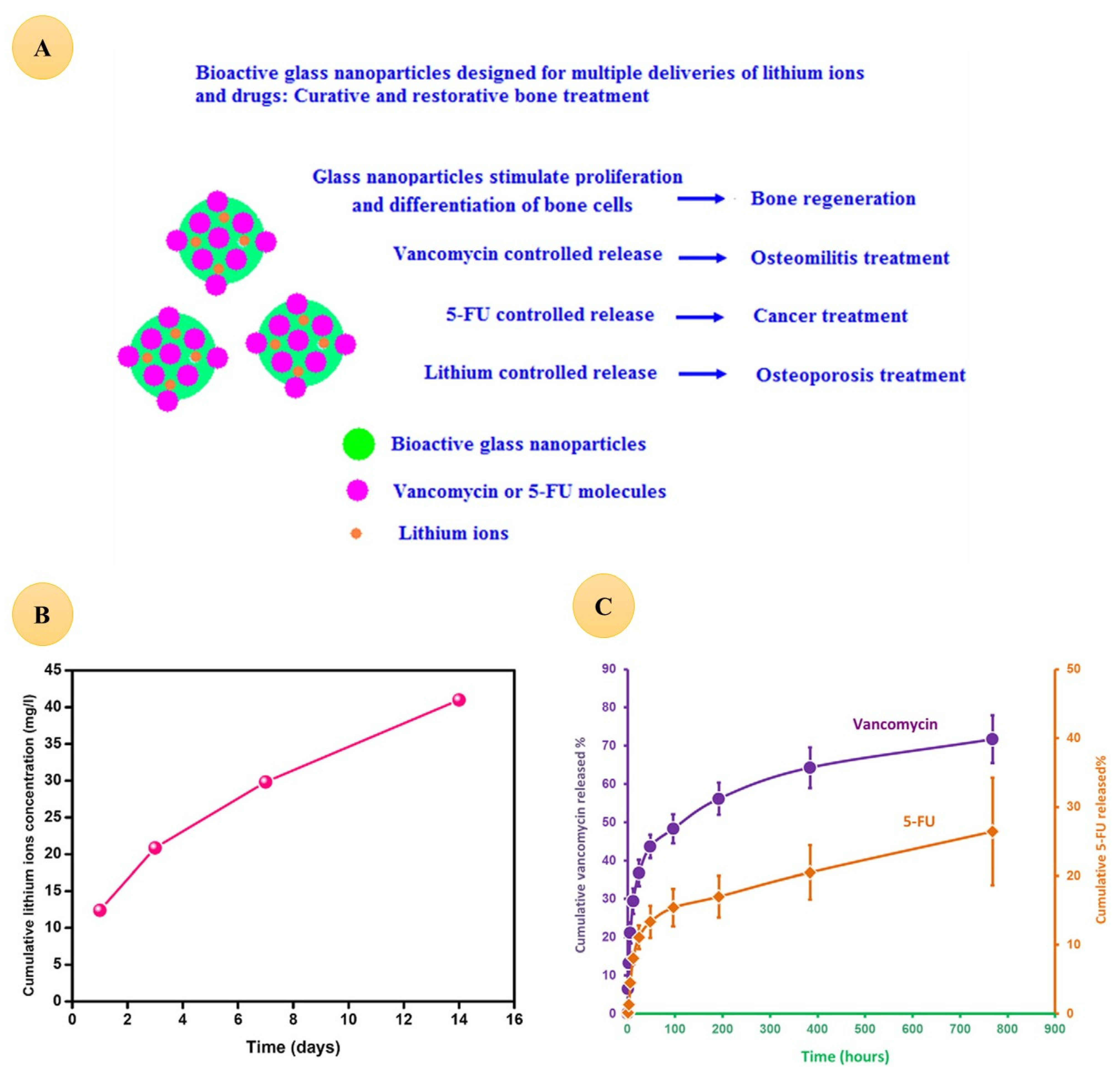
- El-Kady, A.M.; Farag, M.M.; El-Rashedi, A.M. Bioactive glass nanoparticles designed for multiple deliveries of lithium ions and drugs: Curative and restorative bone treatment. Eur. J. Pharm. Sci. 2016, 91, 243–250. [Google Scholar] [CrossRef] [PubMed]
- Yazdanpanah, A.; Moztarzadeh, F.; Arabyazdi, S. A heat-generating lithium-ferrite doped bioactive glass for cancer hyperthermia. Phys. B Condens. Matter 2020, 593, 412298. [Google Scholar] [CrossRef]
- Xie, J.; Lu, Y.-C. A retrospective on lithium-ion batteries. Nat. Commun. 2020, 11, 2499. [Google Scholar] [CrossRef]
- Grey, C.P.; Hall, D.S. Prospects for lithium-ion batteries and beyond—A 2030 vision. Nat. Commun. 2020, 11, 6279. [Google Scholar] [CrossRef]
- Wu, F.; Maier, J.; Yu, Y. Guidelines and trends for next-generation rechargeable lithium and lithium-ion batteries. Chem. Soc. Rev. 2020, 49, 1569–1614. [Google Scholar] [CrossRef]
- Nitta, N.; Wu, F.; Lee, J.T.; Yushin, G. Li-ion battery materials: Present and future. Mater. Today 2015, 18, 252–264. [Google Scholar] [CrossRef]
- Soltani, M.; Beheshti, S.H. A comprehensive review of lithium ion capacitor: Development, modelling, thermal management and applications. J. Energy Storage 2021, 34, 102019. [Google Scholar] [CrossRef]
- Karimi, D.; Behi, H.; Van Mierlo, J.; Berecibar, M. A Comprehensive Review of Lithium-Ion Capacitor Technology: Theory, Development, Modeling, Thermal Management Systems, and Applications. Molecules 2022, 27, 3119. [Google Scholar] [CrossRef] [PubMed]
- Liang, X.; Qi, R.; Zhao, M.; Zhang, Z.; Liu, M.; Pu, X.; Wang, Z.L.; Lu, X. Ultrafast lithium-ion capacitors for efficient storage of energy generated by triboelectric nanogenerators. Energy Storage Mater. 2020, 24, 297–303. [Google Scholar] [CrossRef]
- Zheng, X.; Jin, Y.; Liu, X.; Liu, T.; Wang, W.; Yu, H. Photoactivatable nanogenerators of reactive species for cancer therapy. Bioact. Mater. 2021, 6, 4301–4318. [Google Scholar] [CrossRef]
- Zhang, S.; Bick, M.; Xiao, X.; Chen, G.; Nashalian, A.; Chen, J. Leveraging triboelectric nanogenerators for bioengineering. Matter 2021, 4, 845–887. [Google Scholar] [CrossRef]
- Wang, Y.M.; Zeng, Q.; He, L.; Yin, P.; Sun, Y.; Hu, W.; Yang, R. Fabrication and application of biocompatible nanogenerators. iScience 2021, 24, 102274. [Google Scholar] [CrossRef] [PubMed]
- Sun, M.; Li, Z.; Yang, C.; Lv, Y.; Yuan, L.; Shang, C.; Liang, S.; Guo, B.; Liu, Y.; Li, Z.; et al. Nanogenerator-based devices for biomedical applications. Nano Energy 2021, 89, 106461. [Google Scholar] [CrossRef]
- Yoon, H.-J.; Kim, S.-W. Nanogenerators to Power Implantable Medical Systems. Joule 2020, 4, 1398–1407. [Google Scholar] [CrossRef]
- Liang, L.; Wen, L.; Weng, Y.; Song, J.; Li, H.; Zhang, Y.; He, X.; Zhao, W.; Zhan, M.; Li, Y.; et al. Homologous-targeted and tumor microenvironment-activated hydroxyl radical nanogenerator for enhanced chemoimmunotherapy of non-small cell lung cancer. Chem. Eng. J. 2021, 425, 131451. [Google Scholar] [CrossRef]
- Chowdhury, A.R.; Abdullah, A.M.; Hussain, I.; Lopez, J.; Cantu, D.; Gupta, S.K.; Mao, Y.; Danti, S.; Uddin, M.J. Lithium doped zinc oxide based flexible piezoelectric-triboelectric hybrid nanogenerator. Nano Energy 2019, 61, 327–336. [Google Scholar] [CrossRef]
- Leucht, S.; Helfer, B.; Dold, M.; Kissling, W.; McGrath, J.J. Lithium for schizophrenia. Cochrane Database Syst. Rev. 2015, 2015, CD003834. [Google Scholar] [CrossRef]
- McKnight, R.F.; Chesney, E.; Amit, B.H.; Geddes, J.; Cipriani, A. Lithium for acute mania. Cochrane Database Syst. Rev. 2019, 2019, CD004048. [Google Scholar] [CrossRef]
- Burgess, S.S.; Geddes, J.; Hawton, K.K.; Taylor, M.J.; Townsend, E.; Jamison, K.; Goodwin, G. Lithium for maintenance treatment of mood disorders. Cochrane Database Syst. Rev. 2001, 2001, CD003013. [Google Scholar] [CrossRef]
- Cipriani, A.; Smith, K.A.; Burgess, S.S.; Carney, S.M.; Goodwin, G.; Geddes, J. Lithium versus antidepressants in the long-term treatment of unipolar affective disorder. Cochrane Database Syst. Rev. 2006, 2006, CD003492. [Google Scholar] [CrossRef]
- Cochrane. Cochrane Central Register of Controlled Trials; Cochrane: London, UK, 2022. [Google Scholar]


| Abbreviation | Word or Phrase |
|---|---|
| ALP | Alkaline phosphatase |
| AP-1 | Activator protein-1 |
| BG | Bioactive glass |
| BMSC | Bone marrow mesenchymal stem cell |
| β-TCP | Beta-three calcium phosphate |
| cAMP | Cyclic adenosine monophosphate |
| CNS | Central nervous system |
| CREB | Response element binding protein |
| DEXA | Dual-energy X-ray absorptiometry |
| Dspp | Dentin sialophosphoprotein |
| EPO | Erythrogenin |
| ERK | Extracellular signal-regulated kinase |
| 5-FU | Fluorouracil |
| GAG | Glycosaminoglycan |
| GIONFH | Glucocorticoid-induced osteonecrosis of the femoral head |
| GSK-3β | Glycogen synthase kinase-3 beta |
| HA | Hydroxyapatite |
| Hh | Hedgehog pathways |
| HIF-1α | Hypoxia-inducible factor 1-alpha |
| HSC | Hematopoietic stem cell |
| HUVEC | Human umbilical vein endothelial cell |
| IGF1 | Insulin growth factor 1 |
| iTENG | Ionic triboelectric nanogenerator |
| KEGG | Kyoto Encyclopedia of Genes and Genomes |
| Klk4 | Axin2, Kallikrein 4 |
| LCS | Lithium-doped calcium silicate |
| LD | Lithium disilicate |
| Li-BG | Lithium-doped bioactive glass |
| Li-BBG | Lithium-doped borate-based bioactive glass |
| Li-MBG | Lithium-doped mesoporous bioactive glass |
| LMNS | Lithium-doped mesoporous silica nanosphere |
| Li-nHA/GMs/rhEPO | Gelatin/lithium-doped-hydroxyapatite nanoparticles/gelatin microspheres/rhEPO |
| Li-PBG | Lithium-doped phosphate-based bioactive glass |
| LPPEEK | Lithium-doped silica nanospheres coated on polyetheretherketone surface |
| LSN | Lithium-doped silica nanosphere |
| MACI | Matrix-associated autologous chondrocyte implantation |
| MAPK | Mitogen-activated protein kinase |
| micro-CT | Micro-computed tomography |
| MSC | Mesenchymal stem cell |
| MSN | Mesoporous silica nanosphere |
| NPWT | Negative pressure wound therapy |
| OA | Osteoarthritis |
| OIM | Osteoimmunomodulation |
| p38MAPK | P38mitogen-activated protein kinase |
| PCL | Poly-ε-caprolactone |
| PDA | Polydopamine |
| PEEK | Polyetheretherketone |
| PI3-K | Phosphatidylinositol 3 kinase |
| PNS | Peripheral nerve system |
| qPCR | Quantitative polymerase chain reaction |
| rBMSC | Rabbit mesenchymal stem cell |
| ROS | Reactive oxygen species |
| Runx2 | Runt-related transcription factor 2 |
| SBF | Simulated body fluid |
| SCI | Spinal-cord injury |
| TGF- β | Transforming growth factor beta |
| VEGF | Vascular endothelial growth factor |
| ZLS | Zirconia-reinforced lithium silicate |
Publisher’s Note: MDPI stays neutral with regard to jurisdictional claims in published maps and institutional affiliations. |
© 2022 by the authors. Licensee MDPI, Basel, Switzerland. This article is an open access article distributed under the terms and conditions of the Creative Commons Attribution (CC BY) license (https://creativecommons.org/licenses/by/4.0/).
Share and Cite
Farmani, A.R.; Salmeh, M.A.; Golkar, Z.; Moeinzadeh, A.; Ghiasi, F.F.; Amirabad, S.Z.; Shoormeij, M.H.; Mahdavinezhad, F.; Momeni, S.; Moradbeygi, F.; et al. Li-Doped Bioactive Ceramics: Promising Biomaterials for Tissue Engineering and Regenerative Medicine. J. Funct. Biomater. 2022, 13, 162. https://doi.org/10.3390/jfb13040162
Farmani AR, Salmeh MA, Golkar Z, Moeinzadeh A, Ghiasi FF, Amirabad SZ, Shoormeij MH, Mahdavinezhad F, Momeni S, Moradbeygi F, et al. Li-Doped Bioactive Ceramics: Promising Biomaterials for Tissue Engineering and Regenerative Medicine. Journal of Functional Biomaterials. 2022; 13(4):162. https://doi.org/10.3390/jfb13040162
Chicago/Turabian StyleFarmani, Ahmad Reza, Mohammad Ali Salmeh, Zahra Golkar, Alaa Moeinzadeh, Farzaneh Farid Ghiasi, Sara Zamani Amirabad, Mohammad Hasan Shoormeij, Forough Mahdavinezhad, Simin Momeni, Fatemeh Moradbeygi, and et al. 2022. "Li-Doped Bioactive Ceramics: Promising Biomaterials for Tissue Engineering and Regenerative Medicine" Journal of Functional Biomaterials 13, no. 4: 162. https://doi.org/10.3390/jfb13040162