Article

The New GPI-Anchored Protein, SwgA, Is Involved in Nitrogen Metabolism in the Pathogenic Filamentous Fungus Aspergillus fumigatus

1
Unité des Aspergillus, Département de Mycologie Institut Pasteur, 25-28 rue du Docteur Roux, CEDEX 15, 75724 Paris, France
2
Manchester Fungal Infection Group, Division of Infection, Immunity and Respiratory Medicine, Faculty of Biology, Medicine and Health, University of Manchester, CTF Building, Grafton Street, Manchester M13 9NT, UK
3
Unité Technologie et Service Bioimagerie Ultrastructurale (UTechS UBI), Institut Pasteur, 28 rue du Docteur Roux, 75015 Paris, France
*
Authors to whom correspondence should be addressed.
Current address: Department of Experimental Biology, Masaryk University, Kamenice 5, 62500 Brno, Czech Republic.
Current address: Unité BAPF, Département de Mycologie Institut Pasteur, 25-28 rue du Docteur Roux, CEDEX 15, 75724 Paris, France.
§
Current address: Institut Pasteur Université Paris Cité, INRAE, USC2019, Unité Biologie et Pathogénicité Fongiques, 25-28 rue du Docteur Roux, CEDEX 15, 75724 Paris, France.
J. Fungi 2023, 9(2), 256; https://doi.org/10.3390/jof9020256
Submission received: 3 January 2023 / Revised: 9 February 2023 / Accepted: 10 February 2023 / Published: 15 February 2023
(This article belongs to the Section Fungal Cell Biology, Metabolism and Physiology)

Abstract

:
GPI-anchored proteins display very diverse biological (biochemical and immunological) functions. An in silico analysis has revealed that the genome of Aspergillus fumigatus contains 86 genes coding for putative GPI-anchored proteins (GPI-APs). Past research has demonstrated the involvement of GPI-APs in cell wall remodeling, virulence, and adhesion. We analyzed a new GPI-anchored protein called SwgA. We showed that this protein is mainly present in the Clavati of Aspergillus and is absent from yeasts and other molds. The protein, localized in the membrane of A. fumigatus, is involved in germination, growth, and morphogenesis, and is associated with nitrogen metabolism and thermosensitivity. swgA is controlled by the nitrogen regulator AreA. This current study indicates that GPI-APs have more general functions in fungal metabolism than cell wall biosynthesis.

1. Introduction

The post-translational modification of proteins by the addition of a glycosylphosphatidylinositol (GPI) anchor is an essential process that occurs in all eukaryotic cells. The role of this anchor is to direct newly synthesized proteins to the cell membrane. In fungi, some GPI-anchored proteins (GPI-APs) are also associated with the cell wall. Precursors of proteins that are modified by a GPI anchor share conserved features, including an amino-terminal signal sequence for localization to the endoplasmic reticulum (ER) and a carboxyl-terminal hydrophobic domain for attachment to the ER membrane [1]. Proteins are cleaved near the so-called omega site (Ω), several amino acids before this C-terminal, before ligation to a pre-assembled GPI anchor. These characteristics have been utilized to direct in silico searches of annotated genomes; recently, 86 GPI-APs were identified in the genome of the pathogenic fungus Aspergillus fumigatus [2].
In A. fumigatus, GPI-APs play a major enzymatic role in cell wall biosynthesis [2,3]. For example, the GEL family of β-(1,3)-glucanosyl-transferases (containing seven members) is responsible for the elongation and branching of β-(1,3)-glucans, an activity essential for fungal growth [4,5,6,7]. The DFG family with seven members is involved in the covalent binding of galactomannan (GM) to the β-(1,3)-glucan-chitin core of the cell wall [8]. Moreover, members of GH16 glucanase families are required to establish normal conidial structures [9], and Bgt2 is a predominant protein in the cell wall encoding β-(1,3)-glucan branching activity [10].
Comprehensive analyses of GPI-anchored proteins were undertaken in Candida albicans and Saccharomyces cerevisiae [11,12]. These studies have shown that these proteins are involved in fungal cell wall integrity, viability, virulence, chemical, and environmental stresses [13]. To date, similar studies have not been conducted in filamentous fungi. However, the investigation of a morphogenetic role unrelated to cell wall functioning has now become possible due to the availability of a mutant library based on the deletion of all genes coding GPI-APs. Three groups of GPI-APs have been recognized in A. fumigatus [2]. The first group of proteins is highly conserved in all fungi (yeast as well as filamentous fungi), which are essential in cell wall morphogenesis, most of them belong to multigenic families of proteins (e.g., GEL, DFG, CRH families). The second group of proteins is present only in filamentous fungi, which are mostly involved in the processes of biofilm formation, adhesion, and virulence (CFEM, MP proteins) [14,15]. Finally, the third group of proteins is only present in the Aspergillus species or the related species of mold (ex CSPA) [16,17]. These proteins seem to be required for the normal formation of conidia, but how they carry out this function remains unclear.
During the analysis of the library of GPI-anchored mutants, we identified and characterized a novel GPI-AP that we call SwgA (for SWollen and Germinated conidia), which is involved in germination, growth, and morphogenesis. Moreover, we show that swgA is involved in nitrogen metabolism under the regulatory control of the transcription factor AreA.

2. Materials and Methods

2.1. Strains and Growth Conditions

The A. fumigatus parental strain KU80pyrG+ [18], an A. fumigatus strain derived from the well-characterized clinical isolate A1163 that lacks non-homologous end joining (Δku80), was used as a host for transformation experiments and maintained on 2% malt agar slants at room temperature. For DNA extraction, cultures were grown in Sabouraud liquid medium (2% glucose + 1% mycopeptone). Conidia were collected from agar slants/plates after seven days of growth at room temperature using 0.05% Tween 20 aqueous solution. The minimal medium (MM) used contained 5 mM of ammonium tartrate (NH4+) and 1% glucose as the nitrogen and carbon source, respectively (MM-NH4+). To select the transformant, 150 µg/mL of hygromycin (Sigma, Saint Quentin Fallavier, France) or chlorimuron (100 µg/mL) was added to the medium.

2.2. Protein Sequence Analysis

Protein sequences and AFUA numbers were retrieved from the CADRE Genome Browser (http://fungi.ensembl.org/Aspergillus_fumigatus/Info/Index, accessed on 15 February 2019). Protein sequences were analyzed with SignalP 3.0 (http://www.cbs.dtu.dk/services/SignalP/, accessed on 15 February 2019), the big-PI fungal predictor (http://mendel.imp.ac.at/gpi/fungi_server.html, accessed on 15 February 2019), PredGPI (http://gpcr.biocomp.unibo.it/predgpi/pred.htm, accessed on 15 February 2019), and the TMHMM Server v. 2.0 (http://www.cbs.dtu.dk/services/TMHMM/, accessed on 15 February 2019). Blast analyses were performed using (https://www.yeastgenome.org/blast-fungal, accessed on 20 March 2019) and (https://blast.ncbi.nlm.nih.gov/Blast.cgi, accessed on 20 March 2019). Protein sequences were aligned using ClustalX 2.0.12.

2.3. Construction of the ΔswgA Mutant, and the Revertant Strain ΔswgA::swgA

The construction of the ΔswgA mutant was carried out as described by Zhao et al. [19] and verification of the mutant was performed by the Southern blot analysis by Samalova et al. [2]. Complementation of the ΔswgA mutant was carried out by the reintroduction of the parental copy of the gene flanks and the chlorimuron-ethyl resistance β-recombinase (ChloriR-β-rec), Valsecchi, et al. [20] at a 3′ flanking region as described in Figure S1 (the primers are listed in Table S1). A. fumigatus conidia were transformed by the electroporation method described by Sanchez and Aguirre [21] with subsequent modifications. Conidia were washed with water and 109 conidia were inoculated in 125 mL of YG medium (0.5% yeast extract, 2% glucose), and incubated at 37 °C at 300 rpm for 4 h to obtain swollen conidia. Those conidia were recovered by centrifugation and then inoculated in 12.5 mL of YG medium (1% yeast extract, 1% glucose, 20 mM HEPES, pH 8.0) and incubated for 1 h at 30 °C at 100 rpm. Conidia were centrifuged and resuspended in 1 mL of cold 1 M sorbitol; 10 µg of DNA were added to 50 µL of conidial suspension, incubated for 15 min on ice, and transferred to 0.1 cm electroporation tanks. Electroporation was performed using the Bio-Rad gene pulser (Gene Pulser Xcell, Bio-Rad, Hercules, CA, USA) with the following parameters: voltage, 1 kV; capacitance, 25 microfarads; and resistance, 400 Ω. After transformation, cold YG medium was added to the conidia and incubated on ice for 15 min and then incubated at 30 °C at 100 rpm for 1 h and 30 min. Conidia were plated on minimal media and incubated at 20 °C overnight. Hygromycin (150 µg/mL) was added to the plates before incubation at 37 °C for two days. After cultivation of the revertant on a minimal medium containing 2% xylose, the excision of the chlorimuron-ethyl resistance cassette was conducted. This strain was used in this study.

2.4. Manipulation of DNA, RNA, Southern Blotting

Genomic DNA was extracted as previously described by Girardin et al. [22]. For the Southern blot analysis, 10 µg of digested genomic DNA were subjected to size fractionation on 0.7% agarose and blotted onto a positively charged nylon membrane (Hybond-N+; Amersham, GE Healthcare, Chicago, IL, USA). For DNA extraction, mycelium was grown for 16 h at 37 °C in Sabouraud liquid medium. For RNA extraction, the mycelium of each strain was grown for 27 or 30 h in a liquid medium and then disrupted with 0.5 mm of glass beads in 500 µL, and RNA was then isolated using the phenol–chloroform method. DNase treatment was performed after elution of the RNA from the column with Turbo DNA-free DNase (Ambion®, Foster City, CA, USA). The quality of RNA was controlled on Bioanalyzer RNA 6000 nano Chips (Agilent Technologie, Santa Clara, CA, USA). One microgram of total RNA was reverse-transcribed using a reverse transcriptase Bio-Rad kit (Iscript cDNA synthesis kit, Bio-Rad, Hercules, CA, USA) following the instructions of the manufacturer. The gene expression levels of swgA and areA (AFUA_6G01970) were quantified by PCR using the same amount of cDNA obtained in the different liquid media MM-NH4+, RPMI-1640+Gln (containing L-glutamine), RPMI-1640 (RPMI without L-glutamine) and MM, without ammonium tartrate (NH4+) and supplemented with 10 mM L-glutamine)(MM+Gln10) with the following program: 1 min at 98 °C, 30 cycles of 10 s at 98 °C, 20 s at 65 °C and 40 s at 72 °C; and a final extension of 10 min at 72 °C.

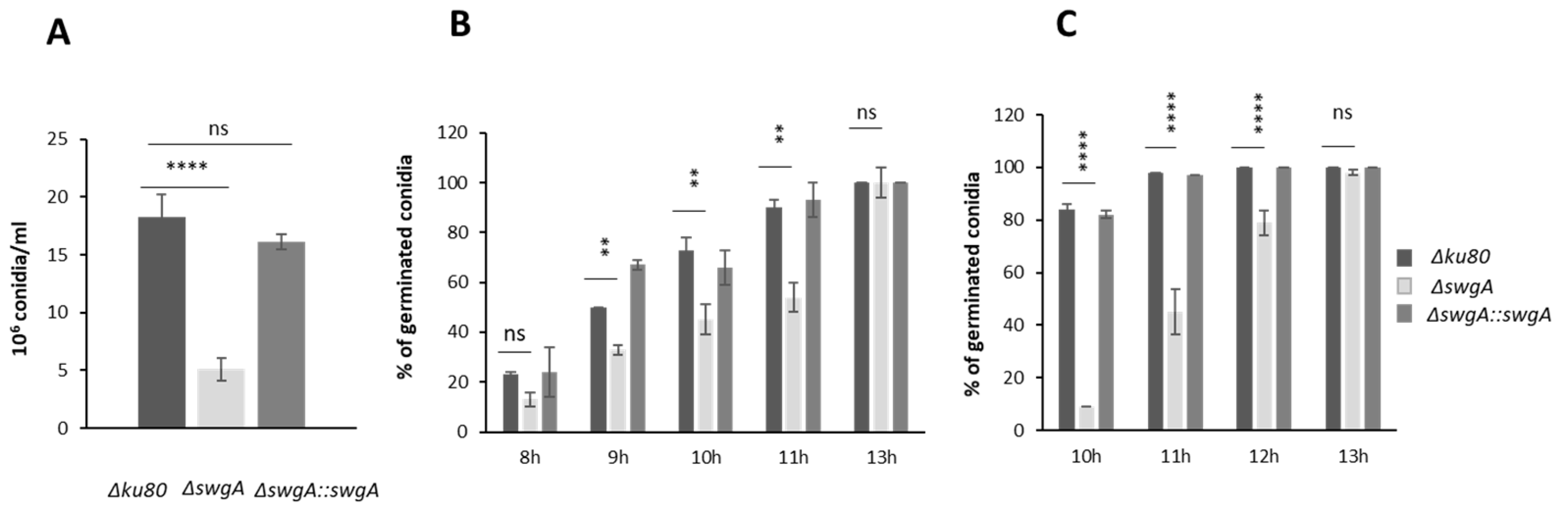
2.5. Conidiation, Conidial Morphology, and Germination

The conidiation rates were estimated following inoculation of 105 conidia onto 3 glass-test tubes (20 mL) containing 2% malt agar (10 mL/tube). After 1 week of growth at 37 °C, conidia were recovered from malt agar slants using a 0.05% Tween 20 aqueous solution. Conidial suspensions were filtered on a 40 µm pore-sized sterile cell strainer (Fisher Scientific, Illkirch, France) and counted using a Luna dual-fluorescence cell counter (Mokascience SARL, La Madelaine, France). After washing with 0.05% Tween 20 water, the permeability of the conidia with respect to FITC was investigated by incubating 200 µL of an aqueous suspension of conidia (2 × 10⁷ conidia/mL) with 30 µL of FITC solution (0.1 mg/mL in 0.1 M Na2CO3, pH 9) overnight at 4 °C in darkness. The conidia were washed three times with 0.05% Tween 20 solution before being subjected to observation under a fluorescence microscope (Evos FL Life Technologies; excitation wavelength [λex], 470/22 nm; emission wavelength [λem], 510/44 nm). For Calcofluor White (CFW) staining, the conidial suspension was incubated with CFW solution (0.5 µg/mL) for 30 min at room temperature and observed with fluorescence microscopy (Evos FL Life Technologies; λex, 357/44 nm; λem, 447/60 nm). Conidial germination was followed microscopically every 30 min after 4 h of growth at 37 °C or 45 °C (inoculation of 10 μL of conidia at 106 cells/mL on a glass slide with 2% agar malt medium). For the viability assays, conidia on agar slants were stored at 37 °C for one and two months, then conidial suspensions were recovered in 0.05% Tween 20 water and the viability of conidia was evaluated following dilutions and plating on 2% malt medium.

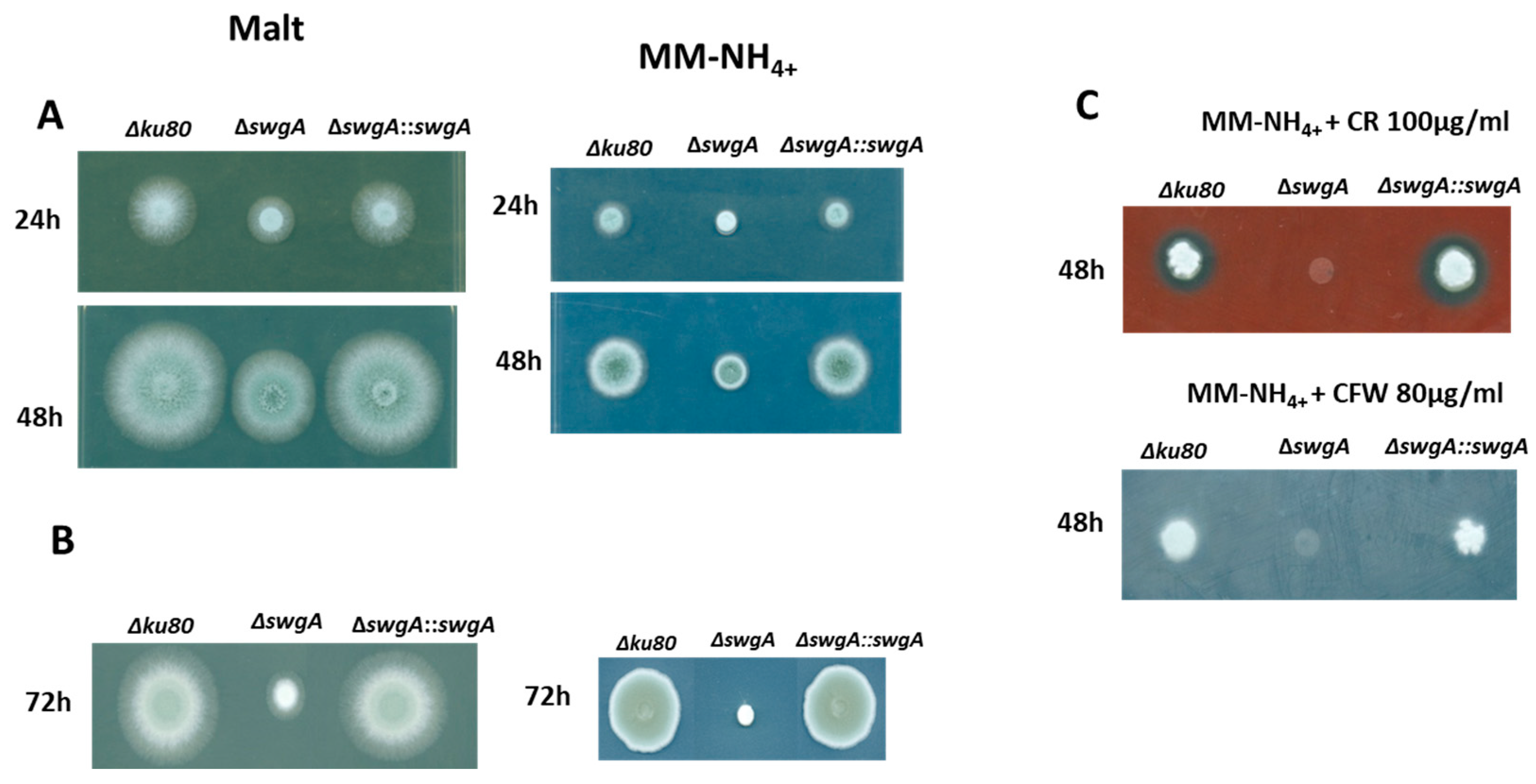
2.6. Growth and Susceptibility of the Mutant Strains to Congo Red and Calcofluor White

Mycelial growth was tested on different types of agar media (Sabouraud, Minimal Medium (MM-NH4+), malt 2%, and glucose 3%-yeast extract 1% (G3%/YE1%)) at 30 °C, 37 °C and 45 °C. For temperatures of 30 °C and 37 °C, 106 conidia (5 µL) of each strain were spotted for up to 72 h. To investigate the colony-forming ability of the ΔswgA mutant at 45 °C, mycelial disks cut out from the perimeter of pre-grown colonies were spotted on malt and MM-NH4+ plates and incubated for 72 h at 45 °C. Growth was also tested in 30 mL of liquid of several types of media containing different nitrogen sources and amino acids: G3%YE1%, MM-NH4+, RPMI-1640, RPMI-1640+Gln, MM without NH4+, and supplemented by either L-glutamine 10 mM (MM+Gln10), glutamate 10 (MM+Glu10), 50 mM (MM+Glu50), or MM-NH4+ supplemented by glutamate 10 mM (MM-NH4++Glu10) and incubated with shaking (150 rpm/min) at 37 °C for 24, 30, or 48 h. The mycelial dry weight was estimated every 24 h after drying at 80 °C until a constant weight was reached. Growth inhibition tests with Congo Red (CR) (50–240 µg/mL) and CFW (50–200 µg/mL) were performed on MM agar plates.

2.7. Hydrophobicity Measurements

One ml of water was added to a three-week-old malt glass-test tube culture and vortexed for 30 s. The water containing hydrophilic conidia was carefully recovered using a Pasteur pipet. One mL of Tween 20 water was then added to the tube, which was vortexed for 30 s, and the Tween 20 water containing hydrophobic conidia was recovered. The percentage of hydrophobic conidia was estimated from the ratio of conidia counted in the Tween 20 water solution vs. the total number of conidia as described by Valsecchi et al. [20].

2.8. Scanning Electron Microscopy (SEM) Analysis

The strains were cultured on 2% malt medium for 24 h at 37 °C and then for 6 days at room temperature. For each strain, 106 conidia were inoculated in liquid glucose 3% yeast extract 1% medium for 5 h and 8 h at 37 °C, 150 rpm. Then dormant, swollen, and germinated conidia were washed in sterile water, centrifuged for 5 min at 2500× g, and then fixed with 2.5% glutaraldehyde and 2.5% paraformaldehyde in PHEM pH 7 buffer (120 mM Pipes, 4 mM MgSO4, 20 mM EDTA and 50 mM HEPES) for 1 h at room temperature and after overnight at 4 °C without agitation. For SEM, they were then sequentially fixed with 1% osmium tetroxide in PHEM pH 7 buffer for 1 h and then dehydrated in ethanol followed by critical-point drying. Samples were then sputter coated with Au-Pd and imaged with a field-emission scanning electron microscope (JSM-6700F, Joel®).

2.9. Adhesion Assays and Biofilm Quantification

The ability of hyphae to adhere to polystyrene was tested by incubating 10 µL of conidia (106/mL) for 24 h in 24-well polystyrene plates (TPP, Thermo Fisher, Illkirch, France) in 1 mL MM-NH4+ supplemented with 0.01% Tween 20 at 37 °C. The plates were then washed three times with PBS 0.01% Tween 20 and the remaining adherent mycelium was quantified by adding 130 µL of crystal violet 0.01% for 20 min at room temperature, washed 3 times with water, and de-stained with 130 µL of 30% acetic acid. Growth was estimated by measuring OD (Optic Density) at 600 nm. Mycelia under aerial static biofilm or planktonic conditions were obtained as previously described [23]. Briefly, 106/mL conidia were inoculated in 20 mL liquid, or agar MM-NH4+ covered by cellophane (DryEase cellophane, Invitrogen, Carlsbad, CA, USA) at 30 °C for 24 h, and the mycelium dry weight was quantified.

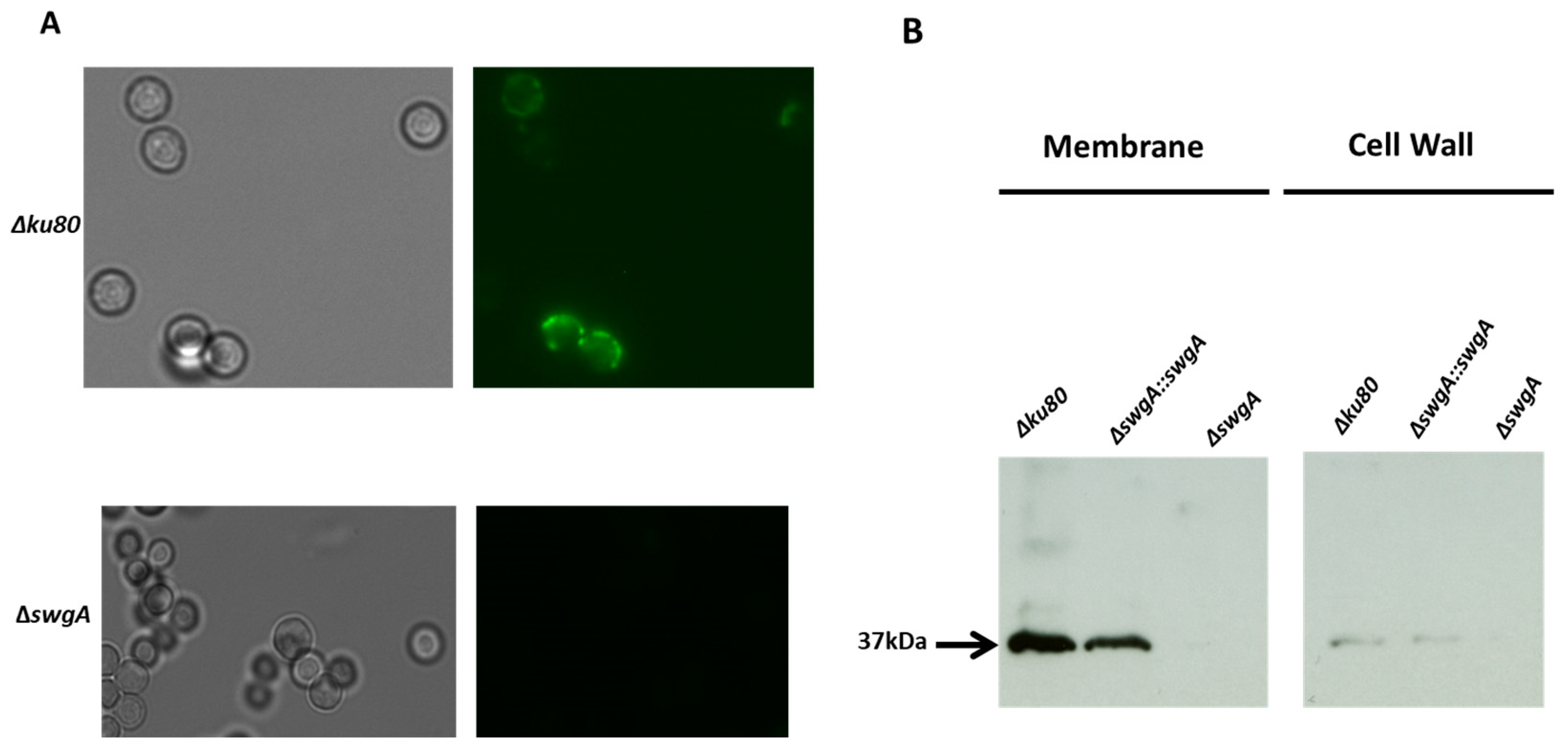
2.10. Anti-SwgA Antibodies

Rabbits were immunized against two different peptides SWLKRDTLKPFQEG-Cys and Cys-STRTRATDTDDVWE designed based on sequence data (ProteoGenix). Coupling of the peptide through cysteine to m-maleimidobenzoyl-N-hydroxy-succinimide ester, immunization of the animal, and titer determination of the antiserum was performed by ProteoGenix SAS (Schiltigheim, France). Immunopurification of the specific antipeptide antibodies was done after coupling the peptide to epoxy-activated Sepharose (Amersham Pharmacia Biotech, Orsay, France) following the instructions of the manufacturer. Immunolabeling of blots was done using the ECL Western blotting detection procedure of Amersham Pharmacia Biotech.

2.11. Immunodetection of SwgA in Cell Wall or Membrane

After 24 h of growth at 37 °C in G3%YE1% liquid medium, the mycelium of the parental strain, the ΔswgA mutant, and the revertant strain were disrupted using 1 mm beads for two minutes at 4 °C using a Fast-Prep cell breaker (MP Biomedical, Illkirch-Graffenstaden, France) in Tris-HCl50 mM buffer pH 7.5, 1 mM EDTA, 0.3 M sucrose, 1% glucose, 2 mM DTT including antiprotease (Complete Protease inhibitor cocktail, Roche, Bale, Swizterland). Cell walls were collected by centrifugation for 10 min at 4000× g and then washed three times with water. The cell lysate supernatants were centrifuged for 1 h at 36,000× g at 4 °C to pellet total cellular membranes which were then suspended in the extraction buffer and stored at −20 °C. Proteins from the cell wall and membrane fractions were extracted by SDS-mercaptoethanol buffer (Tris-HCl 62 mM pH 6.8 containing 2% SDS and 5% β-mercaptoethanol) and were analyzed by SDS-PAGE on a 12% polyacrylamide gel, under reducing conditions (Laemmli, Bio-Rad Mini-Protean Tetra Cell instruction manual). SwgA was detected by Western blot using anti-SwgA serum (1:1000 dilution) and secondary anti-rabbit IgG conjugated to peroxidase (1:2000 dilution; Sigma, Saint Quentin Fallavier, France). The detection was performed with the ECL chemiluminescence method of Amersham (GE Healthcare Life Sciences, Vélizy-Villacoublay, France).

2.12. Immunofluorescence of Cell Wall Polysaccharides

For each strain, 106 conidia were inoculated in liquid G3%YE1% medium for 4 h (swollen conidia) at 37 °C, 150 rpm. After 5 min centrifugation at 4000 rpm, dormant and swollen conidia were washed in 1× PBS-0.05% Tween 20 (PBS-Tw). Briefly, conidia were fixed with p-formaldehyde 2.5% (pFA) overnight at 4 °C and washed with phosphate-buffered saline (PBS) pH 7 containing 0.1 M NH4Cl and then with PBS-Tw. After washing, conidia were resuspended in PBS-Tw supplemented with 5% Goat serum (PBS-Tw-G) for 30 min before adding the primary antibodies or receptors described below. After incubation for 1 h, the conidia were washed and incubated in PBS-Tw-G with the secondary antibodies conjugated to a fluorochrome. After washing in PBS-Tw-G, the samples were observed under a wide-field epifluorescence microscope. The different antibodies tested were the following: for SwgA detection, samples were immunolabeled with the polyclonal antibodies anti-SwgA (dilution 1:500) and a secondary anti-rabbit IgG conjugated to Alexa488 (anti-rabbit IgG-A488; Sigma). For chitin and glycoproteins detection, chitin and glycoproteins were labeled with wheat germ agglutinin coupled with tetramethylrhodamine (WGA-TRITC) and concanavalin A labeled with fluorescein isothiocyanate (ConA-FITC), respectively, (Sigma) at 0.1 mg/mL. The α-(1,3)-glucan was detected by immunolabeling with the anti-α-(1,3)-glucan IgA monoclonal antibody B-4.1 [kind gift of J. Kearney, University of Alabama, Birmingham, USA or with the MOPC IgM [Sigma, 8 μg/mL [24], and revealed by the anti-mouse IgG-A488. For the Anti-SwgA, the primary antibody was diluted 1/50, and the secondary FITC rabbit 1/200. Fluorescent images were collected using an EVOS FL epifluorescence microscope (Life Technology®) with a 1.3-megapixel charge-coupled-device (CCD) camera (Sony ICX455 monochrome).

2.13. Carbohydrate Analysis of the Cell Wall Fractions and Supernatant

The amount of 5 × 10⁷ conidia of the parental strain, ∆swgA mutant, and the revertant strain were incubated for 24 h in 40 mL MM-NH4+ at 37 °C, 150 rpm. The mycelium was then collected by filtration under vacuum and washed with water and the culture supernatant was precipitated with 2.5 volume of ethanol overnight at 4 °C. The cell wall was prepared as described by Millet et al. [25]. Total neutral sugar content was estimated by the phenol sulfuric method [26] and glucosamines and galactosamines were quantified by high-performance liquid chromatography (HPLC) after acid hydrolysis with 6 N HCl at 100 °C for 6 h [27].

2.14. In Vivo Experiments in a Murine Model of Invasive Aspergillosis

Parental, ∆swgA mutant, and ∆swgA::swgA A. fumigatus strains were cultured in 2% malt agar in glass-test tubes. For virulence assays, twenty-four-week-old OF1 female mice (each 18–20 g, Charles River Laboratory, (L’Arbresle, France)) were immunosuppressed with Kenacort® Retard (Acetonide de Triamcinolone; Bristol-Myers Squibb) injected subcutaneously on day −1 (40 mg Kenacort per kg of the mouse). Before conidial inhalation (day 0) mice were anesthetized with an intraperitoneal injection of 0.2 mL of a solution containing 10 mg/mL ketamine (Imalgène® 1000, Lyon, France), 1 mg/mL xylazine (Rompun®, Sigma, Saint Quentin Fallavier, France) per mouse. By means of an automatic pipetting device, each mouse was inoculated intranasally with 2 × 10⁶ conidia (20 µL per mouse of 0.05% Tween 20 conidial suspension at 108 conidia per mL). Non-infected control immunosuppressed mice only received 20 µL of PBS plus 0.05% Tween 20. Daily monitoring of the body weight was done and a loss greater than 25% of the initial body weight was considered as a limit of pain not to be exceeded. Any animal that passed the critical weight loss would be immediately euthanized. The simple survival analysis was done by the Kaplan–Meier test. The statistical significance of the comparative survival values was calculated using the Mantel–Cox and Gehan–Breslow–Wilcoxon log-rank analyses, and the Prism statistical analysis package.

2.15. Statistical Analysis

Statistical data analysis was performed with GraphPad Prism. Unpaired, two-tailed Student’s t tests or one-way analyses of variance (ANOVA) were used to determine statistically significant differences in growth-related, conidiation, or germination phenotypes. Differences between the groups were considered significant at a p-value of ≤0.05. Throughout the article, significance is denoted as follows: * p < 0.0332; ** p < 0.00221; *** p < 0.0002; **** p < 0.0001, ns, nonsignificant.

3. Results

3.1. A Unique GPI-Anchored Protein Common to Aspergillus Section Clavati

Our preliminary screen of the library of putative GPI-anchored proteins of unknown function in A. fumigatus showed that only one of them encoded by the gene AFUA_8G01170 was involved in fungal growth [2]. Its morphogenetic function was confirmed after the construction of the complemented strain using the chlorimuron β-rec/six cassette [20] (Figure S1). AFUA_8G01170 (AFUB_084830) coded for a putative GPI-anchored protein of 281 amino acid residues including a signal peptide of 23 amino acids, and a 27 amino acid hydrophobic C-terminus with the amino acid trimer SGA providing the Ω cleavage site (http://gpcr.biocomp.unibo.it/predgpi/pred.htm, accessed on 15 February 2019). Blast analysis revealed that orthologs of this protein were restricted to Aspergillus section Clavati. Alignment of the orthologues showed that the N-terminal 120 amino acids are highly conserved (61 to 90% identity) and rich in cysteine residues (17 conserved cysteine residues) (Figure S2). The characteristics of the protein encoded by AFUA_8G01170 are given in Tables S2 and S3. Although no significant homology was found with any known proteins, it was classified as a hydrophobin-like protein because of the presence of distinctive patterns of conserved cysteines and hydrophobic residues [28]. Analysis of the protein using the fungal adhesins and adhesin-like protein prediction tool (http://bioinfo.icgeb.res.in/faap/query.html, accessed on 15 February 2019) indicated that it was likely to be an adhesin [29].
Analysis of previously published RNA-seq data designed to assess transcription during conidial germination showed that AFUA_8G01170 is not expressed in dormant conidia, but its expression level was increased during the swelling and germination process (from 4 h to 8 h of growth) [9].
For this reason, the gene was named swgA for Swollen and Germinated conidia. The use of an anti-SwgA antibody has confirmed the gene expression during germination, SwgA was detected by immunofluorescence as a dot at the cell surface (Figure 1A). Western blots using the anti-SwgA on protein extracts from membrane and cell wall fractions of the wild type, revertant strain, and ΔswgA mutant conidia, as well as hyphae, confirmed that SwgA was mainly located in the membrane fraction. The protein migrated with a molecular marker of 37 kDa. The expected size of this protein was 31 kDa, suggesting that this protein could be glycosylated (Figure 1B).

3.2. SwgA Is Not Involved in Cell Wall Synthesis

No difference in hydrophobicity was observed for the ΔswgA mutant compared to the parental or the revertant strain (data not shown). CFW and FITC labeling can be used as a proxy to check the permeability of cell wall mutants [30] and revealed that the permeability of ΔswgA dormant and swollen conidia was similar to the parental and revertant strains (Figure S3). Since the role of GPI-anchored proteins has been often associated with cell wall biosynthesis [2], the cell wall of parental and ΔswgA mutant strains was analyzed. However, no discernable differences in hexose, N-acetylglucosamine, or N-acetylgalactosamine levels were observed (Table 1). Immunolabeling of swollen conidia of the parental and ΔswgA mutant was performed with antibodies specifically recognizing the polysaccharides of the cell wall and showed no differences in the localization of α-(1-3)-glucan and chitin in parental and ΔswgA mutant strains (Figure S4). Moreover, no significant modifications of the outside layer of the cell wall were observed in SEM (Figure S5). Altogether, these results indicated that the SwgA protein is not involved in cell wall organization.

3.3. The Growth Phenotype of the ∆swgA Mutant Is Temperature Sensitive

Important morphogenetic alterations occurred in the ∆swgA mutant. First, a decrease in conidiation was observed for the mutant (35% ± 1) compared to the parental and the revertant strains (Figure 2A). Germination was delayed in the mutant: after 8 h of growth, 13% ± 3 of conidia had germinated in the mutant and 23% ± 1 in the parental strain, but all conidia were able to germinate (100% after 13 h of growth for both mutant and parental strains) (Figure 2B). This delay increased with temperature. At 45 °C, after 10 h of growth, only 9% ± 1 of mutant conidia germinated compared to 84% ± 2 for the parental or the revertant strain (Figure 2C).
The growth kinetics of the parental, the ΔswgA mutant, and the revertant strains were investigated on MM-NH4+ and malt agar media. After 48 h at 37 °C, a two-fold reduction of growth was observed for the mutant independently of the agar medium used (Figure 3A). The growth defect was more severe at 45 °C (>three times reduction of growth) (Figure 3B), confirming the temperature-sensitive phenotype observed for the mutant during conidiation and germination. Moreover, we observed a strong sensitivity to CR and CFW (Figure 3C). Considering that we did not observe any difference in permeability or cell wall composition (see Figure S3 and Table 1) for the ΔswgA mutant, this strong sensitivity suggested cell wall structural differences with the parental strain.
A significant reduction of growth of the mutant compared to the parental strain was also observed in liquid glucose 3%/Yeast extract 1% medium, and in MM-NH4+ medium, at 37 °C (27% reduction after 48 h of growth for both media), however, surprisingly no reduction of growth was observed in RPMI-1640+Gln liquid medium (Figure 4). At 45 °C after 24 h, the growth defect was more pronounced, except for RPMI-1640+Gln medium, where no reduction of growth was observed (Figure S6). All these results suggested that the growth defect of the ΔswgA mutant was related to the nutrient composition of the medium. The ability of hyphae to adhere to polystyrene was also tested after 24 h of growth in liquid MM-NH4+ medium at 37 °C, the adherent mycelium was quantified by Crystal violet and the OD were observed at 600 nm. A growth decrease ratio of three times was observed for the ΔswgA mutant compared to the parental strain similar to the difference observed when the growth kinetics of both strains were analyzed indicating that the ΔswgA mutant did not show differences in adhesion (Figure 4 and Figure S7). Accordingly, no difference in biofilm formation was observed (data not shown).

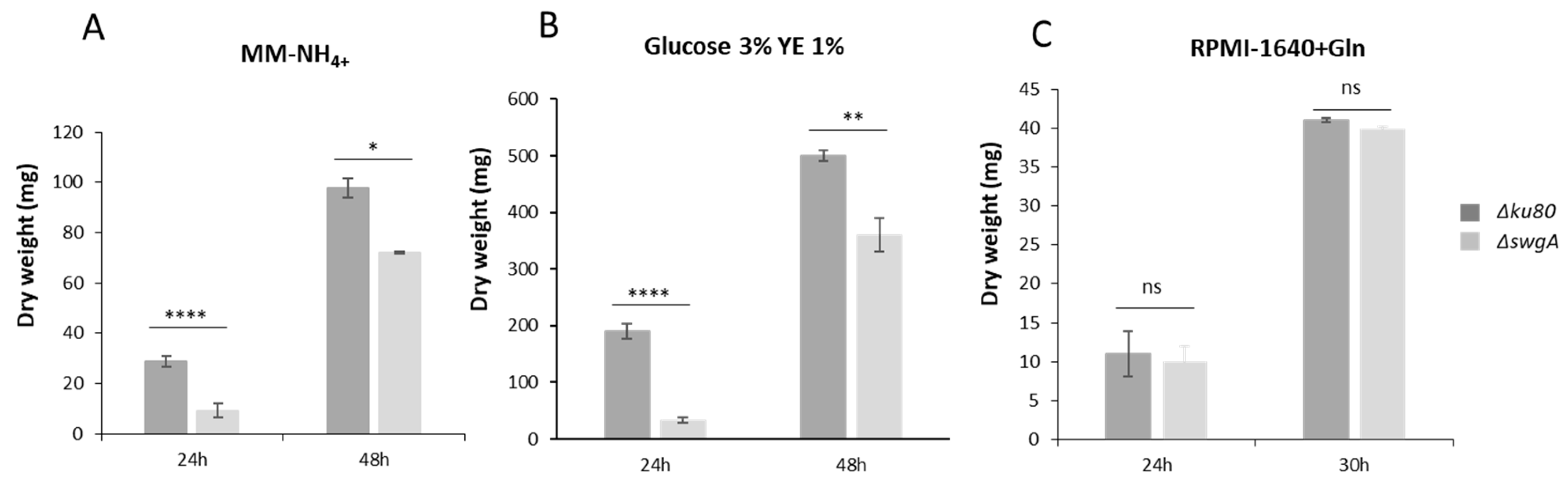
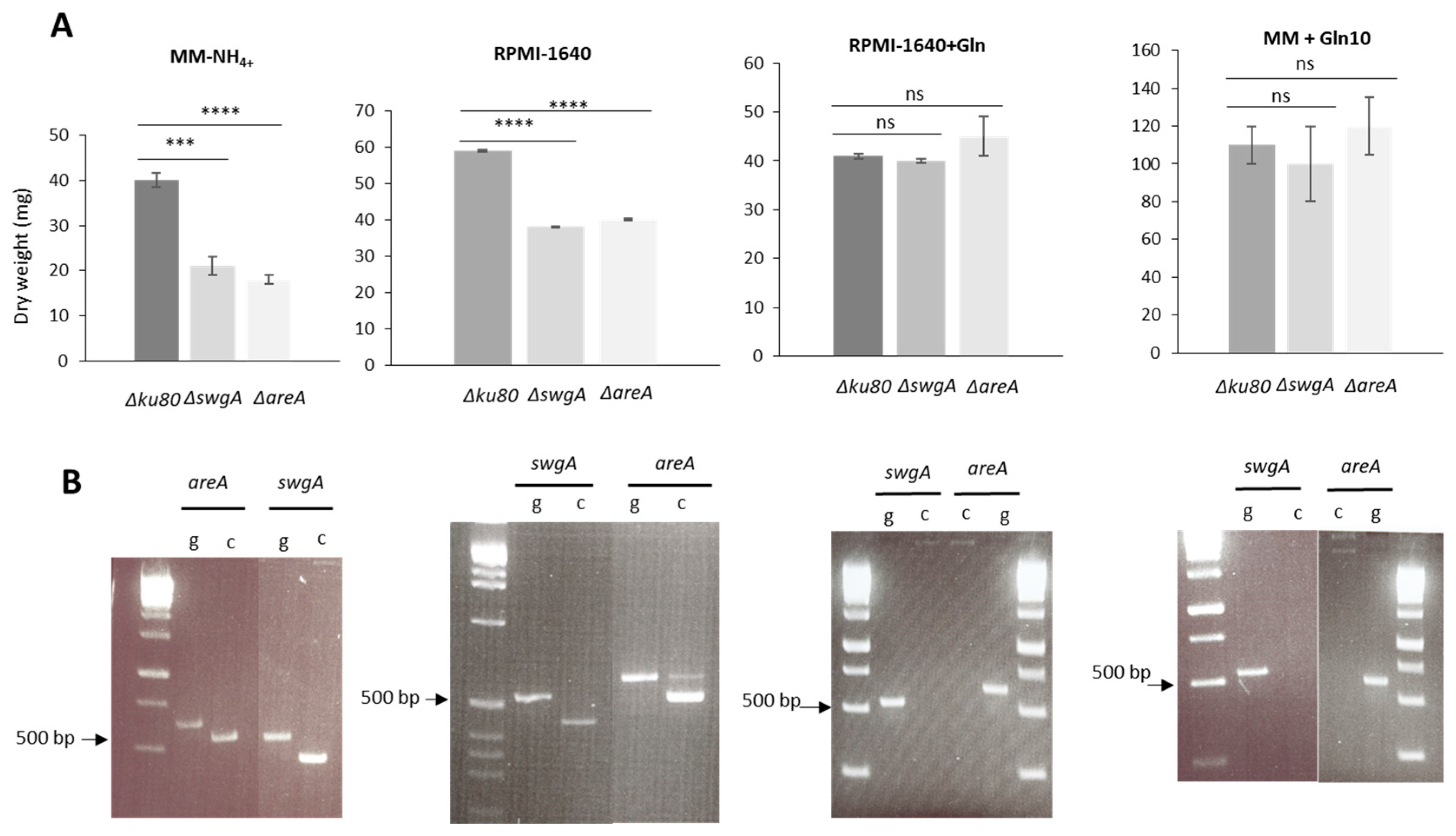
3.4. swgA Is Involved in Nitrogen Metabolism and Is Regulated by the Transcription Factor AreA (AFUA_6G01970)

To investigate the putative function of SwgA in nutritional metabolism, the growth of the mutants was compared in several different liquid media at 37 °C after 27 h or 30 h of growth with different nitrogen sources: MM(without NH4+)(MM) and supplemented by either L-glutamine 10 mM (MM+Gln10), or glutamate 10 ((MM+Glu10) or 50 mM (MM+Glu50), also MM-NH4+ supplemented by glutamate 10 mM (MM-NH4++Glu10), and RPMI-1640 without L-glutamine (RPMI-1640). Depending on the nitrogen source, differences in the growth of the ΔswgA mutant in comparison to the parental strain were observed (Figure 5A). A decrease of 35 to 50% of growth was observed for the ΔswgA mutant in comparison to the parental strain when MM-NH4+ or RPMI-1640 were used. In contrast, when MM+ Gln10 or RPMI-1640+Gln were used, no difference in growth was observed for the ΔswgA mutant (Figure 5A). In MM+Glu10 or MM+Glu50, the growth of ΔswgA was significantly decreased. In contrast, in MM-NH4++Glu10, no growth defect was observed indicating that the phenotype was not due to a defect of glutamine synthase activity (Figure S8). The ∆areA mutant was also included as a control [31] as AreA is a major regulator of nitrogen metabolism. Similar results were observed for the ∆areA mutant indicating that the ΔswgA and ∆areA mutants behave similarly to the wild type in a medium containing L-glutamine as the nitrogen source. The expressions of both genes were investigated in all of these media in the parental strain, and the ∆areA mutant. In the parental strain, swgA and areA were not expressed in a medium containing L-glutamine (RPMI-1640+Gln and MM+Gln10 (Figure 5B). In contrast, these two genes were expressed in a medium without L-glutamine (MM-NH4+, RPMI-1640). No swgA gene expression was detected in the ∆areA mutant (data not shown) independently of the medium used, indicating that the transcription factor AreA regulated swgA expression. Moreover, a consensus sequence CGATA is present in 5′ position 313 nucleotides upstream of the start codon of swgA supporting the fact that swgA is one of the many genes regulated by the transcription factor AreA.
All of these results showed that swgA is involved in nitrogen metabolism and is regulated by the transcription factor AreA.

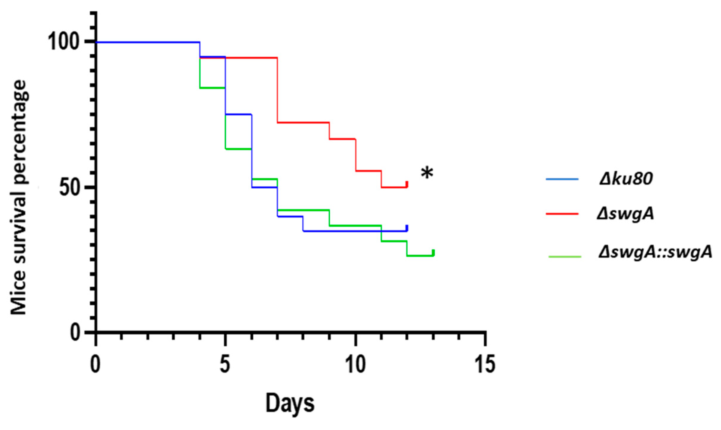
3.5. Virulence of the ΔswgA Mutant Is Affected

The virulence of the ΔswgA mutant was tested in a mouse model of invasive aspergillosis. The parental and the revertant strains showed the same survival curve. However, statistical analysis showed that the virulence of the ΔswgA mutant was significantly reduced (p-value < 0.5 (Figure 6).

4. Discussion

Our preliminary analysis of the A. fumigatus mutant library of all the GPI-AP encoding (identified via an in silico analysis) has shown that three mutants have significant reduction in the growth on malt and MM media at 37 °C [2]. Our screening was validated by the fact that GEL2 and DFG3 mutants of the library [5,8] had reduced growth. Only one mutant with a drastic reduction of growth, corresponding to the gene AFUA_8G01170 had not been investigated previously.
In this study, we characterized a new GPI-anchored protein that we named SwgA. Although no significant homology has been found with any known proteins, it has been classified as a hydrophobin-like protein because of the presence of distinctive patterns of conserved cysteines and hydrophobic residues as proposed by Littlejohn et al. [28]. Hydrophobins are low molecular weight proteins secreted by filamentous fungi and are divided into two classes (class I form functional amyloid fibers organized in layers with rodlet morphology, while class II hydrophobin layers show no defined morphology). In general, hydrophobins have low sequence conservation, and they are characterized by their hydrophobicity profiles and an idiosyncratic pattern of eight conserved cysteine residues that form four disulfate linkages. Seven hydrophobins have been characterized and studied, RODA-RODG in A. fumigatus [20,32]. It has been shown that only RodA is responsible for rodlet formation, sporulation conidial hydrophobicity, and immunological inertia of the conidia. Our data suggested that SwgA should not be considered a hydrophobin-like protein. For the ΔswgA mutant, we have not observed differences in rodlet formation or conidial hydrophobicity (data not shown).
Other proteins containing conserved cysteine residues, such as proteins with CFEM (common in fungal extracellular membrane) domains, are shown to be involved in host–pathogen interactions and virulence. In C. albicans, deletion of the three GPI-anchored CFEM-encoding genes in the genome (RBT5/RBT51/CSA1) resulted in an increased sensitivity to cell wall damaging agents and reduced ability to form a biofilm [33,34]. In the genome of A. fumigatus, three GPI-APs, CFEM (A-C), containing fungal-specific CFEM domains characterized by spaced cysteine residues are present [35]. Vaknin et al. [14] showed that these proteins did not play any role in cell wall morphogenesis or virulence. However, we did not observe any sequence similarity with CFEM proteins and SwgA.
The adhesion of A. fumigatus conidia to host tissues is an essential event leading to the persistence and infection of the host target lung. Several A. fumigatus molecules are shown to serve as host cell adhesion factors in the swollen conidia, germling, and hyphal states [36,37]. Takahashi-Nakaguchi et al. [38] identified 31 genes as putative candidates for conidial adhesion factors. The classification of SwgA as a hydrophobin-like protein, and its corresponding gene expression during hyphal morphogenesis, suggested a putative role in fungal adherence of this protein as predicted by Faapred’s prediction [29]. However, we demonstrated here that SwgA is not involved in adhesion and no difference in biofilm formation has been observed (data not shown).
In contrast, germination and growth of the ΔswgA mutant were strongly affected depending on the culture medium. We demonstrated that the growth of the ΔswgA mutant depends on the nitrogen composition of the medium. No differences in growth were observed between the ΔswgA mutant and the wild type in a liquid medium containing L-glutamine.
Nitrogen is an essential requirement for growth and fungi are able to use a wide variety of compounds as nitrogen sources. However, the use of different nitrogen compounds is selective, and readily assimilated nitrogen sources such as ammonium and glutamine are preferentially used [39]. In Aspergillus, the areA gene encodes the major nitrogen regulatory protein, which activates the transcription of many structural gene-encoding enzymes for nitrogen source catabolism under nitrogen-limiting conditions [40]. We showed that swgA expression is under the control of the transcription factor AreA. Indeed, we showed that swgA is not expressed in the ΔareA mutant whatever the medium used. Moreover, the AreA transcription factor, such as the GATA factor in fungi, share a common DNA binding motif that recognizes a core 5′-GATA-3′ sequence [41].
The growth of the ΔareA mutant in A. nidulans, A. fumigatus, and A. oryzae, in different nitrogen media were investigated [42,43,44]. The growth of these ΔareA mutants was identical to the wild type in presence of glutamine and the growth was affected by other nitrogen sources.
In our study, several nitrogen sources were tested and we showed that the ΔswgA mutant phenocopies the ΔareA mutant, i.e., in the presence of glutamine as the sole nitrogen source (RPMI-1640+Gln or MM+Gln), the growth of these mutants were not affected in contrast to the medium, which did not contain glutamine, such as the MM-NH4+ or RPMI-1640 medium. In Aspergillus oryzae and Aspergillus nidulans, the glutamine synthetase levels were not affected [42] by the areA deletion. The restoration of the growth phenotype of the ΔswgA mutant in the presence of NH4+ and glutamate suggested that the glutamine synthetase level, such as for ΔareA mutants, was also not affected in this A. fumigatus (Figure S8).
Consistent with the reduction in virulence observed for the ΔswgA mutant, loss of areA in A. fumigatus was shown to contribute to virulence but was not essential for virulence in a neutropenic murine model of pulmonary aspergillosis, with the deletion mutants presenting a delayed growth phenotype during infection [44]. In vivo data analysis showed that the gene expression level of swgA is 80 times higher than in vitro (Beau, Latgé, unpublished data) suggesting the lack of availability of preferred nitrogen source in the lung. This could explain the reduction of virulence of the ΔswgA. The mechanism involved in this nitrogen metabolism for SwgA is still unknown, but it was recently shown in Pichia pastoris that a GPI-anchored protein of unknown function, Gcw13, was able to regulate a permease GAP1 resulting in decreasing Gap1-dependent uptake of amino acids [45]. Moreover, in C. albicans, several permeases, such as Csy1p [46] and Gap2p [47], are shown to participate in glutamine assimilation. To date, several permeases are shown to be present in the A. fumigatus genome, but until now, not one has been characterized [48].

5. Conclusions

In this study, we showed that a new GPI-anchored protein called SwgA is involved in nitrogen metabolism; this protein is regulated by the transcription factor AreA. Our data showed that GPI-anchored proteins are not only involved in cell wall biosynthesis but may be key actors in the regulation of the general fungal metabolism.

Supplementary Materials

The following supporting information can be downloaded at: https://www.mdpi.com/article/10.3390/jof9020256/s1, Figure S1: Southern blot analysis and validation of the constructed strains; Figure S2: Sequence alignments of AFUA_8G01170 (SwgA) protein and orthologs in Aspergillus species; Figure S3: Permeability and Calcofluor White staining of the dormant and swollen conidia of the parental Δku80, the ΔswgA mutant and the ΔswgA::swgA; Figure S4: Polysaccharide cell surface immunolabelling of the parental Δku80 and ΔswgA mutant strains of swollen conidia after 4 h of growth in Glucose 3% Yeast Extract 1% liquid medium; Figure S5: SEM of the dormant and germinated conidia of the parental Δku80 and the ΔswgA mutant strains; Figure S6: Growth kinetics of the parental Δku80 strain and the ΔswgA mutant strains in MM, Glucose 3% Yeast Extract 1% and RPMI-1640+Gln liquid medium after 24 h of growth at 45 °C; Figure S7: Adherence assays to polystyrene after 24 h in MM medium + 0.01 % Tween 20 using crystal violet assay; Figure S8: Growth kinetics of the parental Δku80 and the ΔswgA mutant strains in MM-NH4++Glu10 (with NH4) supplemented with 10 mM glutamate, and MM without NH4 and supplemented with 10 mM or 50 mM glutamate (MM+ Glu10 or MM+Glu50) liquid medium after 27 h of growth at 37 °C. Table S1: Primers used in this study; Table S2: Characteristics of the protein encoded by the AFUA_8G01170 gene and orthologs in others species; Table S3: Pourcentage of identities between the different proteins: XP_001275113.1-A.clavatus; AFUA_8G01170-A.fumigatus; XP_001261813.1-A.fisheri.

Author Contributions

Conceptualization, I.M. and J.-P.L.; methodology, M.S., P.F., R.B., M.B., T.F. and M.M.-N.; validation, formal analysis, investigation; resources, writing—original draft preparation, M.S., J.-P.L. and I.M. All authors have read and agreed to the published version of the manuscript.

Funding

This research was funded by l’Agence Nationale pour la Recherche (Afulnf ANR-16-CE92-0039), la Fondation pour la Recherche Médicale (DEQ20150331722), Laboratoire d’Excellence “Integrative Biology of Emerging Infectious Diseases” (grant ANR-10-LABX-62-IBEID), and an AIC grant. This work was also supported by the Wellcome trust grant 208396/Z/17/Z.

Institutional Review Board Statement

Not applicable.

Informed Consent Statement

Not applicable.

Data Availability Statement

Not applicable.

Conflicts of Interest

The authors declare no conflict of interest. The funders had no role in the design of the study; in the collection, analyses, or interpretation of data; in the writing of the manuscript, or in the decision to publish the results.

References

  1. Kinoshita, T.; Fujita, M. Biosynthesis of GPI-Anchored Proteins: Special Emphasis on GPI Lipid Remodeling. J. Lipid Res. 2016, 57, 6–24. [Google Scholar] [CrossRef] [PubMed] [Green Version]
  2. Samalova, M.; Carr, P.; Bromley, M.; Blatzer, M.; Moya-Nilges, M.; Latgé, J.-P.; Mouyna, I. GPI Anchored Proteins in Aspergillus Fumigatus and Cell Wall Morphogenesis. Curr. Top. Microbiol. Immunol. 2020, 425, 167–186. [Google Scholar] [PubMed]
  3. Gastebois, A.; Clavaud, C.; Aimanianda, V.; Latgé, J.-P. Aspergillus Fumigatus: Cell Wall Polysaccharides, Their Biosynthesis and Organization. Future Microbiol. 2009, 4, 583–595. [Google Scholar] [CrossRef] [PubMed] [Green Version]
  4. Mouyna, I.; Fontaine, T.; Vai, M.; Monod, M.; Fonzi, W.A.; Diaquin, M.; Popolo, L.; Hartland, R.P.; Latgé, J.P. Glycosylphosphatidylinositol-Anchored Glucanosyltransferases Play an Active Role in the Biosynthesis of the Fungal Cell Wall. J. Biol. Chem. 2000, 275, 14882–14889. [Google Scholar] [CrossRef] [Green Version]
  5. Mouyna, I.; Morelle, W.; Vai, M.; Monod, M.; Léchenne, B.; Fontaine, T.; Beauvais, A.; Sarfati, J.; Prévost, M.-C.; Henry, C.; et al. Deletion of GEL2 Encoding for a Beta(1-3)Glucanosyltransferase Affects Morphogenesis and Virulence in Aspergillus Fumigatus. Mol. Microbiol. 2005, 56, 1675–1688. [Google Scholar] [CrossRef]
  6. Gastebois, A.; Fontaine, T.; Latgé, J.-P.; Mouyna, I. Beta(1-3)Glucanosyltransferase Gel4p Is Essential for Aspergillus Fumigatus. Eukaryot. Cell 2010, 9, 1294–1298. [Google Scholar] [CrossRef] [Green Version]
  7. Aimanianda, V.; Simenel, C.; Garnaud, C.; Clavaud, C.; Tada, R.; Barbin, L.; Mouyna, I.; Heddergott, C.; Popolo, L.; Ohya, Y.; et al. The Dual Activity Responsible for the Elongation and Branching of β-(1,3)-Glucan in the Fungal Cell Wall. MBio 2017, 8, e00619-17. [Google Scholar] [CrossRef] [Green Version]
  8. Muszkieta, L.; Fontaine, T.; Beau, R.; Mouyna, I.; Vogt, M.S.; Trow, J.; Cormack, B.P.; Essen, L.-O.; Jouvion, G.; Latgé, J.-P. The Glycosylphosphatidylinositol-Anchored DFG Family Is Essential for the Insertion of Galactomannan into the β-(1,3)-Glucan-Chitin Core of the Cell Wall of Aspergillus Fumigatus. mSphere 2019, 4, e00397-19. [Google Scholar] [CrossRef] [Green Version]
  9. Mouyna, I.; Aimanianda, V.; Hartl, L.; Prevost, M.-C.; Sismeiro, O.; Dillies, M.-A.; Jagla, B.; Legendre, R.; Coppee, J.-Y.; Latgé, J.-P. GH16 and GH81 Family β-(1,3)-Glucanases in Aspergillus Fumigatus Are Essential for Conidial Cell Wall Morphogenesis. Cell. Microbiol. 2016, 18, 1285–1293. [Google Scholar] [CrossRef] [Green Version]
  10. Gastebois, A.; Mouyna, I.; Simenel, C.; Clavaud, C.; Coddeville, B.; Delepierre, M.; Latgé, J.-P.; Fontaine, T. Characterization of a New Beta(1-3)-Glucan Branching Activity of Aspergillus Fumigatus. J. Biol. Chem. 2010, 285, 2386–2396. [Google Scholar] [CrossRef]
  11. Caro, L.H.; Tettelin, H.; Vossen, J.H.; Ram, A.F.; van den Ende, H.; Klis, F.M. In Silicio Identification of Glycosyl-Phosphatidylinositol-Anchored Plasma-Membrane and Cell Wall Proteins of Saccharomyces Cerevisiae. Yeast 1997, 13, 1477–1489. [Google Scholar] [CrossRef]
  12. Plaine, A.; Walker, L.; Da Costa, G.; Mora-Montes, H.M.; McKinnon, A.; Gow, N.A.R.; Gaillardin, C.; Munro, C.A.; Richard, M.L. Functional Analysis of Candida Albicans GPI-Anchored Proteins: Roles in Cell Wall Integrity and Caspofungin Sensitivity. Fungal Genet. Biol. 2008, 45, 1404–1414. [Google Scholar] [CrossRef] [PubMed] [Green Version]
  13. Timmermans, B.; De Las Peñas, A.; Castaño, I.; Van Dijck, P. Adhesins in Candida Glabrata. J. Fungi 2018, 4, 60. [Google Scholar] [CrossRef] [PubMed] [Green Version]
  14. Vaknin, Y.; Shadkchan, Y.; Levdansky, E.; Morozov, M.; Romano, J.; Osherov, N. The Three Aspergillus Fumigatus CFEM-Domain GPI-Anchored Proteins (CfmA-C) Affect Cell-Wall Stability but Do Not Play a Role in Fungal Virulence. Fungal Genet. Biol. 2014, 63, 55–64. [Google Scholar] [CrossRef]
  15. Woo, P.C.Y.; Lau, S.K.P.; Lau, C.C.Y.; Tung, E.T.K.; Au-Yeung, R.K.H.; Cai, J.-P.; Chong, K.T.K.; Sze, K.H.; Kao, R.Y.; Hao, Q.; et al. Mp1p Homologues as Virulence Factors in Aspergillus Fumigatus. Med. Mycol. 2018, 56, 350–360. [Google Scholar] [CrossRef] [Green Version]
  16. Levdansky, E.; Kashi, O.; Sharon, H.; Shadkchan, Y.; Osherov, N. The Aspergillus Fumigatus CspA Gene Encoding a Repeat-Rich Cell Wall Protein Is Important for Normal Conidial Cell Wall Architecture and Interaction with Host Cells. Eukaryot. Cell 2010, 9, 1403–1415. [Google Scholar] [CrossRef] [Green Version]
  17. Valsecchi, I.; Sarikaya-Bayram, Ö.; Wong Sak Hoi, J.; Muszkieta, L.; Gibbons, J.; Prevost, M.-C.; Mallet, A.; Krijnse-Locker, J.; Ibrahim-Granet, O.; Mouyna, I.; et al. MybA, a Transcription Factor Involved in Conidiation and Conidial Viability of the Human Pathogen Aspergillus Fumigatus. Mol. Microbiol. 2017, 105, 880–900. [Google Scholar] [CrossRef] [Green Version]
  18. Fraczek, M.G.; Bromley, M.; Buied, A.; Moore, C.B.; Rajendran, R.; Rautemaa, R.; Ramage, G.; Denning, D.W.; Bowyer, P. The Cdr1B Efflux Transporter Is Associated with Non-Cyp51a-Mediated Itraconazole Resistance in Aspergillus Fumigatus. J. Antimicrob. Chemother. 2013, 68, 1486–1496. [Google Scholar] [CrossRef]
  19. Zhao, C.; Fraczek, M.G.; Dineen, L.; Lebedinec, R.; Macheleidt, J.; Heinekamp, T.; Delneri, D.; Bowyer, P.; Brakhage, A.A.; Bromley, M. High-Throughput Gene Replacement in Aspergillus Fumigatus. Curr. Protoc. Microbiol. 2019, 54, e88. [Google Scholar] [CrossRef] [Green Version]
  20. Valsecchi, I.; Dupres, V.; Stephen-Victor, E.; Guijarro, J.I.; Gibbons, J.; Beau, R.; Bayry, J.; Coppee, J.-Y.; Lafont, F.; Latgé, J.-P.; et al. Role of Hydrophobins in Aspergillus Fumigatus. J. Fungi 2017, 4, 2. [Google Scholar] [CrossRef]
  21. Sánchez, O.; Aguirre, J. Efficient Transformation of Aspergillus nidulans by Electroporation of Germinated Conidia. Fungal Genet. Rep. 1996, 43, 48–51. [Google Scholar] [CrossRef] [Green Version]
  22. Girardin, H.; Latgé, J.P.; Srikantha, T.; Morrow, B.; Soll, D.R. Development of DNA Probes for Fingerprinting Aspergillus Fumigatus. J. Clin. Microbiol. 1993, 31, 1547–1554. [Google Scholar] [CrossRef] [PubMed] [Green Version]
  23. Beauvais, A.; Schmidt, C.; Guadagnini, S.; Roux, P.; Perret, E.; Henry, C.; Paris, S.; Mallet, A.; Prévost, M.-C.; Latgé, J.P. An Extracellular Matrix Glues Together the Aerial-Grown Hyphae of Aspergillus Fumigatus. Cell. Microbiol. 2007, 9, 1588–1600. [Google Scholar] [CrossRef]
  24. Beauvais, A.; Bozza, S.; Kniemeyer, O.; Formosa, C.; Formosa, C.; Balloy, V.; Henry, C.; Roberson, R.W.; Dague, E.; Chignard, M.; et al. Deletion of the α-(1,3)-Glucan Synthase Genes Induces a Restructuring of the Conidial Cell Wall Responsible for the Avirulence of Aspergillus Fumigatus. PLoS Pathog. 2013, 9, e1003716. [Google Scholar] [CrossRef]
  25. Millet, N.; Latgé, J.-P.; Mouyna, I. Members of Glycosyl-Hydrolase Family 17 of A. fumigatus Differentially Affect Morphogenesis. J. Fungi 2018, 4, 18. [Google Scholar] [CrossRef] [PubMed] [Green Version]
  26. Dubois, M.; Gilles, K.; Hamilton, J.K.; Rebers, P.A.; Smith, F. Colorimetric Method for Determination of Sugars and Related Substances. Anal. Chem. 1956, 28, 350–356. [Google Scholar] [CrossRef]
  27. Stalhberger, T.; Simenel, C.; Clavaud, C.; Eijsink, V.G.H.; Jourdain, R.; Delepierre, M.; Latgé, J.-P.; Breton, L.; Fontaine, T. Chemical Organization of the Cell Wall Polysaccharide Core of Malassezia Restricta. J. Biol. Chem. 2014, 289, 12647–12656. [Google Scholar] [CrossRef] [Green Version]
  28. Littlejohn, K.A.; Hooley, P.; Cox, P. Bioinformatics Predicts Diverse Aspergillus Hydrophobins with Novel Properties. Food Hydrocoll. 2012, 27, 503–516. [Google Scholar] [CrossRef]
  29. Chaudhuri, R.; Ansari, F.A.; Raghunandanan, M.V.; Ramachandran, S. FungalRV: Adhesin Prediction and Immunoinformatics Portal for Human Fungal Pathogens. BMC Genom. 2011, 12, 192. [Google Scholar] [CrossRef] [Green Version]
  30. Henry, C.; Fontaine, T.; Heddergott, C.; Robinet, P.; Aimanianda, V.; Beau, R.; Beauvais, A.; Mouyna, I.; Prevost, M.-C.; Fekkar, A.; et al. Biosynthesis of Cell Wall Mannan in the Conidium and the Mycelium of Aspergillus Fumigatus. Cell. Microbiol. 2016, 18, 1881–1891. [Google Scholar] [CrossRef]
  31. Liu, Z.; Raj, S.; van Rhijn, N.; Fraczek, M.; Michel, J.-P.; Sismeiro, O.; Legendre, R.; Varet, H.; Fontaine, T.; Bromley, M.; et al. Functional Genomic and Biochemical Analysis Reveals Pleiotropic Effect of Congo Red on Aspergillus Fumigatus. mBio 2021, 12, e00863-21. [Google Scholar] [CrossRef] [PubMed]
  32. Aimanianda, V.; Bayry, J.; Bozza, S.; Kniemeyer, O.; Perruccio, K.; Elluru, S.R.; Clavaud, C.; Paris, S.; Brakhage, A.A.; Kaveri, S.V.; et al. Surface Hydrophobin Prevents Immune Recognition of Airborne Fungal Spores. Nature 2009, 460, 1117–1121. [Google Scholar] [CrossRef] [PubMed]
  33. Pérez, A.; Pedrós, B.; Murgui, A.; Casanova, M.; López-Ribot, J.L.; Martínez, J.P. Biofilm Formation by Candida Albicans Mutants for Genes Coding Fungal Proteins Exhibiting the Eight-Cysteine-Containing CFEM Domain. FEMS Yeast Res. 2006, 6, 1074–1084. [Google Scholar] [CrossRef] [Green Version]
  34. Pérez, A.; Ramage, G.; Blanes, R.; Murgui, A.; Casanova, M.; Martínez, J.P. Some Biological Features of Candida Albicans Mutants for Genes Coding Fungal Proteins Containing the CFEM Domain. FEMS Yeast Res. 2011, 11, 273–284. [Google Scholar] [CrossRef] [PubMed] [Green Version]
  35. Kulkarni, R.D.; Kelkar, H.S.; Dean, R.A. An Eight-Cysteine-Containing CFEM Domain Unique to a Group of Fungal Membrane Proteins. Trends Biochem. Sci. 2003, 28, 118–121. [Google Scholar] [CrossRef] [PubMed]
  36. Al Abdallah, Q.; Choe, S.-I.; Campoli, P.; Baptista, S.; Gravelat, F.N.; Lee, M.J.; Sheppard, D.C. A Conserved C-Terminal Domain of the Aspergillus Fumigatus Developmental Regulator MedA Is Required for Nuclear Localization, Adhesion and Virulence. PLoS ONE 2012, 7, e49959. [Google Scholar] [CrossRef] [Green Version]
  37. Sheppard, D.C. Molecular Mechanism of Aspergillus Fumigatus Adherence to Host Constituents. Curr. Opin. Microbiol. 2011, 14, 375–379. [Google Scholar] [CrossRef] [Green Version]
  38. Takahashi-Nakaguchi, A.; Sakai, K.; Takahashi, H.; Hagiwara, D.; Toyotome, T.; Chibana, H.; Watanabe, A.; Yaguchi, T.; Yamaguchi, M.; Kamei, K.; et al. Aspergillus Fumigatus Adhesion Factors in Dormant Conidia Revealed through Comparative Phenotypic and Transcriptomic Analyses. Cell. Microbiol. 2018, 20, e12802. [Google Scholar] [CrossRef] [Green Version]
  39. Perez-Cuesta, U.; Guruceaga, X.; Cendon-Sanchez, S.; Pelegri-Martinez, E.; Hernando, F.L.; Ramirez-Garcia, A.; Abad-Diaz-de-Cerio, A.; Rementeria, A. Nitrogen, Iron and Zinc Acquisition: Key Nutrients to Aspergillus Fumigatus Virulence. J. Fungi 2021, 7, 518. [Google Scholar] [CrossRef]
  40. Tudzynski, B. Nitrogen Regulation of Fungal Secondary Metabolism in Fungi. Front. Microbiol. 2014, 5, 656. [Google Scholar] [CrossRef]
  41. Scazzocchio, C. The Fungal GATA Factors. Curr. Opin. Microbiol. 2000, 3, 126–131. [Google Scholar] [CrossRef] [PubMed]
  42. Christensen, T.; Hynes, M.J.; Davis, M.A. Role of the Regulatory Gene AreA of Aspergillus Oryzae in Nitrogen Metabolism. Appl. Environ. Microbiol. 1998, 64, 3232–3237. [Google Scholar] [CrossRef] [PubMed] [Green Version]
  43. Langdon, T.; Sheerins, A.; Ravagnani, A.; Gielkens, M.; Caddick, M.X.; Arst, H.N. Mutational Analysis Reveals Dispensability of the N-Terminal Region of the Aspergillus Transcription Factor Mediating Nitrogen Metabolite Repression. Mol. Microbiol. 1995, 17, 877–888. [Google Scholar] [CrossRef]
  44. Hensel, M.; Arst, H.N.; Aufauvre-Brown, A.; Holden, D.W. The Role of the Aspergillus Fumigatus AreA Gene in Invasive Pulmonary Aspergillosis. Mol. Gen. Genet. 1998, 258, 553–557. [Google Scholar] [CrossRef] [PubMed]
  45. Zou, C.; Wang, P.; Liang, S.; Han, S.; Zheng, S.; Lin, Y. Deletion of the GCW13 Gene Derepresses Gap1-Dependent Uptake of Amino Acids in Pichia Pastoris Grown on Methanol as the Sole Carbon Source. Biochem. Biophys. Res. Commun. 2018, 501, 226–231. [Google Scholar] [CrossRef]
  46. Brega, E.; Zufferey, R.; Mamoun, C.B. Candida Albicans Csy1p Is a Nutrient Sensor Important for Activation of Amino Acid Uptake and Hyphal Morphogenesis. Eukaryot. Cell 2004, 3, 135–143. [Google Scholar] [CrossRef] [Green Version]
  47. Kraidlova, L.; Van Zeebroeck, G.; Van Dijck, P.; Sychrová, H. The Candida Albicans GAP Gene Family Encodes Permeases Involved in General and Specific Amino Acid Uptake and Sensing. Eukaryot. Cell 2011, 10, 1219–1229. [Google Scholar] [CrossRef] [Green Version]
  48. Ries, L.N.A.; Beattie, S.; Cramer, R.A.; Goldman, G.H. Overview of Carbon and Nitrogen Catabolite Metabolism in the Virulence of Human Pathogenic Fungi. Mol. Microbiol. 2018, 107, 277–297. [Google Scholar] [CrossRef]
Figure 1. (A) SwgA localization by immunolabeling with the anti-SwgA antibodies (1/250) in swollen conidia (after 4 h of growth in glucose 3% yeast extract 1% liquid medium) of the parental and the ΔswgA mutant strains. (B) Western blot of the cell wall and membrane of the parental, complemented strain and ΔswgA mutant strains labeled with the anti-SwgA antibody (1/500). The membrane and cell wall were prepared after 24 h of growth in Glucose 3% Yeast Extract 1% medium.
Jof 09 00256 g001
Figure 2. Conidiation and germination assay. (A) Conidiation of the parental, the revertant, and the ΔswgA mutant strains after one week at room temperature on malt medium. Kinetics of conidial germination: percentage (%) of germinated conidia of the parental strain, the revertant, and the ΔswgA mutant (B) at 37 °C on agar malt medium (C) at 45 °C on agar malt medium. The error bars represent the standard deviation from the mean values of three different experiments. p value are indicated: asterisks indicated that it is significant ** p < 0.00221; **** p < 0.0001 and ns, nonsignificant.
Jof 09 00256 g002
Figure 3. Growth of the parental, the revertant, and the ΔswgA mutant strains on malt agar and MM-NH4+ after 24 h, 48 h, and 72 h (A) at 37 °C (B) at 45 °C and (C) on agar MM-NH4+ medium after 48 h of growth at 37 °C with the addition of CR (100 µg/mL) or CFW (80 µg/mL).
Jof 09 00256 g003
Figure 4. Growth kinetics of the parental and the ΔswgA mutant strains in (A) MM-NH4+, (B) glucose 3% yeast extract 1% (C) RPMI-1640+Gln liquid media after 24 h, 30 h, or 48 h of growth at 37 °C. p value are indicated: asterisks indicated that it is significant: * p < 0.0332; ** p < 0.00221; **** p < 0.0001 and ns, nonsignificant.
Jof 09 00256 g004
Figure 5. (A) Growth kinetics of the parental, ΔswgA, and ΔareA strains at 37 °C after 27 h in several different nitrogen sources: MM-NH4+, RPMI-1640, RPMI-1640+Gln, and MM+Gln10 and (B) gene expression levels of areA and swgA in the parental control strain in the different media shown above in panel A. g: genomic control, c: cDNA. p value are indicated: asterisks indicated that it is significant: *** p < 0.0002; **** p < 0.0001, ns, nonsignificant.
Jof 09 00256 g005
Figure 6. Mice survival percentage. Twenty-four-week-old OF1 female mice/strains were immunosuppressed with Kenacort. Before conidial inhalation (day 0), mice were anesthetized intraperitoneally with 10 mg/mL of ketamine and 1 mg/mL of xylazine per mouse. Inoculation was conducted intranasally with 2 × 10⁶ of conidia. Non-infected control immunosuppressed mice only received PBS (1X; * p-value Wilcoxon test < 0.5.
Jof 09 00256 g006
Table 1. Percentage (%) of polysaccharide composition of the alkali-insoluble and the alkali-soluble fraction of the cell wall of the parental Δku80 strain and the ΔswgA mutant. The cell wall alkali-insoluble fraction (AI) was purified from mycelium grown for 24 h in a liquid medium at 37 °C (expressed as a percentage of total hexoses plus hexosamines). Each value represents an average of data from three independent replicates. Hexose; GlcNAc, N-acetylglucosamine; GalNAc, N-acetylgalactosamine.
HexoseGlcNAcGalNAc
Δku80-AI66.85 ± 2.528.17 ± 1.94.98 ± 0.7
ΔswgA-AI64.95 ± 530.31 ± 4.64.74 ± 0.54
Δku80-AS42.63 ± 12.6057.37 ± 12.7
ΔswgA-AS27.54 ± 8.2072.46 ± 8.2
Disclaimer/Publisher’s Note: The statements, opinions and data contained in all publications are solely those of the individual author(s) and contributor(s) and not of MDPI and/or the editor(s). MDPI and/or the editor(s) disclaim responsibility for any injury to people or property resulting from any ideas, methods, instructions or products referred to in the content.

