Article

Ultrarare Loss-of-Function Mutations in the Genes Encoding the Ionotropic Glutamate Receptors of Kainate Subtypes Associated with Schizophrenia Disrupt the Interaction with PSD95

1
Department of Psychiatry, Yuli Branch, Taipei Veterans General Hospital, Hualien 98142, Taiwan
2
Department of Future Studies and LOHAS Industry, Fo Guang University, Jiaosi, Yilan County 26247, Taiwan
*
Author to whom correspondence should be addressed.
J. Pers. Med. 2022, 12(5), 783; https://doi.org/10.3390/jpm12050783
Submission received: 7 April 2022 / Revised: 28 April 2022 / Accepted: 10 May 2022 / Published: 12 May 2022
(This article belongs to the Special Issue Recent Advances in Precision Psychiatry)

Abstract

:
Schizophrenia is a complex mental disorder with a genetic component. The GRIK gene family encodes ionotropic glutamate receptors of the kainate subtype, which are considered candidate genes for schizophrenia. We screened for rare and pathogenic mutations in the protein-coding sequences of the GRIK gene family in 516 unrelated patients with schizophrenia using the ion semiconductor sequencing method. We identified 44 protein-altered variants, and in silico analysis indicated that 36 of these mutations were rare and damaging or pathological based on putative protein function. Notably, we identified four truncating mutations, including two frameshift deletion mutations (GRIK1p.Phe24fs and GRIK1p.Thr882fs) and two nonsense mutations (GRIK2p.Arg300Ter and GRIK4p.Gln342Ter) in four unrelated patients with schizophrenia. They exhibited minor allele frequencies of less than 0.01% and were absent in 1517 healthy controls from Taiwan Biobank. Functional analysis identified these four truncating mutants as loss-of-function (LoF) mutants in HEK-293 cells. We also showed that three mutations (GRIK1p.Phe24fs, GRIK1p.Thr882fs, and GRIK2p.Arg300Ter) weakened the interaction with the PSD95 protein. The results suggest that the GRIK gene family harbors ultrarare LoF mutations in some patients with schizophrenia. The identification of proteins that interact with the kainate receptors will be essential to determine kainate receptor-mediated signaling in the brain.

Graphical Abstract

1. Introduction

Schizophrenia is a chronic debilitating mental disorder that affects approximately 1% of the population worldwide, with symptoms appearing in early adulthood or late adolescence. The heritability of schizophrenia is estimated at 80% on average [1]. Evidence from genetic, neuropathological, and meta-analysis studies indicates that a reduction in neuronal processes and synaptic dysfunction is involved in the pathogenesis of schizophrenia, and patients exhibit synaptic degeneration in the brain [2,3,4,5,6]. The pathogenesis of schizophrenia may arise from mutations of synapse-associated genes; thus, identifying such mutations in schizophrenia will provide insight into the cause of schizophrenia and lead to new treatments. Glutamate receptors regulate neurotransmission in the mammalian central nervous system and play an essential role in synaptic plasticity, neurodevelopment, and cognitive function [7]. Glutamate receptor ion channels, including the N-methyl-d-aspartic acid (NMDA) receptors, the amino-3-hydroxy-5-methyl-4-isoxazole (AMPA) receptors, and the kainate receptors, are the major excitatory neurotransmitter receptors in the brain. These contribute to various normal neurophysiologic processes [8,9,10].
The kainate receptors, encoded by the GRIK gene family, are composed of four subunits and function as ligand-activated ion channels [11]. Rare pathologic mutations of the GRIK gene family may play a role in conferring susceptibility to schizophrenia for the following reasons: first, the kainate receptors are expressed in the brain and contribute to synaptic plasticity, transmission, learning, memory, and neuropsychiatric disorders, such as autism and schizophrenia [9,12,13,14]; second, several studies have demonstrated abnormal kainate receptor expression in the brains of subjects with schizophrenia [15,16,17,18,19,20,21,22,23]; third, although several studies have demonstrated that no significant associations of genetic markers of the GRIK gene family are associated with schizophrenia [24,25,26,27,28,29,30], some have identified novel rare mutations of the GRIK gene family, suggesting a potential role for rare and significant effects of mutations of the GRIK gene family in the susceptibility to schizophrenia [24,31,32,33,34].
The genetic basis of schizophrenia may involve rare deleterious mutations with high penetrance or multiple common polymorphisms with low penetrance [35,36]. In particular, rare mutations in a group of genes linked to synaptic development, function, and plasticity were identified in patients diagnosed with schizophrenia, suggesting that rare high-risk genetic mutations with result in the dysregulation of synaptic transmission and contribute to the pathophysiology of schizophrenia [37,38,39,40]. Therefore, we sequenced the exonic regions of five kainate receptor genes (GRIK1, GRIK2, GRIK3, GRIK4, and GRIK5) to identify rare pathogenic variants in patients with schizophrenia using the ion semiconductor sequencing method. We analyzed protein function by immunoblotting, immunocytochemistry, and protein–protein interactions of four protein-truncating mutations using a bioluminescent resonance energy transfer (BRET) assay.

2. Materials and Methods

2.1. Human Subjects

All subjects were Han Chinese from Taiwan: 516 unrelated patients with schizophrenia (258 males, mean age = 43.0 ± 9.3 years, mean age of onset = 22.0 ± 6.7 years; 258 females, mean age = 48.8 ± 10.2 years, mean age of onset = 22.0 ± 7.6 years) were screened for mutations. Blood samples from patients with schizophrenia was obtained from the Yuli Branch, Taipei Veterans General Hospital. Patients fulfilling the diagnostic criteria for schizophrenia defined by the Diagnostic and Statistical Manual of Mental Disorders-Fourth Edition Tex Revision were recruited for the study. The diagnosis of schizophrenia was based on clinical interviews and the review of medical records by senior psychiatrists with consensus. Organic brain syndrome cases, intellectual disability, substance-related psychosis, and mood disorder with psychotic features were excluded. The study was approved by the institutional review board from the Antai Medical Care Cooperation Antai-Tian-Sheng Memorial Hospital (Approval number: 18-144-A). All patients received a full explanation of the study and provided signed written informed consent. Genomic DNA was isolated from peripheral blood cells using the Gentra Genomic DNA Purification kit (QIAGEN, USA) and stored at −20 °C until use.

2.2. Ion Semiconductor Sequencing, PCR Reaction, and Fluorescence-Based Cycle Sequencing

Amplification primers were designed to cover the exonic regions of five genes, GRIK1 (GeneID:2897), GRIK2 (GeneID:2898), GRIK3 (GeneID:2899), GRIK4 (GeneID:2900), and GRIK5 (GeneID:2901), with the Ion AmpliSeq™ Designer software (Thermo Fisher Scientific Inc., Waltham, MA, USA). Ion semiconductor sequencing, PCR reactions, and fluorescence-based cycle sequencing were performed following previously described methods [41].

2.3. In Silico Analysis of Amino Acid Substitutions

The potential functional consequences of amino acid substitution were predicted using the Polyphen-2 (http://genetics.bwh.harvard.edu/pph2/, accessed on 18 July 2019), SIFT (https://sift.bii.a-star.edu.sg/, accessed on 18 July 2019), Pmut (http://mmb.pcb.ub.es/PMut/, accessed on 18 July 2019), PROVEAN (http://provean.jcvi.org/index.php, accessed on 18 July 2019), and CADD (https://cadd.gs.washington.edu/, accessed on 26 April 2022) software tools. Mutations were checked whether they were documented in the NCBI dbSNP database (http://www.ncbi.nlm.nih.gov/SNP/, accessed on 18 July 2019), the Genome Aggregation Database (gnomAD, https://gnomad.broadinstitute.org/, accessed on 18 July 2019), 1000 Genomes Project (https://www.internationalgenome.org/, accessed on 26 April 2022) and the Taiwan BioBank (https://www.twbiobank.org.tw/new_web/index.php, accessed on 18 July 2019). Protein–protein interactions were predicted by the STRING database (version 11.5, https://string-db.org/, accessed on 1 September 2021).

2.4. RNA Preparation and Semi-Quantitative RT-PCR

Peripheral blood cells from a non-psychotic control (a lab technician) were used in RNA purification and lymphoblastoid cell line preparation. Lymphoblastoid cell lines were prepared according to a standard protocol established in our laboratory [42]. Total cellular RNA was purified using TRIzol reagent, according to the manufacturer’s protocol (Invitrogen Life Technologies, Carlsbad, CA, USA). Human brain Marathon-Ready cDNA was purchased from Clontech Laboratories, Inc., Mountain View, CA, USA. Complementary DNA was synthesized using SuperScript III™ Reverse Transcriptase (Invitrogen), according to the manufacturer’s instructions. Semi-quantitative RT-PCR was used for the quantitation of GRIK gene family expression. The primer sequences are listed in Supplementary Table S1. The PCR products were electrophoresed on 2% agarose gels, stained with ethidium bromide, and photographed. The intensity of the gene band was assessed by NIH ImageJ software (http://rsb.info.nih.gov/nih-image/, accessed on 26 April 2022) and normalized by the intensity of the GAPDH gene. Each experiment was repeated three times. The differences in gene expression activity between the two groups were analyzed using Student’s t-test. p < 0.05 was considered significant for all tests.

2.5. Gene Constructs and Transient Transfection of Cell Lines

pCMV6-Myc-DDK-tagged GRIK1 (RC222898), GRIK2 (RC222369), and DLG4 genes (RC215178) were purchased from Origene Technologies, Inc., Rockville, MD, USA. The cDNA encoding GRIK3, GRIK4, and GRIK5 were synthesized by MDBio, Inc. (Taiwan) and subcloned into the pCMV6-Myc-DDK vector (Origene). The open reading frames derived from the pCMV6-Myc-DDK vector were cloned into pCMV6-AC-GFP (Origene) with a C-terminal tGFP tag using the SgfI and MluI enzymes (New England Biolabs, Ipswich, MA, USA). The DLG4 cDNA was cloned into pFN31K-Nluc-CMV-neo Flexi®, whereas the GRIK1, GRIK2, and GRIK4 cDNAs were cloned into pFN21A-HaloTag-CMV-Flexi by Flexi® Vector Systems (Promega, Madison, WI, USA), according to the manufacturer’s protocol. The mutant-type constructs were generated using the QuikChange® Lightning Site-Directed Mutagenesis kit (Agilent Technologies, Santa Clara, CA, USA), according to the manufacturer’s instructions. The authenticity of the cloned sequences was verified by qualitative restriction enzyme digestion and Sanger sequencing.
Human embryonic kidney cells (HEK-293 cells, ATCC® CRL1573™) were purchased from the Bioresource Collection and Research Center, Food Industry Research and Development Institute, Taiwan, and cultured in the complete minimum essential medium supplemented with 10% FBS and 1% antibiotics at 37 °C in a 5% CO2 environment. The cells were cultured in 6-well plates for immunoblot analysis and transfected with constructs using Lipofectamine™ 3000 (Invitrogen, Waltham, MA, USA).

2.6. Immunoblot Assay

HEK-293 cells were homogenized in 500 μL lysis solution containing 100 mM Tris-HCl (pH = 9.0), 100 mM NaCl, 0.5% (v/v) Triton X-100, and a protease inhibitor cocktail (Roche Diagnostics GmbH, Mannheim, Germany). The homogenates were centrifuged at 13,000 rpm for 60 min at 4 °C, and the supernatants were stored at −80 °C until use. Protein concentrations were determined by the Coomassie protein assay (Pierce Biotechnology Inc., Rockford, IL, USA). Immunoblotting analysis was performed according to standard protocols with the following primary antibodies: rabbit anti-GRIK1 (ARG55272, arigo Biolaboratories Corp, Taiwan), rabbit anti-GRIK2 (tcua3837, Taiclone, Taiwan), rabbit anti-GRIK3 (A10701, ABclonal, Woburn, MA, USA), rabbit anti-GRIK4 (tcba3836, Taiclone), mouse anti-tGFP (TA150041, Origene), and mouse anti-GAPDH (G8795, Sigma-Aldrich, Saint Louis, MO, USA). Anti-mouse IgG and goat anti-rabbit IgG conjugated with horseradish peroxidase were used as the secondary antibodies. The chemiluminescence signal was visualized using an ECL detection system (GE Healthcare Bio-Sciences AB, Uppsala, Sweden). The intensity of the immunoblot was assessed with NIH ImageJ software (http://rsb.info.nih.gov/nih-image/, accessed on 1 April 2019).

2.7. Immunocytochemistry

Cultured cells were fixed in 4% paraformaldehyde in PBS (pH 7.4) for 20 min at room temperature, washed three times with PBS containing 0.1% Triton X-100, and blocked for 40 min in PBS containing 1% bovine serum albumin and 0.1% Triton X-100. The samples were stained with Alexa Fluor® (Invitrogen, Illkirch-Graffenstaden, France) 594 wheat germ agglutinin (WGA) (Invitrogen) and 4′,6-diamidino-2-phenylindole (DAPI). Images were acquired with an Axio Vert.A1 (Zeiss, Jana, Germany) fluorescence microscope and processed with ZEN 2 software (Zeiss).

2.8. BRET Assay

The NanoBRET™ assay was performed in white 96-well plates, according to the manufacturers’ protocols (Promega). Briefly, both NanoLuc® and HaloTag® fusion vectors were transfected into HEK-293 cells. After a 24 h incubation, the cells were seeded into white 96-well plates with and without NanoBRET™ HaloTag® 618 Ligand. Following a 24 h incubation, NanoBRET NanoGlo substrate (Promega) was added (0.1 μL/well) and readings were performed for 0.3 s using NanoLuc® emission (460 nm) to measure the donor signal and NanoBRET™ ligand emission (620 nm) for the acceptor signal using a Varioskan Flash instrument (Thermo Scientific). BRET was calculated as the ratio of the emission at 620 nm/460 nm. Each experiment was performed at least six times.

3. Results

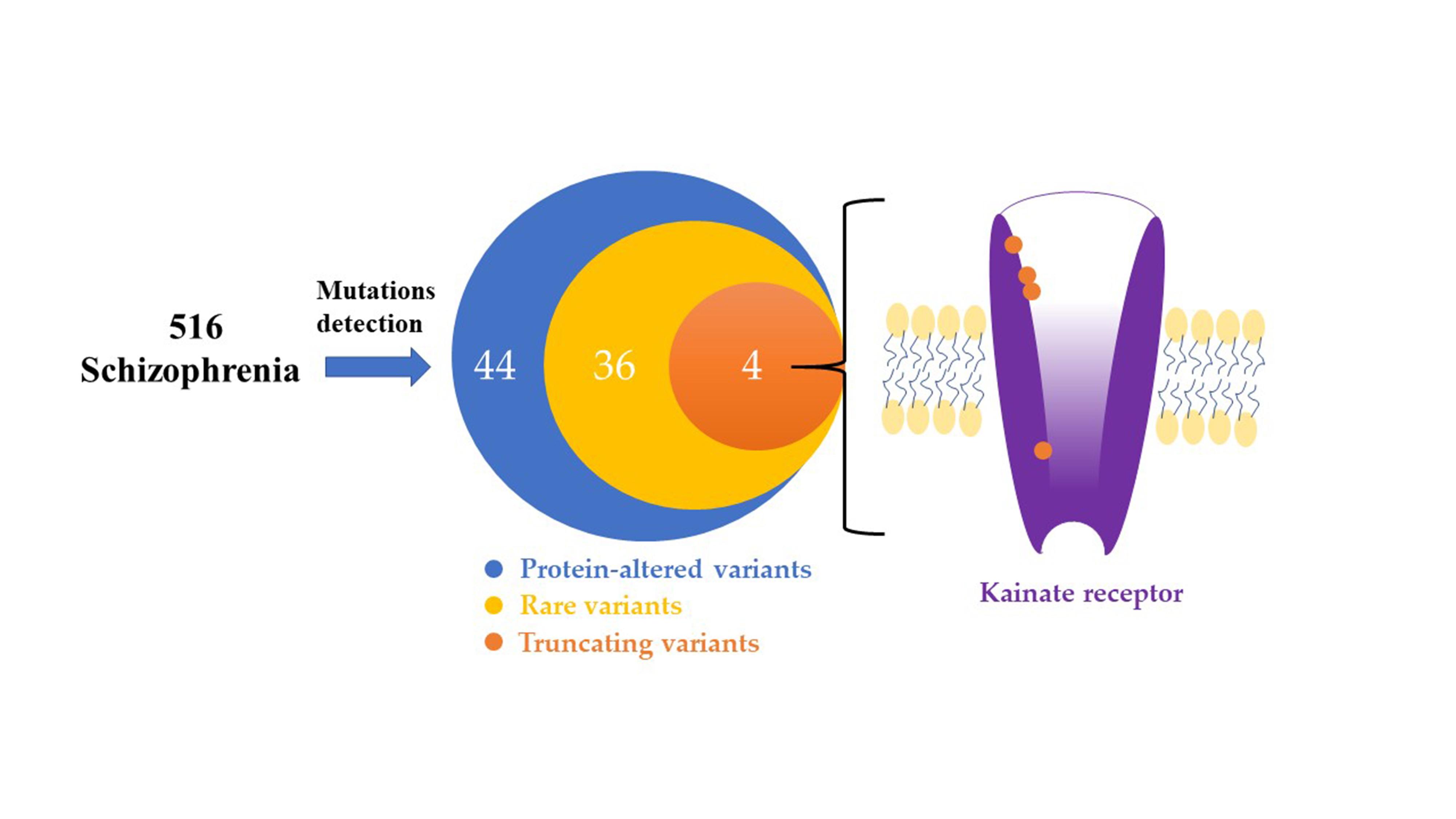
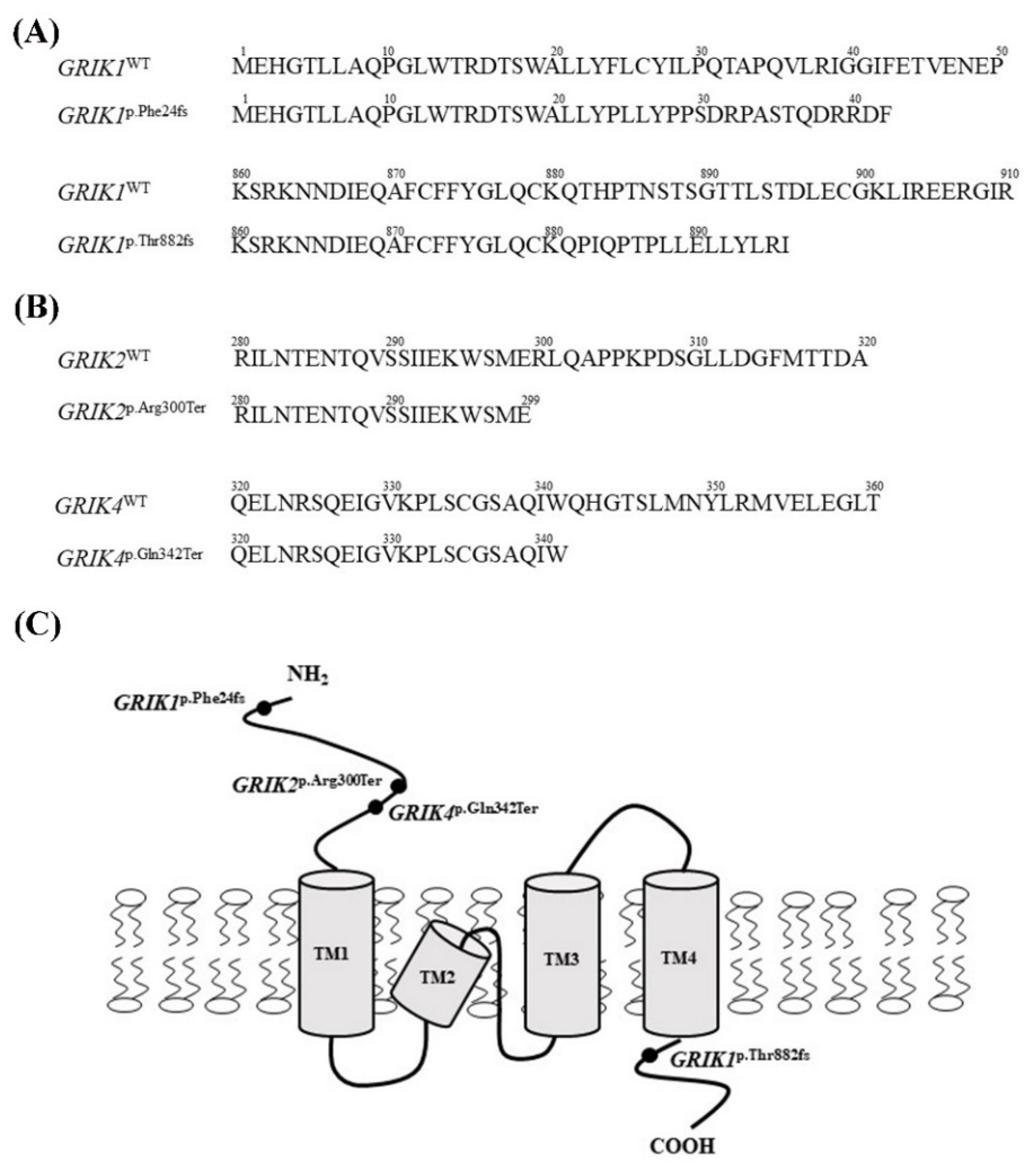
3.1. Identification of the Rare and Protein-Altering Variants of the GRIK Gene Family in Patients with Schizophrenia

We designed 158 amplicons (97.4% overall coverage) to cover the exons of GRIK1, GRIK2, GRIK3, GRIK4, and GRIK5. After resequencing 516 unrelated patients with schizophrenia, 44 protein-altering variants were identified, including 12 in the GRIK1, 5 in the GRIK2, 7 in the GRIK3, 13 in the GRIK4, and 7 in the GRIK5 (Table 1, Supplementary Figure S1). Of these, 36 missense mutations had minor allele frequencies (MAFs) less than 0.5% in the gnomAD database and Taiwan BioBank. Five variants (GRIK1p.Phe24fs, GRIK1p.Arg203Leu, GRIK1p.His336Arg, GRIK4p.Gln342Ter, and GRIK4p.His403Gln) were not observed among 1517 healthy controls from the Taiwan BioBank, 1000 Genomes Project, and the gnomAD database. In silico analysis revealed that the five mutations (GRIK1p.Arg203Leu, GRIK1p.Arg558Gln, GRIK2p.His253Tyr, GRIK4p.Arg507Trp, and GRIK4p.Arg760Pro) were predicted to be pathogenic using the Polyphen-2, SIFT, Pmut, and PROVEAN prediction programs (Table 1, bold text). We identified two heterozygous frameshift deletion mutations (GRIK1p.Phe24fs and GRIK1p.Thr882fs) in two unrelated patients with schizophrenia and two heterozygous nonsense mutations (GRIK2p.Arg300Ter and GRIK4p.Gln342Ter) in two unrelated patients with schizophrenia. The two mutants (GRIK1p.Phe24fs and GRIK1 p.Thr882fs) were predicted to shift the reading frames, resulting in a completely different protein translation from the original GRIK1 protein (Figure 1A), whereas the two mutants (GRIK2p.Arg300Ter and GRIK4p.Gln342Ter) were predicted to introduce a stop codon in the coding region (Figure 1B). Three mutations (GRIK1p.Phe24fs, GRIK2p.Arg300Ter, and GRIK4p.Gln342Ter) resulted in a truncated protein lacking a predicted gene transmembrane domain of the kainate receptor (Figure 1C).

3.2. GRIK Gene Family mRNA Expressions in Peripheral Blood Cells, Lymphoblastoid Cells, and Human Brain

We measured the expression of the GRIK1, GRIK2, GRIK3, GRIK4, and GRIK5 genes in peripheral blood cells and lymphoblastoid cells of a non-psychotic control and human brain specimens using semi-quantitative RT-PCR. The GRIK1, GRIK2, GRIK3, GRIK4, and GRIK5 genes were detected in human brain tissue, but not significantly expressed in peripheral blood cells and lymphoblastoid cells compared with human brain tissue (Supplementary Figure S2).

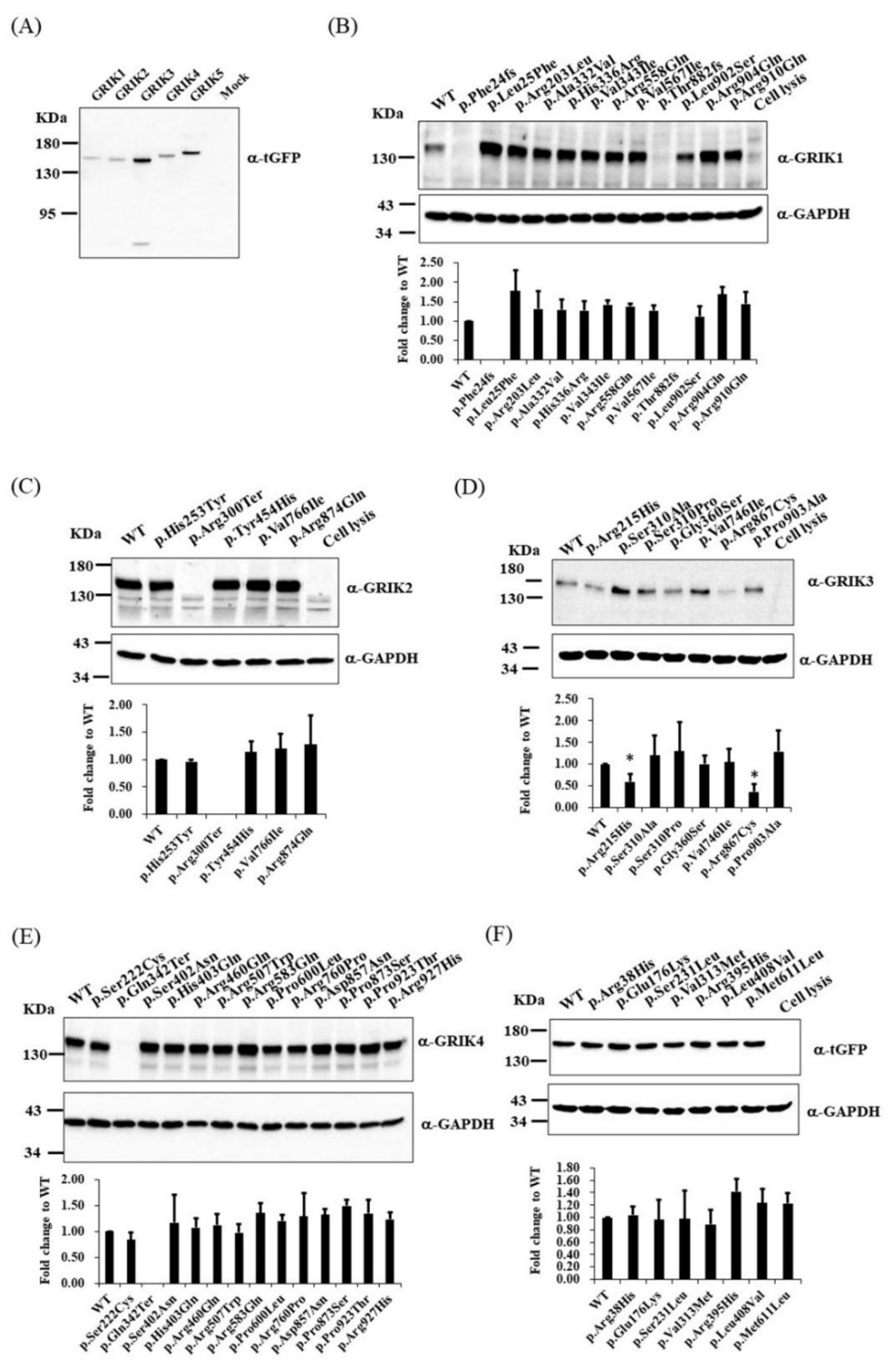
3.3. Immunoblotting and Immunocytochemistry Analysis of GRIK Gene Family Mutants in Cultured Cells

To understand the effects of protein-altering mutations on GRIK protein expression and translocation, we examined the expression and subcellular localization of the wild-type and mutant GRIK proteins in HEK-293 cells. We cloned the wild-type cDNAs of the GRIK gene family into a mammalian vector with a C-terminal tGFP tag, pCMV6-AC-GFP. Immunoblotting was used to confirm GRIK gene family protein overexpression in lysates using anti-tGFP (Figure 2A). We determined the effects of GRIK gene family mutants on protein expression using an immunoblot assay. HEK-293 cells were transfected with wild-type (WT) or mutant constructs. After 24 h of transfection, the four mutant-type constructs (GRIK1p.Phe24fs, GRIK1p.Thr882fs, GRIK2p.Arg300Ter, and GRIK4p.Gln342Ter) did not express GRIK-tGFP fusion proteins in HEK-293 cells compared with the WT constructs (Figure 2B,C,E). Immunoblotting of the cell lysates from the HEK-293 cells carrying GRIK3p.Arg215His and GRIK3p.Thr867Cys revealed significantly decreased GRIK3-tGFP fusion proteins in the mutants relative to the WT cells (Figure 2D). HEK-293 cells carrying GRIK5 mutants demonstrated similar levels of GRIK5-tGFP fusion protein expression as the WT cells (Figure 2F). Immunocytochemical analysis revealed the four mutant-type constructs (GRIK1p.Phe24fs, GRIK1p.Thr882fs, GRIK2p.Arg300Ter, and GRIK4p.Gln342Ter) did not express the recombinant protein in HEK-293 cells (Supplementary Figure S3).

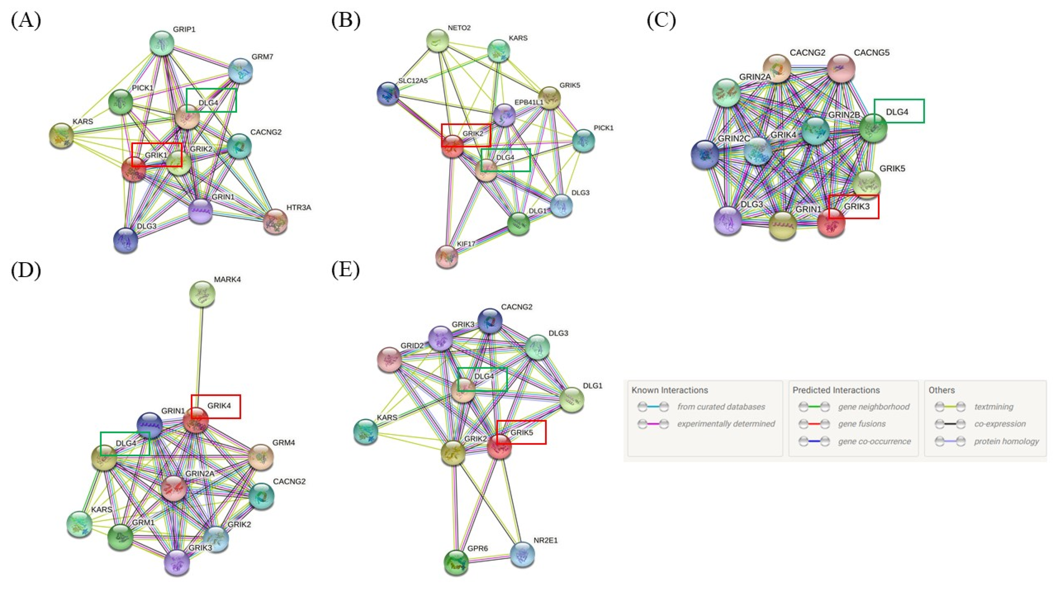
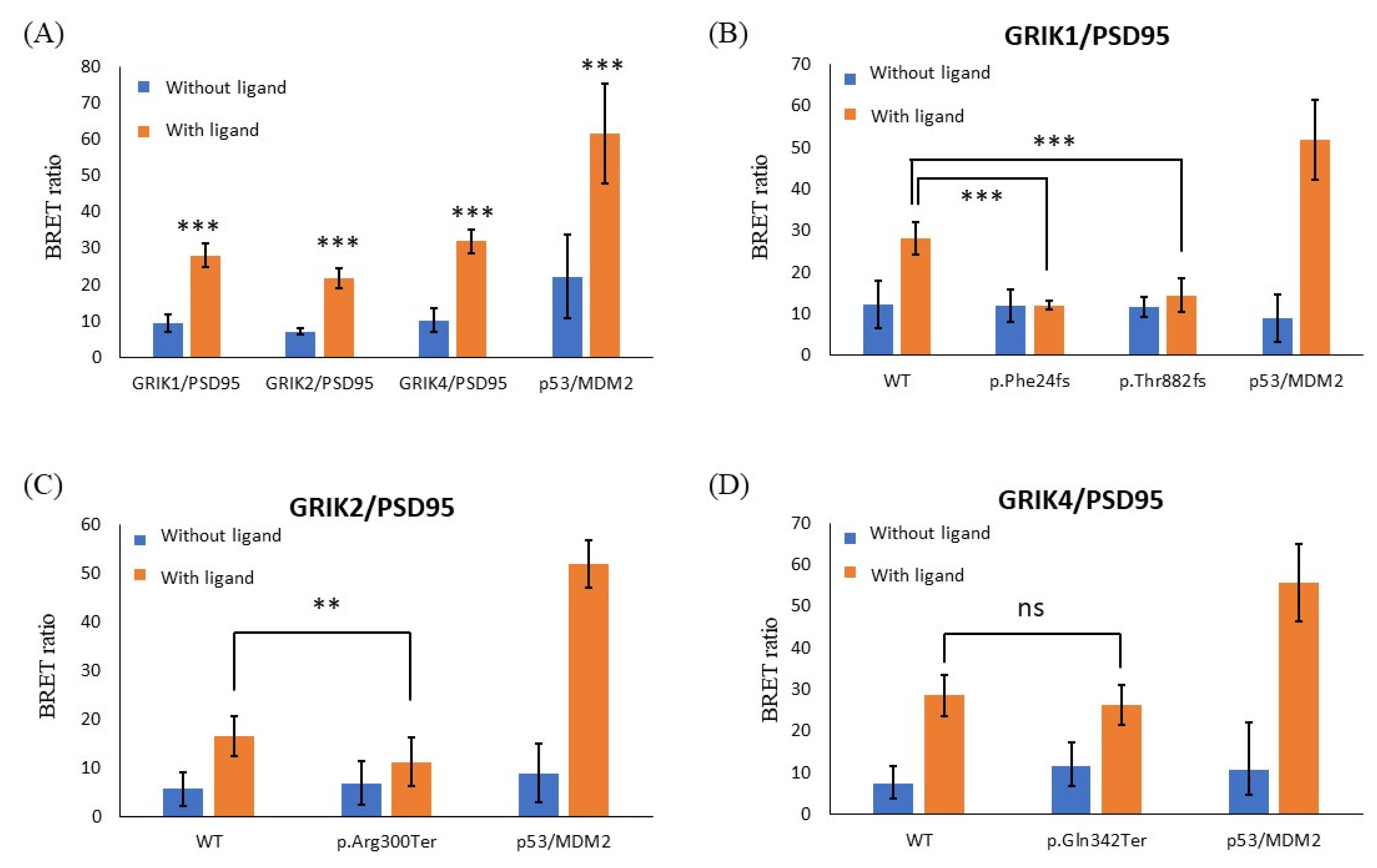
3.4. Protein-Truncating Mutations in the GRIK Gene Family Disrupt the Interaction with the Post-Synaptic Density Protein 95 (PSD95) Protein

The STRING database was used to identify interactions of the GRIK gene family and to map the protein–protein interaction network with an integrated confidence score of 0.400. The STRING results revealed that all GRIK gene members (red frame) interact with the DLG4 gene encoding the PSD95 protein (green frame, Figure 3). The PSD95 protein was used for in vitro protein–protein interaction analysis based on the STRING results and a literature review.
NanoBRET assay used a NanoLuc® fusion protein as the energy donor and a fluorescently labeled HaloTag™ fusion protein as the energy acceptor. First, we used the NanoBRET assay to detect GRIK gene family/PSD95 interactions in live cells. We found that GRIK1, GRIK2, and GRIK4 proteins were associated with PSD95 by measuring the energy transfer from a bioluminescent protein donor to a fluorescent protein acceptor (Figure 4A). Next, we evaluated the impact of four ultrarare truncated GRIK mutations (GRIK1p.Phe24fs, GRIK1p.Thr882fs, GRIK2p.Arg300Ter, and GRIK4p.Gln342Ter) on the interaction among themselves and with the PSD95 protein. We observed that the interactions of the three truncated GRIK mutant proteins (GRIK1p.Phe24fs, GRIK1p.Thr882fs, and GRIK2p.Arg300Ter) were significantly weakened or even abolished with PSD95, whereas the interaction of wild-type GRIK protein with PSD95 protein was preserved (Figure 4B–D).

4. Discussion

We identified 4 truncating mutants and 36 rare missense mutations with MAFs less than 0.5% in the GRIK gene family from 516 patients with schizophrenia, including 5 variants (GRIK1p.Phe24fs, GRIK1p.Arg203Leu, GRIK1p.His336Arg, GRIK4p.Gln342Ter, and GRIK4p.His403Gln) that were not observed among 1517 healthy controls from the Taiwan BioBank, the gnomAD database, and 1000 Genomes Project. We presumed that these are ultrarare variants, which may contribute to the pathogenesis of schizophrenia. To interpret the clinical relevance of the protein-altering mutations, we planned to directly evaluate the expression changes in the GRIK gene family in peripheral blood cells and lymphoblastoid cells of patients. We measured the expression of the GRIK gene family members in peripheral blood cells and lymphoblastoid cells from a non-psychotic control; however, they were not significantly expressed, suggesting that the GRIK gene family is not expressed in these cell types. Thus, we did not perform expression assays in peripheral blood cells and lymphoblastoid cells from patients with schizophrenia. We confirmed the four loss-of-function (LoF) mutants (GRIK1p.Phe24fs, GRIK1p.Thr882fs, GRIK2p.Arg300Ter, and GRIK4p.Gln342Ter) in HEK-293 cells as they did not express GRIK-tGFP fusion proteins compared with the wild-type constructs, and two missense mutants (GRIK3p.Arg215His and GRIK3p.Thr867Cys) were significantly decreased in expression in HEK-293 cells. Notably, the four protein-truncating mutations (GRIK1p.Phe24fs, GRIK1p.Thr882fs, GRIK2p.Arg300Ter, and GRIK4p.Gln342Ter) and two missense mutations (GRIK3p.Arg215His and GRIK3p.Thr867Cys) identified in this study represent ultrarare variants that have not been reported in the literature, to the best of our knowledge. We presumed that these ultrarare mutations impair protein function and alter biological processes of synaptic function in patients with schizophrenia. However, in silico analysis of these two missense mutations (GRIK3p.Arg215His and GRIK3p.Thr867Cys) denied this possibility. Thus, the effects of these two missense mutations on the pathophysiology of schizophrenia still need to be fully elucidated. Given the premature termination of protein synthesis or reduction in the transcript levels by nonsense and frameshift-mediated decay [43,44], the three predicted mutations (GRIK1p.Phe24fs, GRIK2p.Arg300Ter, and GRIK4p.Gln342Ter) lack the adjacent transmembrane domain, which results in a complete loss of function of the kainate receptor. Taken together, we presumed that the rare protein-altering mutations, especially LoF mutations, in genes encoding kainate receptors are associated with the molecular pathology of schizophrenia.
Several synapse-associated proteins, especially PSD95 protein, were identified as interacting proteins for the kainate receptors [45,46,47]. For example, co-immunoprecipitations using rat brain extracts showed that GRIK2 and GRIK5 proteins were associated with the PSD95 protein [46]. In addition, the results from the STRING database indicated that all GRIK gene family members interact with the PSD95 protein. PSD95 is involved in the maturation of synapse formation and genetic studies have indicated that genetic variants of PSD95 are associated with increased susceptibility to schizophrenia [48,49]. Therefore, we speculated that the four LoF mutations (GRIK1p.Phe24fs, GRIK1p.Thr882fs, GRIK2p.Arg300Ter, and GRIK4p.Gln342Ter) that we identified from patients with schizophrenia may affect the binding of the kainate receptors with PSD95 protein. Based on the results from our NanoBRET assay, GRIK1, GRIK2, and GRIK4 bind to PSD95. Moreover, we experimentally confirmed that two frameshift deletion mutations (GRIK1p.Phe24fs and GRIK1p.Thr882fs) and one nonsense mutation (GRIK2p.Arg300Ter) identified in patients with schizophrenia weaken or even disrupt the interaction between GRIK and PSD95 proteins. Thus, we speculated that three out of these four ultrarare LoF mutations (GRIK1p.Phe24fs, GRIK1p.Thr882fs, and GRIK2p.Arg300Ter) may affect the interaction of PSD95 protein with kainate receptors in the brain and consequently affect synaptic function in patients with schizophrenia. Further analysis of these mutations is needed to confirm our hypothesis.
Several studies have demonstrated shared predisposing genetic factors and common mechanisms in neurodevelopmental pathways between different psychiatric disorders [50,51,52]. Cordoba et al. identified a nonsense mutation (GRIK2p.Arg198*) in the GRIK2 gene as a basis for the etiology of intellectual disabilities [53]. In addition, recurrent de novo variants (GRIK2p.Ala657Thr, GRIK2p.Thr660Lys, and GRIK2p.Thr660Arg) in the GRIK2 gene were identified in various neurodevelopmental disorders [54,55]. In addition, several studies found that the GRIK2 gene may be associated with autism spectrum disorder [56,57,58]. For example, Jamain et al. identified a missense mutation, GRIK2p.Met867Ile, in a highly conserved domain of the intracytoplasmic C-terminal region of the GRIK2 protein in patients with autism [58]. A case report of a girl with severe developmental delay revealed a 2.6 Mb microdeletion in 1p34.3 involving the GRIK3 gene [59]. Recently, another study found that damaging coding variants within the kainate receptor ion channel genes confers increased risk for schizophrenia, autism, and intellectual disabilities [60]. In this study, we identified several ultrarare missense variants in the GRIK gene family from patients with schizophrenia and demonstrated altered protein expression and weakened interactions with the PSD95 protein. These studies and our results support the existence of shared biological pathways in schizophrenia, autism, and intellectual disability.
In summary, the GRIK gene family may harbor ultrarare variants, particularly those deleterious to protein expression in certain patients with schizophrenia, supporting the idea that rare coding variants may contribute to the genetic architecture of schizophrenia. Furthermore, identifying proteins that interact with kainate receptors is essential to unravel kainate-receptor-mediated signaling in the brain. Future proteomic studies using cell models carrying these rare deleterious mutations are necessary to understand the role of kainate-receptor-interacting proteins and how they contribute to the etiology of schizophrenia.

Supplementary Materials

The following supporting information can be downloaded at: https://www.mdpi.com/article/10.3390/jpm12050783/s1, Supplementary Table S1: Primer sequences of semi-qPCR and size of PCR products; Supplementary Figure S1: Sequence electropherograms of the protein-altering variants of the GRIK gene family found in patients with schizophrenia; Supplementary Figure S2: The mRNA expression of the GRIK1, GRIK2, GRIK3, GRIK4, and GRIK5 gene in peripheral blood cells, lymphoblastoid cells, and the human brain; Supplementary Figure S3: Localization analysis of GRIK gene family mutants in HEK-293 cells.

Author Contributions

Conceptualization, M.-C.C. and T.-M.H.; methodology, M.-C.C., T.-M.H., C.-L.W., S.-H.H., H.-Y.T., and F.-Y.C.; formal analysis, M.-C.C., S.-H.H., H.-Y.T., and F.-Y.C.; investigation, M.-C.C., C.-L.W., S.-H.H., H.-Y.T., and F.-Y.C.; resources, M.-C.C. and T.-M.H.; writing—original draft preparation, M.-C.C.; writing—review and editing, M.-C.C.; funding acquisition, M.-C.C. All authors have read and agreed to the published version of the manuscript.

Funding

This research was funded by the Ministry of Science and Technology, Taiwan, grant number MOST107-2314-B-480-001 and MOST108-2314-B-480-001-MY2, and the Yuli Branch, Taipei Veterans General Hospital, Taiwan, grant number VHYL110-008.

Institutional Review Board Statement

The study was conducted in accordance with the Declaration of Helsinki, and approved by the Institutional Review Board of the Antai Medical Care Cooperation Antai-Tian-Sheng Memorial Hospital (Approval number: 18-144-A).

Informed Consent Statement

Informed consent was obtained from all subjects involved in the study.

Data Availability Statement

The raw data are available upon request of the corresponding author.

Conflicts of Interest

The authors declare no conflict of interest. The funders had no role in the design of the study; in the collection, analyses, or interpretation of data; in the writing of the manuscript, or in the decision to publish the results.

References

  1. Freedman, R. Schizophrenia. N. Engl. J. Med. 2003, 349, 1738–1749. [Google Scholar] [CrossRef] [PubMed]
  2. Weinberger, D.R. Implications of normal brain development for the pathogenesis of schizophrenia. Arch. Gen. Psychiatry 1987, 44, 660–669. [Google Scholar] [CrossRef] [PubMed]
  3. McGlashan, T.H.; Hoffman, R.E. Schizophrenia as a disorder of developmentally reduced synaptic connectivity. Arch. Gen. Psychiatry 2000, 57, 637–648. [Google Scholar] [CrossRef] [PubMed]
  4. Stephan, K.E.; Baldeweg, T.; Friston, K.J. Synaptic plasticity and dysconnection in schizophrenia. Biol. Psychiatry 2006, 59, 929–939. [Google Scholar] [CrossRef] [PubMed]
  5. Fatemi, S.H.; Folsom, T.D. The neurodevelopmental hypothesis of schizophrenia, revisited. Schizophr. Bull. 2009, 35, 528–548. [Google Scholar] [CrossRef]
  6. van Berlekom, A.B.; Muflihah, C.H.; Snijders, G.; MacGillavry, H.D.; Middeldorp, J.; Hol, E.M.; Kahn, R.S.; de Witte, L.D. Synapse pathology in schizophrenia: A meta-analysis of postsynaptic elements in postmortem brain studies. Schizophr. Bull. 2020, 46, 374–386. [Google Scholar] [CrossRef] [Green Version]
  7. Belsham, B. Glutamate and its role in psychiatric illness. Hum. Psychopharmacol. 2001, 16, 139–146. [Google Scholar] [CrossRef]
  8. Traynelis, S.F.; Wollmuth, L.P.; McBain, C.J.; Menniti, F.S.; Vance, K.M.; Ogden, K.K.; Hansen, K.B.; Yuan, H.; Myers, S.J.; Dingledine, R. Glutamate receptor ion channels: Structure, regulation, and function. Pharmacol. Rev. 2010, 62, 405–496. [Google Scholar] [CrossRef] [Green Version]
  9. Bortolotto, Z.A.; Clarke, V.R.; Delany, C.M.; Parry, M.C.; Smolders, I.; Vignes, M.; Ho, K.H.; Miu, P.; Brinton, B.T.; Fantaske, R.; et al. Kainate receptors are involved in synaptic plasticity. Nature 1999, 402, 297–301. [Google Scholar] [CrossRef]
  10. Hollmann, M.; Heinemann, S. Cloned glutamate receptors. Annu. Rev. Neurosci. 1994, 17, 31–108. [Google Scholar] [CrossRef]
  11. Collingridge, G.L.; Olsen, R.W.; Peters, J.; Spedding, M. A nomenclature for ligand-gated ion channels. Neuropharmacology 2009, 56, 2–5. [Google Scholar] [CrossRef] [PubMed] [Green Version]
  12. Kamboj, R.K.; Schoepp, D.D.; Nutt, S.; Shekter, L.; Korczak, B.; True, R.A.; Rampersad, V.; Zimmerman, D.M.; Wosnick, M.A. Molecular cloning, expression, and pharmacological characterization of humEAA1, a human kainate receptor subunit. J. Neurochem. 1994, 62, 1–9. [Google Scholar] [CrossRef] [PubMed]
  13. Lowry, E.R.; Kruyer, A.; Norris, E.H.; Cederroth, C.R.; Strickland, S. The GluK4 kainate receptor subunit regulates memory, mood, and excitotoxic neurodegeneration. Neuroscience 2013, 235, 215–225. [Google Scholar] [CrossRef] [PubMed] [Green Version]
  14. Arora, V.; Pecoraro, V.; Aller, M.I.; Roman, C.; Paternain, A.V.; Lerma, J. Increased Grik4 gene dosage causes imbalanced circuit output and human disease-related behaviors. Cell Rep. 2018, 23, 3827–3838. [Google Scholar] [CrossRef]
  15. Mueller, H.T.; Haroutunian, V.; Davis, K.L.; Meador-Woodruff, J.H. Expression of the ionotropic glutamate receptor subunits and NMDA receptor-associated intracellular proteins in the substantia nigra in schizophrenia. Mol. Brain Res. 2004, 121, 60–69. [Google Scholar] [CrossRef]
  16. Scarr, E.; Beneyto, M.; Meador-Woodruff, J.H.; Dean, B. Cortical glutamatergic markers in schizophrenia. Neuropsychopharmacology 2005, 30, 1521–1531. [Google Scholar] [CrossRef] [Green Version]
  17. Beneyto, M.; Kristiansen, L.V.; Oni-Orisan, A.; McCullumsmith, R.E.; Meador-Woodruff, J.H. Abnormal glutamate receptor expression in the medial temporal lobe in schizophrenia and mood disorders. Neuropsychopharmacology 2007, 32, 1888–1902. [Google Scholar] [CrossRef]
  18. Meador-Woodruff, J.H.; Davis, K.L.; Haroutunian, V. Abnormal kainate receptor expression in prefrontal cortex in schizophrenia. Neuropsychopharmacology 2001, 24, 545–552. [Google Scholar] [CrossRef] [Green Version]
  19. Porter, R.H.; Eastwood, S.L.; Harrison, P.J. Distribution of kainate receptor subunit mrnas in human hippocampus, neocortex and cerebellum, and bilateral reduction of hippocampal GluR6 and KA2 transcripts in schizophrenia. Brain Res. 1997, 751, 217–231. [Google Scholar] [CrossRef]
  20. Sokolov, B.P. Expression of NMDAR1, GluR1, GluR7, and KA1 glutamate receptor mrnas is decreased in frontal cortex of “neuroleptic-free” schizophrenics: Evidence on reversible up-regulation by typical neuroleptics. J. Neurochem. 1998, 71, 2454–2464. [Google Scholar] [CrossRef]
  21. Ibrahim, H.M.; Hogg, A.J., Jr.; Healy, D.J.; Haroutunian, V.; Davis, K.L.; Meador-Woodruff, J.H. Ionotropic glutamate receptor binding and subunit mRNA expression in thalamic nuclei in schizophrenia. Am. J. Psychiatry 2000, 157, 1811–1823. [Google Scholar] [CrossRef] [PubMed]
  22. Garey, L.J.; Von Bussmann, K.A.; Hirsch, S.R. Decreased numerical density of kainate receptor-positive neurons in the orbitofrontal cortex of chronic schizophrenics. Exp. Brain Res. 2006, 173, 234–242. [Google Scholar] [CrossRef] [PubMed]
  23. Benes, F.M.; Todtenkopf, M.S.; Kostoulakos, P. GluR5,6,7 subunit immunoreactivity on apical pyramidal cell dendrites in hippocampus of schizophrenics and manic depressives. Hippocampus 2001, 11, 482–491. [Google Scholar] [CrossRef] [PubMed]
  24. Shibata, H.; Joo, A.; Fujii, Y.; Tani, A.; Makino, C.; Hirata, N.; Kikuta, R.; Ninomiya, H.; Tashiro, N.; Fukumaki, Y. Association study of polymorphisms in the glur5 kainate receptor gene (GRIK1) with schizophrenia. Psychiatr. Genet. 2001, 11, 139–144. [Google Scholar] [CrossRef] [PubMed]
  25. Hirata, Y.; Zai, C.C.; Souza, R.P.; Lieberman, J.A.; Meltzer, H.Y.; Kennedy, J.L. Association study of GRIK1 gene polymorphisms in schizophrenia: Case-control and family-based studies. Hum. Psychopharmacol. 2012, 27, 345–351. [Google Scholar] [CrossRef]
  26. Shibata, H.; Aramaki, T.; Sakai, M.; Ninomiya, H.; Tashiro, N.; Iwata, N.; Ozaki, N.; Fukumaki, Y. Association study of polymorphisms in the glur7, ka1 and ka2 kainate receptor genes (GRIK3, GRIK4, GRIK5) with schizophrenia. Psychiatry Res. 2006, 141, 39–51. [Google Scholar] [CrossRef]
  27. Li, Z.; He, Z.; Tang, W.; Tang, R.; Huang, K.; Xu, Z.; Xu, Y.; Li, L.; Li, X.; Feng, G.; et al. No genetic association between polymorphisms in the kainate-type glutamate receptor gene, GRIK4, and schizophrenia in the chinese population. Prog. Neuropsychopharmacol. Biol. Psychiatry 2008, 32, 876–880. [Google Scholar] [CrossRef]
  28. Ren, D.; Xu, F.; Bi, Y.; Niu, W.; Zhang, R.; Hu, J.; Guo, Z.; Cao, Y.; Huang, X.; Wu, X.; et al. No association of GRIK4 polymorphisms with schizophrenia in the chinese han population. Psychiatr. Genet. 2017, 27, 159–160. [Google Scholar] [CrossRef]
  29. Shibata, H.; Shibata, A.; Ninomiya, H.; Tashiro, N.; Fukumaki, Y. Association study of polymorphisms in the glur6 kainate receptor gene (GRIK2) with schizophrenia. Psychiatry Res. 2002, 113, 59–67. [Google Scholar] [CrossRef]
  30. Lai, I.C.; Liou, Y.J.; Chen, J.Y.; Wang, Y.C. No association between the ionotropic glutamate receptor kainate 3 gene ser310ala polymorphism and schizophrenia. Neuropsychobiology 2005, 51, 211–213. [Google Scholar] [CrossRef]
  31. Begni, S.; Popoli, M.; Moraschi, S.; Bignotti, S.; Tura, G.B.; Gennarelli, M. Association between the ionotropic glutamate receptor kainate 3 (GRIK3) ser310ala polymorphism and schizophrenia. Mol. Psychiatry 2002, 7, 416–418. [Google Scholar] [CrossRef] [PubMed] [Green Version]
  32. Djurovic, S.; Kahler, A.K.; Kulle, B.; Jonsson, E.G.; Agartz, I.; Le Hellard, S.; Hall, H.; Jakobsen, K.D.; Hansen, T.; Melle, I.; et al. A possible association between schizophrenia and GRIK3 polymorphisms in a multicenter sample of scandinavian origin (SCOPE). Schizophr. Res. 2009, 107, 242–248. [Google Scholar] [CrossRef] [PubMed]
  33. Pickard, B.S.; Malloy, M.P.; Christoforou, A.; Thomson, P.A.; Evans, K.L.; Morris, S.W.; Hampson, M.; Porteous, D.J.; Blackwood, D.H.; Muir, W.J. Cytogenetic and genetic evidence supports a role for the kainate-type glutamate receptor gene, GRIK4, in schizophrenia and bipolar disorder. Mol. Psychiatry 2006, 11, 847–857. [Google Scholar] [CrossRef] [PubMed] [Green Version]
  34. Greenwood, T.A.; Lazzeroni, L.C.; Calkins, M.E.; Freedman, R.; Green, M.F.; Gur, R.E.; Gur, R.C.; Light, G.A.; Nuechterlein, K.H.; Olincy, A.; et al. Genetic assessment of additional endophenotypes from the consortium on the genetics of schizophrenia family study. Schizophr. Res. 2016, 170, 30–40. [Google Scholar] [CrossRef] [Green Version]
  35. McClellan, J.; King, M.C. Genomic analysis of mental illness: A changing landscape. JAMA 2010, 303, 2523–2524. [Google Scholar] [CrossRef]
  36. Purcell, S.M.; Wray, N.R.; Stone, J.L.; Visscher, P.M.; O’Donovan, M.C.; Sullivan, P.F.; Sklar, P. Common polygenic variation contributes to risk of schizophrenia and bipolar disorder. Nature 2009, 460, 748–752. [Google Scholar]
  37. Kirov, G.; Pocklington, A.J.; Holmans, P.; Ivanov, D.; Ikeda, M.; Ruderfer, D.; Moran, J.; Chambert, K.; Toncheva, D.; Georgieva, L.; et al. De novo cnv analysis implicates specific abnormalities of postsynaptic signalling complexes in the pathogenesis of schizophrenia. Mol. Psychiatry 2012, 17, 142–153. [Google Scholar] [CrossRef] [Green Version]
  38. Gauthier, J.; Champagne, N.; Lafrenière, R.G.; Xiong, L.; Spiegelman, D.; Brustein, E.; Lapointe, M.; Peng, H.; Côté, M.; Noreau, A.; et al. De novo mutations in the gene encoding the synaptic scaffolding protein SHANK3 in patients ascertained for schizophrenia. Proc. Natl. Acad. Sci. USA 2010, 107, 7863–7868. [Google Scholar] [CrossRef] [Green Version]
  39. Genovese, G.; Fromer, M.; Stahl, E.A.; Ruderfer, D.M.; Chambert, K.; Landen, M.; Moran, J.L.; Purcell, S.M.; Sklar, P.; Sullivan, P.F.; et al. Increased burden of ultra-rare protein-altering variants among 4,877 individuals with schizophrenia. Nat. Neurosci. 2016, 19, 1433–1441. [Google Scholar] [CrossRef]
  40. Fromer, M.; Pocklington, A.J.; Kavanagh, D.H.; Williams, H.J.; Dwyer, S.; Gormley, P.; Georgieva, L.; Rees, E.; Palta, P.; Ruderfer, D.M.; et al. De novo mutations in schizophrenia implicate synaptic networks. Nature 2014, 506, 179–184. [Google Scholar] [CrossRef] [Green Version]
  41. Hu, T.M.; Wang, Y.C.; Wu, C.L.; Hsu, S.H.; Tsai, H.Y.; Cheng, M.C. Multiple rare risk coding variants in postsynaptic density-related genes associated with schizophrenia susceptibility. Front. Genet. 2020, 11, 524258. [Google Scholar] [CrossRef] [PubMed]
  42. Hu, T.M.; Chen, S.J.; Hsu, S.H.; Cheng, M.C. Functional analyses and effect of DNA methylation on the EGR1 gene in patients with schizophrenia. Psychiatry Res. 2019, 275, 276–282. [Google Scholar] [CrossRef]
  43. Khajavi, M.; Inoue, K.; Lupski, J.R. Nonsense-mediated mRNA decay modulates clinical outcome of genetic disease. Eur. J. Hum. Genet. 2006, 14, 1074–1081. [Google Scholar] [CrossRef] [PubMed] [Green Version]
  44. Hentze, M.W.; Kulozik, A.E. A perfect message: RNA surveillance and nonsense-mediated decay. Cell 1999, 96, 307–310. [Google Scholar] [CrossRef] [Green Version]
  45. Mehta, S.; Wu, H.; Garner, C.C.; Marshall, J. Molecular mechanisms regulating the differential association of kainate receptor subunits with SAP90/PSD-95 and SAP97. J. Biol. Chem. 2001, 276, 16092–16099. [Google Scholar] [CrossRef] [PubMed] [Green Version]
  46. Garcia, E.P.; Mehta, S.; Blair, L.A.; Wells, D.G.; Shang, J.; Fukushima, T.; Fallon, J.R.; Garner, C.C.; Marshall, J. SAP90 binds and clusters kainate receptors causing incomplete desensitization. Neuron 1998, 21, 727–739. [Google Scholar] [CrossRef] [Green Version]
  47. Suzuki, E.; Kamiya, H. PSD-95 regulates synaptic kainate receptors at mouse hippocampal mossy fiber-CA3 synapses. Neurosci. Res. 2016, 107, 14–19. [Google Scholar] [CrossRef] [Green Version]
  48. Xing, J.; Kimura, H.; Wang, C.; Ishizuka, K.; Kushima, I.; Arioka, Y.; Yoshimi, A.; Nakamura, Y.; Shiino, T.; Oya-Ito, T.; et al. Resequencing and association analysis of six PSD-95-related genes as possible susceptibility genes for schizophrenia and autism spectrum disorders. Sci. Rep. 2016, 6, 27491. [Google Scholar] [CrossRef]
  49. Cheng, M.C.; Lu, C.L.; Luu, S.U.; Tsai, H.M.; Hsu, S.H.; Chen, T.T.; Chen, C.H. Genetic and functional analysis of the DLG4 gene encoding the post-synaptic density protein 95 in schizophrenia. PLoS ONE 2010, 5, e15107. [Google Scholar] [CrossRef]
  50. Lee, S.H.; Ripke, S.; Neale, B.M.; Faraone, S.V.; Purcell, S.M.; Perlis, R.H.; Mowry, B.J.; Thapar, A.; Goddard, M.E.; Witte, J.S.; et al. Genetic relationship between five psychiatric disorders estimated from genome-wide SNPs. Nat. Genet. 2013, 45, 984–994. [Google Scholar]
  51. Waltereit, R.; Banaschewski, T.; Meyer-Lindenberg, A.; Poustka, L. Interaction of neurodevelopmental pathways and synaptic plasticity in mental retardation, autism spectrum disorder and schizophrenia: Implications for psychiatry. World J. Biol. Psychiatry 2014, 15, 507–516. [Google Scholar] [CrossRef] [PubMed]
  52. Rees, E.; Creeth, H.D.J.; Hwu, H.G.; Chen, W.J.; Tsuang, M.; Glatt, S.J.; Rey, R.; Kirov, G.; Walters, J.T.R.; Holmans, P.; et al. Schizophrenia, autism spectrum disorders and developmental disorders share specific disruptive coding mutations. Nat. Commun. 2021, 12, 5353. [Google Scholar] [CrossRef] [PubMed]
  53. Cordoba, M.; Rodriguez, S.; Moron, D.G.; Medina, N.; Kauffman, M.A. Expanding the spectrum of Grik2 mutations: Intellectual disability, behavioural disorder, epilepsy and dystonia. Clin. Genet. 2015, 87, 293–295. [Google Scholar] [CrossRef] [PubMed]
  54. Guzman, Y.F.; Ramsey, K.; Stolz, J.R.; Craig, D.W.; Huentelman, M.J.; Narayanan, V.; Swanson, G.T. A gain-of-function mutation in the GRIK2 gene causes neurodevelopmental deficits. Neurol Genet. 2017, 3, e129. [Google Scholar] [CrossRef] [Green Version]
  55. Stolz, J.R.; Foote, K.M.; Veenstra-Knol, H.E.; Pfundt, R.; Ten Broeke, S.W.; de Leeuw, N.; Roht, L.; Pajusalu, S.; Part, R.; Rebane, I.; et al. Clustered mutations in the GRIK2 kainate receptor subunit gene underlie diverse neurodevelopmental disorders. Am. J. Hum. Genet. 2021, 108, 2206. [Google Scholar] [CrossRef]
  56. Griswold, A.J.; Ma, D.; Cukier, H.N.; Nations, L.D.; Schmidt, M.A.; Chung, R.H.; Jaworski, J.M.; Salyakina, D.; Konidari, I.; Whitehead, P.L.; et al. Evaluation of copy number variations reveals novel candidate genes in autism spectrum disorder-associated pathways. Hum. Mol. Genet. 2012, 21, 3513–3523. [Google Scholar] [CrossRef] [Green Version]
  57. Casey, J.P.; Magalhaes, T.; Conroy, J.M.; Regan, R.; Shah, N.; Anney, R.; Shields, D.C.; Abrahams, B.S.; Almeida, J.; Bacchelli, E.; et al. A novel approach of homozygous haplotype sharing identifies candidate genes in autism spectrum disorder. Hum. Genet. 2012, 131, 565–579. [Google Scholar] [CrossRef] [Green Version]
  58. Jamain, S.; Betancur, C.; Quach, H.; Philippe, A.; Fellous, M.; Giros, B.; Gillberg, C.; Leboyer, M.; Bourgeron, T. Linkage and association of the glutamate receptor 6 gene with autism. Mol. Psychiatry 2002, 7, 302–310. [Google Scholar] [CrossRef] [Green Version]
  59. Takenouchi, T.; Hashida, N.; Torii, C.; Kosaki, R.; Takahashi, T.; Kosaki, K. 1p34.3 deletion involving GRIK3: Further clinical implication of grik family glutamate receptors in the pathogenesis of developmental delay. Am. J. Med. Genet. A 2014, 164, 456–460. [Google Scholar] [CrossRef]
  60. Koromina, M.; Flitton, M.; Blockley, A.; Mellor, I.R.; Knight, H.M. Damaging coding variants within kainate receptor channel genes are enriched in individuals with schizophrenia, autism and intellectual disabilities. Sci. Rep. 2019, 9, 19215. [Google Scholar] [CrossRef]
Figure 1. Four schizophrenia-associated and rare protein-truncating mutations identified in this study. (A) Schematic representation of the two frameshift mutants (GRIK1p.Phe24fs and GRIK1p.Thr882fs) of the GRIK1. (B) Schematic representation of the two nonsense mutants (GRIK2p.Arg300Ter and GRIK4p.Gln342Ter) in GRIK2 and the GRIK4, respectively. (C) Schematic representation of the kainate receptor structure and locations of the four rare protein-truncating mutations of the GRIK gene family. TM, transmembrane.
Jpm 12 00783 g001
Figure 2. Immunoblotting of lysates extracted from HEK-293 transiently co-transfected with plasmids expressing either WT GRIK cDNA or mutant cDNAs. (A) Immunoblotting validated GRIK gene family protein overexpression in lysates using anti-tGFP. (B) GRIK1 WT versus 12 mutants. (C) GRIK2 WT versus five mutants. (D) GRIK3 WT versus seven mutants. (E) GRIK4 WT versus 13 mutants. (F) GRIK5 WT versus seven mutants. For normalization, the lysates were analyzed in parallel by anti-GAPDH immunoblotting. The results are representative of three independent experiments. WT, wild-type. Graphs represent means ± standard deviation. * p < 0.05.
Jpm 12 00783 g002
Figure 3. Graphical representation of the GRIK gene family network. (A) GRIK1: number of nodes: 11; PPI enrichment p-value: 5.66 × 10⁻¹⁴. (B) GRIK2: number of nodes: 11; PPI enrichment p-value: 7.15 × 10⁻¹¹. (C) GRIK3: number of nodes: 11; PPI enrichment p-value: <1.0 × 10⁻¹⁶. (D) GRIK4: number of nodes: 11; PPI enrichment p-value: 3.79 × 10⁻¹². (E) GRIK5: number of nodes: 11; PPI enrichment p-value: 8.92 × 10⁻¹⁰.
Jpm 12 00783 g003
Figure 4. NanoBRET assay to detect the interaction between GRIK and PSD95 protein in live cells. (A) GRIK1, GRIK2, and GRIK4 proteins were associated with PSD95 protein by measuring energy transfer from a bioluminescent protein donor to a fluorescent protein acceptor. The p53/MDM2 was a positive control. Differences in BRET ratio between with and without NanoBRET™ HaloTag® 618 Ligands were assessed using Student’s t-tests. (B,C) The three mutants (GRIK1p.Phe24fs, GRIK1p.Thr882fs, and GRIK2p.Arg300Ter) significantly weakened or even abolished the interaction among themselves and with the PSD95 protein, whereas the interaction of GRIK WT proteins with PSD95 was preserved. (D) The interactions of GRIK4 WT and mutant-type (GRIK4p.Gln342Ter) proteins with PSD95 were preserved. Graphs represent means ± standard deviation. *** p < 0.001, ** p < 0.01, ns means nonsignificant.
Jpm 12 00783 g004
Table 1. Bioinformatic analysis of the protein-altering mutations identified from 516 patients with schizophrenia in the semiconductor sequencing stage.
Genomic
Position
Variant (Amino Acid Change)Patient NumberdbSNP IDMAFIn Silico Analysis for Amino Acid Substitution
gnomAD (Non-Neuro) 1000GTaiwan BiobankPolyphen-2SIFTPmutPROVEANCADD Raw/PHRED
GRIK1
chr21:31311747c.70_71delTT (p.Phe24fs)1N/RN/RN/RN/RN/AN/AN/AN/AN/A
chr21:31311746c.73C > T (p.Leu25Phe)2rs2002620140.00020.00080.0011BenignToleratedNeutralNeutral2.15/21.1
chr21:31045421c.608G > T (p.Arg203Leu)1N/RN/RN/RN/RProbably damagingDamagingDiseaseDeleterious4.27/30
chr21:31015249c.995C > T (p.Ala332Val)1rs1432521170.00010.00040.0007BenignDamagingDiseaseDeleterious3.56/25.2
chr21:31015237c.1007A > G (p.His336Arg)1rs756781607N/RN/RN/RBenignToleratedNeutralNeutral2.51/22.6
chr21:31015217c.1027G > A (p.Val343Ile)1rs1459657790<0.0001N/RN/RBenignToleratedNeutralNeutral2.33/22.1
chr21:30959806c.1673G > A (p.Arg558Gln)1rs776162409<0.0001N/RN/RProbably damagingDamagingDiseaseDeleterious3.90/26.9
chr21:30959780c.1699G > A (p.Val567Ile)1rs5368901890.00010.0006N/RProbably damagingDamagingDiseaseNeutral3.63/25.5
chr21:30925988c.2644delA (p.Thr882fs)1rs867634999<0.0001N/RN/RN/AN/AN/AN/AN/A
chr21:30925928c.2705T > C (p.Leu902Ser)41rs3635040.06940.10980.0639BenignToleratedNeutralNeutral2.00/19.76
chr21:30925922c.2711G > A (p.Arg904Gln)1rs769191901<0.0001N/RN/RBenignDamagingNeutralNeutral2.62/22.8
chr21:30925904c.2729G > A (p.Arg910Gln)3rs3721064160.00020.00020.0023BenignToleratedNeutralNeutral2.55/22.6
GRIK2
chr6:102130461c.757C > T (p.His253Tyr)4rs770586258<0.0001N/R0.0007Probably damagingDamagingDiseaseDeleterious4.09/28.5
chr6:102134175c.898C > T (p.Arg300Ter)1rs1351417917<0.0001N/RN/RN/AN/AN/AN/AN/A
chr6:102307204c.1360T > C (p.Tyr454His)8rs186727716<0.00010.00060.0020BenignToleratedNeutralNeutral2.29/22
chr6:102483426c.2296G > A (p.Val766Ile)7rs32136080.00340.006390.0026BenignToleratedNeutralNeutral2.23/21.6
chr6:102516280c.2621G > A (p.Arg874Gln)2rs267600750<0.0001N/R0.0003Possibly damagingDamagingNeutralNeutral3.23/24.1
GRIK3
chr1:37337877c.644G > A (p.Arg215His)1rs755366301<0.0001N/RN/RProbably damagingDamagingNeutralDeleterious3.90/26.9
chr1:37325477c.928T > G (p.Ser310Ala)49rs66918400.27470.306110.0455BenignToleratedNeutralNeutral0.54/9.832
chr1:37325477c.928T > C (p.Ser310Pro)2rs66918400.0001N/R0.0017BenignToleratedNeutralNeutral0.60/10.34
chr1:37324735c.1078G > A (p.Gly360Ser)2rs5515259260.00020.00040.0049BenignToleratedNeutralNeutral2.79/23.1
chr1:37271783c.2236G > A (p.Val746Ile)1rs142411639<0.0001N/R0.0003BenignToleratedNeutralNeutral1.41/15.77
chr1:37267613c.2599C > T (p.Arg867Cys)2rs758481140<0.0001N/R0.0003Probably damagingToleratedNeutralDeleterious3.26/24.2
chr1:37267505c.2707C > G (p.Pro903Ala)1rs777066849<0.0001N/R0.0010BenignDamagingNeutralNeutral2.96/23.5
GRIK4
chr11:120702714c.665C > G (p.Ser222Cys)1rs747558889<0.0001N/R0.0004BenignDamagingNeutralDeleterious3.06/23.7
chr11:120744892c.1024C > T (p.Gln342Ter)1N/RN/RN/RN/RN/AN/AN/AN/AN/A
chr11:120769281c.1205G > A (p.Ser402Asn)1rs776598317<0.0001N/RN/RBenignToleratedNeutralNeutral1.82/18.37
chr11:120769285c.1209C > A (p.His403Gln)1N/RN/RN/RN/RBenignToleratedNeutralNeutral0.89/12.66
chr11:120776105c.1379G > A (p.Arg460Gln)1rs777568733<0.0001N/R0.0003BenignDamagingNeutralNeutral2.16/21.1
chr11:120811098c.1519C > T (p.Arg507Trp)1rs747648319<0.0001N/R0.0003Probably damagingDamagingDiseaseDeleterious4.41/29.0
chr11:120827536c.1748G > A (p.Arg583Gln)1rs367593579<0.0001N/R0.0003Probably damagingToleratedNeutralNeutral3.79/26.2
chr11:120827587c.1799C > T (p.Pro600Leu)1rs766921998<0.0001N/RN/RProbably damagingToleratedDiseaseDeleterious4.16/29.2
chr11:120837916c.2279G > C (p.Arg760Pro)1rs199658262<0.00010.0002N/RProbably damagingDamagingDiseaseDeleterious4.42/32
chr11:120856667c.2569G > A (p.Asp857Asn)8rs5365589550.00040.00080.0100BenignToleratedNeutralNeutral2.87/23.3
chr11:120856715c.2617C > T (p.Pro873Ser)3rs1485463780<0.0001N/R0.0005BenignToleratedNeutralNeutral1.92/19.14
chr11:120856865c.2767C > A (p.Pro923Thr)1rs772831685N/RN/R0.0005Possibly damagingToleratedNeutralNeutral2.87/23.3
chr11:120856878c.2780G > A (p.Arg927His)2rs537835045<0.00010.00020.0015Probably damagingDamagingNeutralNeutral4.45/32
GRIK5
chr19:42569506c.113G > A (p.Arg38His)3rs1430682690.00040.0004N/RProbably damagingDamagingNeutralNeutral4.13/28.9
chr19:42563662c.526G > A (p.Glu176Lys)3rs2017242970.00020.00100.0030BenignToleratedNeutralNeutral3.04/23.7
chr19:42561126c.692C > T (p.Ser231Leu)1rs557315821<0.00010.00020.0023Probably damagingToleratedNeutralNeutral2.83/23.2
chr19:42558591c.937G > A (p.Val313Met)8rs746539777<0.0001N/R0.0023Probably damagingToleratedNeutralNeutral3.18/24.0
chr19:42557839c.1184G > A (p.Arg395His)2rs1387591620.00030.00060.0010Possibly damagingToleratedNeutralNeutral2.97/23.5
chr19:42557801c.1222C > G (p.Leu408Val)1rs749428580<0.0001N/RN/RBenignToleratedNeutralNeutral1.33/15.37
chr19:42525493c.1831A > T (p.Met611Leu)1rs772424091<0.0001N/R0.0010BenignToleratedNeutralNeutral2.27/21.9
Reference version: GRCh37; GRIK1: NM_000830.4; GRIK2: NM_021956.4; GRIK3: NM_000831.3; GRIK4: NM_014619.4; GRIK5: NM_002088.4; N/R: not registered; N/A: not available; Polyphen-2: http://genetics.bwh.harvard.edu/pph2/, accessed on 18 July 2019; SIFT: https://sift.bii.a-star.edu.sg/, accessed on 18 July 2019; Pmut: http://mmb.irbbarcelona.org/PMut/, accessed on 18 July 2019; PROVEAN: http://provean.jcvi.org/index.php, accessed on 18 July 2019; CADD: https://cadd.gs.washington.edu/, accessed on 26 April 2022.
Publisher’s Note: MDPI stays neutral with regard to jurisdictional claims in published maps and institutional affiliations.

