Article

A 3D Coordination Polymer Based on Syn-Anti Bridged [Mn(RCOO)2]n Chains Showing Spin-Canting with High Coercivity and an Ordering Temperature of 14 K

by
Soumen Kumar Dubey
1,
Maxcimilan Patra
1,
Kajal Gupta
1,
Subham Bhattacharjee
1,*,
Rajat Saha
1,2,* and
Carlos J. Gómez-García
2,*
1
Department of Chemistry, Kazi Nazrul University, Asansol 713340, West Bengal, India
2
Departamento de Química Inorgánica, Universidad de Valencia, 46100 Burjasot, Valencia, Spain
*
Authors to whom correspondence should be addressed.
Magnetochemistry 2023, 9(2), 55; https://doi.org/10.3390/magnetochemistry9020055
Submission received: 6 January 2023 / Revised: 2 February 2023 / Accepted: 8 February 2023 / Published: 10 February 2023

Abstract

:
A new 3D manganese(II) coordination polymer, formulated as [Mn3(HL)6] (1) (where H2L = 6-hydroxypicolinic acid), has been hydrothermally synthesized and characterized by single-crystal X-ray crystallographic analysis along with other spectroscopic and magnetic techniques. Structural analysis shows that the compound crystallizes in the monoclinic C2/c space group and is a non-porous 3D coordination polymer formed by three different Mn(II) centres connected by 6-hydroxypicolinic acid ligands in their keto form. Each Mn(II) centre shows a distorted octahedral coordination environment. Neighbouring Mn(II) centres are connected by two different syn-anti bridging carboxylate groups to form regular coordination chains. There are two different [Mn2(RCOO)2] units along the chain, formed by two crystallographically independent Mn centres (Mn1 and Mn2). These chains are further connected by HL ligands to form a 3D coordination network. Interestingly, both the hydroxy and the carboxylate groups of the ligands are deprotonated and coordinated to the metal centres, whereas the pyridyl group is protonated and uncoordinated, although it participates in strong hydrogen bonding interactions with oxygen atoms of the HL ligand, as shown by the Hirshfeld surface analysis. Both the absorption and emission spectra of the compound have also been measured. Variable temperature magnetic studies reveal the presence of a spin-canted antiferromagnetic behaviour with a high coercivity of 40 mT at 2 K and an ordering temperature of 14 K.

Graphical Abstract

1. Introduction

The design of metal-organic coordination polymers has gained much attention for their tuneable structural features and enormous functionalities [1,2,3,4,5]. The search for different molecule-based magnets that show spontaneous magnetization below a critical temperature has also been an attractive field of research [6,7,8,9,10,11]. Thus, many types of magnetic coordination polymers with different topologies and dimensionalities have been developed, and their magnetic properties have been studied [12,13,14,15]. In most cases, mixed N,O-donor ligands are used to create a magnetic exchange pathway between the paramagnetic metal centres in those polymeric materials [16,17,18,19]. Albeit, it is still very difficult to control and predict the geometry and magnetic behaviour of the polymers due to the diverse coordination modes of many ligands and the sensitivity of the magnetic interactions to slight structural modifications [20,21,22]. Among the different strategies used to design magnetic materials, the most successful ones are: (i) the use of isolated magnetic building blocks to design extended structures [23,24], (ii) the use of radical-based bridging ligands [25,26,27], and (iii) the synthesis of magnetic molecules inside the cavity of metal-organic frameworks and covalent organic frameworks [28,29]. Moreover, the presence of three paramagnetic metal centres in the basic building block of homometallic complexes and coordination polymers may induce interesting magnetic properties due to an imbalance of the overall magnetic moments [30,31,32].
Picolinic acids bearing hydroxy groups are very versatile ligands as their coordination behaviour is dependent on their keto-enolic tautomerism [33,34]. In the keto form, they coordinate with the carboxylate and keto groups, while the protonated pyridyl N atom remains uncoordinated [35]. In the enolic form, they can use their three different coordination sites (hydroxy, carboxylate, and pyridyl). In 6-hydroxypicolinic acid, the proximity of the hydroxy group and the basic pyridyl N atom facilitates the migration of the hydroxy proton to the pyridyl N atom, resulting in the deprotonation of the OH group and protonation of the pyridyl N atom (keto form). However, the coordination modes of the ligand and, therefore, the resulting nuclearities and dimensionalities are difficult to control [36]. A search in the CCDC database shows that there is a total of 29 reported structures with the ligand 6-hydroxypicolinic acid. In 16 of these structures, the ligand is totally deprotonated (L2−). In nine structures, the ligand is present as the enol (O-H) tautomer, whereas only in four structures, it presents its keto (N-H) form (Table S1). In the literature, there are some examples of metal complexes with 6-OHpicH with both transition metals and lanthanoids, and most of them are monomer or discrete metal clusters [37,38,39]. In fact, among the 29 structures, there are three with lanthanoids (DyIII, ErIII, and YbIII), two with alkaline metals (Cs+ and Na+), and 24 with transition (MnIII, CoII, NiII, CuII, RuII, ReII, ReIII, ReIV, and ReV) and post-transition (ZnII and CdII) metal ions. Surprisingly, there is no example of any compound with this ligand and manganese(II) (Table S1). From the structural point of view, 23 of these 29 compounds are discrete complexes (there are 11 monomers, 9 dimers, and 3 trimers), and only 6 are polymers (one 1D, three 2D, and two 3D). The two only reported 3D examples are obtained with Na+ and Cs+. Compound 1 is, therefore, the first 3D polymer with a transition metal and the first one with manganese(II) and 6-hydroxypicolinic acid. Furthermore, compound 1 is one of the very few compounds with this ligand in its keto tautomer (only 4 out of 29 to date).
Here we report the first coordination polymer obtained with the ligand 6-hydroxypicolinic acid (H2L) and MnII: [Mn3(HL)6] (1). This compound is a 3D coordination polymer formed by MnII centres connected by the keto form of the deprotonated 6-hydroxypyridine carboxylate ligand (Scheme 1). As the pyridyl N atom is protonated, only the deprotonated carboxylate and hydroxy groups are coordinated with the MnII atoms. There are two different MnII centres (Mn1 and Mn2) that form two different dimeric units: one contains one Mn1 and one Mn2 atom (Mn1-Mn2), whereas the other contains two Mn2 atoms (Mn2-Mn2). In both dimeric units, the Mn atoms are connected by syn-anti carboxylate groups giving rise to [Mn2(RCOO)2] dimers, which are interconnected by carboxylate bridges to generate alternating zigzag chains of the type [Mn(RCOO)2]n. These chains are further connected by four HL ligands through their keto groups to generate a 3D coordination polymer. We also present the magnetic properties that show that compound 1 is a spin-canted antiferromagnet with an ordering temperature of ca. 14 K and a coercive field of 49 mT at 2 K. The Hirshfeld surface analysis has also been performed to understand the intramolecular interactions mainly originated by the protonated pyridyl groups.

2. Materials and Methods

Mn(NO3)2·6H2O and 6-hydroxypicolinic acid were purchased from Sigma Aldrich chemical company and were used as received. All other chemicals were purchased from commercial sources and used without further purification. Elemental analysis (C, H, N) was carried out using a Perkin-Elmer 240C elemental analyser. IR spectra were recorded via the ATR method using a SHIMADZU IRA 1S WL instrument within the range of 500-4000 cm−1. Absorption spectra were recorded on SHIMADZU UV 1800 instrument in solution. Emission spectra were recorded by using SHIMADZU RF 1000 instrument in solution. Powder X-ray diffraction patterns were recorded by using Cu-Kα radiation (Bruker D8; 40 kV, 40 mA).
Synthesis of [Mn3(HL)6] (1). 0.2 mmol (80.8 mg) of Mn(NO3)3·6H2O and 0.2 mmol (27.2 mg) of 6-hydroxypicolinic acid were dissolved in 5 mL of DMF via ultrasonication. The solution was poured into a 15 mL sealed glass vial and heated at 120 °C for 72 h to yield rhombic-shaped colourless crystals suitable for single-crystal X-ray diffraction. The reaction mixture was allowed to cool naturally to room temperature, and crystals were isolated by filtration. Yield: 30%. Anal. Calcd. For C36H24Mn3N6O18: C = 43.48%, H = 2.41% and N = 8.45%. Found: C = 43.50%, H = 2.40% and N = 8.45%. IR (cm−1): 3400 (N-H), 1660 (C=O, keto), 1652 (νasym -COO), 1634 (C-N), 1614 (N-H), 1590 (C=O, carboxylate), 1550 (C=O, keto), 1469, 1410, 1378 (νsym -COO), 1337 (νsym -COO), 1259, 1105, and 1000. The X-ray powder diffractogram is identical to the simulated one, confirming the phase purity of compound 1 (Figure S1).
Single crystal data collection and structure refinement: Suitable single crystal of 1 was mounted on Bruker APEX II diffractometer having graphite monochromator and Cu-Kα (λ = 1.54 Å) radiation. Unit cell parameters were calculated by using the APEX2 program [40]. Data reduction was done by the SAINT program [40] and absorption correction was performed by using the SADABS program [40]. The structure was solved by using Patterson method through SHELXS-2018/3 [41] using WINGX software package [42] and refined by using SHELXL-2018/3 [43]. Difference Fourier synthesis and least-square refinement have pointed out the positions of the non-hydrogen atoms. The non-hydrogen atoms were refined with independent anisotropic displacement parameters. All the hydrogen atoms were placed in their calculated positions and their displacement parameters were fixed to be 1.2 times larger than the attached non-hydrogen atom. The hydrogen atoms attached to the pyridyl groups are located from the Fourier map and refined freely. Figures were drawn by PLATON [44] and ORTEP [45]. Data collection, structure refinement parameters and crystallographic data of 1 are given in Table 1.
Variable temperature magnetic measurements: Variable temperature susceptibility measurements were carried out in the temperature range 2–300 K with an applied magnetic field of 0.1 T on a polycrystalline sample with a mass of 13.598 mg using a Quantum Design MPMS-XL-5 SQUID magnetometer. The isothermal magnetization was measured at 2 K with magnetic fields in the −5 to 5 T range on the same sample. AC susceptibility measurements were performed in the temperature range of 2–18 K on the same sample with an alternating field of 0.4 mT oscillating at 10 and 110 Hz. The susceptibility data were corrected for the sample holder previously measured using the same conditions and for the diamagnetic contribution of the samples as deduced by using Pascal’s constant tables [46].
Hirshfeld surface analysis: Hirshfeld surface analysis can depict and quantify the intermolecular interactions present within a crystal system. It is described from the electron distribution around a molecular entity which gives the clear visualization of the intermolecular interactions present in the crystal. 2D fingerprint plots are calculated from the HS analysis to identify and calculate the relative contributions to the total Hirshfeld surface of different intermolecular interactions. Every point on the HS is calculate by di (distance from the nearest nucleus internal to the surface) and de (distance from the nearest nucleus external to the surface).
d n o r m = d i r i v d W r i v d W + d e r e v d W r e v d W
where r i v d W and r e v d W are the van der Waals radii of the appropriate atoms internal and external to the HS. Hirshfeld surfaces and their corresponding 2D fingerprint plots were calculated over the constituent ionic and molecular geometries using CRYSTALEXPLORER 17.5 software package [47,48]. The properties such as normalized contact distance dnorm, shape index, curvedness, and fragment patch were mapped over the Hirshfeld surface and plotted with the appropriate colour scale. The 2D fingerprint plots were presented as de vs. di.

3. Results and Discussion

3.1. Synthesis

The solvothermal reaction between Mn(NO3)2 and 6-hydroxypicolinc acid (H2L) in a 1:1 mole ratio in DMF leads to the formation of a 3D coordination polymer with the molecular formula [Mn3(HL)6] (1) having a metal:ligand ratio of 1:2. Within the structure, the ligand is in its keto form, and the pyridyl group remains protonated (Scheme 1). IR spectroscopic analysis also supports the presence of the protonated pyridyl group within the structure (Figure S2). The presence of broad peaks in the 3300–3600 cm−1 and 1614 cm−1 regions indicates the presence of a protonated pyridyl group within the structure [49]. This section may be divided into subheadings. It should provide a concise and precise description of the experimental results, their interpretation, as well as the experimental conclusions that can be drawn.

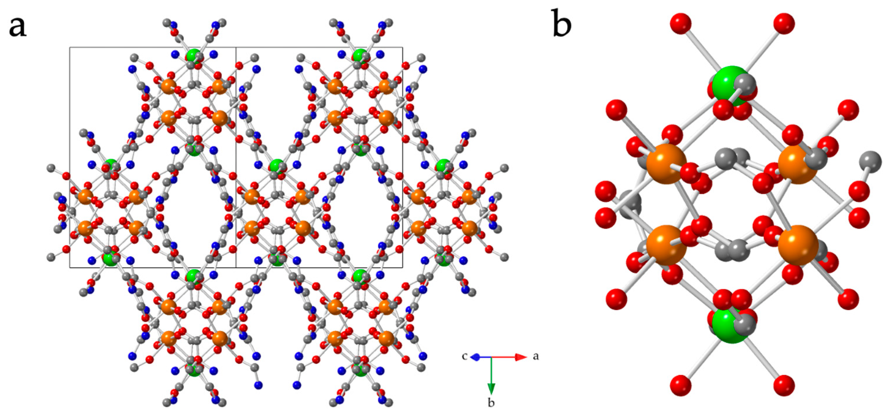
3.2. Structure of [Mn3(HL)6] (1)

Single-crystal X-ray analysis reveals that compound 1 is a 3D non-porous coordination polymer showing the (3,6)-c rtl topology [50] that crystallizes in the monoclinic space group C2/c. The asymmetric unit contains two independent Mn atoms: Mn1 (with half occupancy) and Mn2 (with full occupancy), and three HL ligands with a protonated pyridyl group and deprotonated hydroxy and carboxylate groups. The ligand-to-metal ratio is, therefore, 2:1, resulting in the formula [Mn3(HL)6] for compound 1. Both Mn centres show distorted octahedral geometry (Figure 1). The coordination environments of both Mn atoms are identical: the equatorial positions around are occupied by three carboxylate oxygen atoms (O1, O1*, and O8* for Mn1 and O4, O5, and O7 for Mn2) and one hydroxy oxygen atom (O9* for Mn1 and O6 for Mn2) from four different HL ligands. The axial positions are occupied by one carboxylate (O8 for Mn1 and O5* for Mn2) and one hydroxy oxygen atom (O9 for Mn1 and O6* for Mn2) from two different HL ligands. So, both Mn atoms are surrounded by six different HL ligands. All the Mn-O bond distances fall in the range of 2.126(3)–2.224(3) Å, and all cisoid and transoid angles fall in the range of 79.25(10)–102.60(10)° and 165.00(10)–176.70(10)°, respectively (Table S2).
Within the structure, all three HL ligands of the asymmetric unit act as non-chelating µ3-bridging ligands (Figure S3). Each ligand coordinates to two different Mn atoms using two carboxylate oxygen atoms in a syn-anti mode and coordinates to a third Mn atom through the deprotonated hydroxy group, giving rise to a 3D structure (see below). The syn-anti carboxylate bridges connect two Mn centres to generate a chain with alternating Mn1-Mn2 (A-type) and Mn2-Mn2 (B-type) [Mn2(RCOO)2] units (Figure 2). These chains run parallel to the [101] direction. Along the chain, there are two A-type (Mn1-Mn2) and one B-type (Mn2-Mn2) units following the sequence -A-A-B- (Figure 2). These chains are further connected by four HL ligands through their keto groups in two other directions to form a 3D network (Figure 3). Interestingly, the pyridyl groups are protonated and do not coordinate with the metal centres, although they participate in N-H···Ohydroxy hydrogen bonding interactions within the structure and form two different R22(6) hydrogen-bonded supramolecular rings (Figure S4). All the hydrogen bond dimensions are given in Table S3.

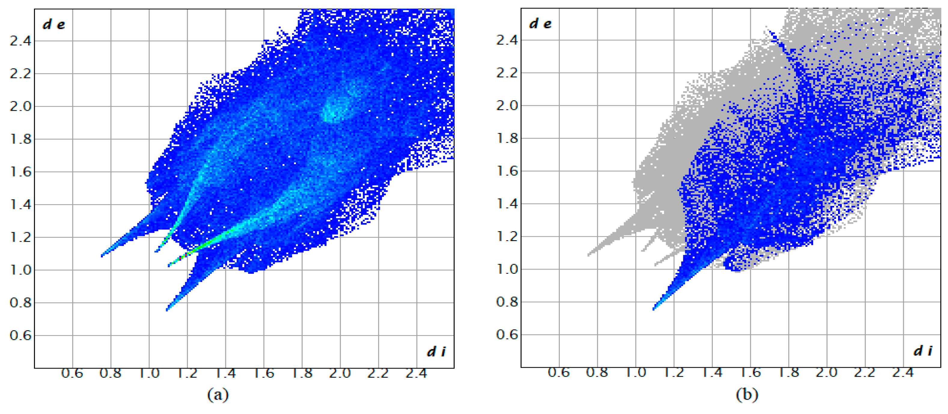
3.3. Hirshfeld Surface Analysis

Hirshfeld surface (HS) analysis has been carried out to understand the intermolecular interactions present in compound 1. The asymmetric unit has been used to calculate the Hirshfeld surface (such as dnorm, shape index, curvedness, and fragment patch) (Figure 4) and the subsequent Finger print plots (Figure 5 and Figure S5) to provide the qualitative and quantitative contribution of the noncovalent contacts present within the crystal system. The red, blue, and white colours used in dnorm indicate the interatomic distances closer, longer, or equal to van der Waals separations, respectively. On the other hand, valuable parameters of curvature, namely shape index and curvedness, have been included to offer further chemical insight into molecular shaping. A high curvedness is highlighted as dark blue edges in the shape index (Figure 4b) with ‘bumps and hollows’ depicted in blue and red, respectively, to indicate the flatness of the surface. The curvedness usually indicates large regions of green (relatively flat) separated by dark blue edges (large positive curvature) [48]. Figure 4 depicts the dnorm mapped surface showing light red spots near the N-H where the deep spots are close to the O atoms, indicating the presence of O-H/H-O interactions. The 2D Finger Print plot (Figure 5) supports the N-H···O interactions between the protonated pyridyl groups and the oxygen atoms as the predominant interaction (17.10%). In addition, O···H/H···O hydrogen bonding interactions show a sharp spike at (di = 1.10 and de = 0.75 Å) and at (di = 0.75 Å and de = 1.10 Å). An additional contribution to the HS comes from the O-O (7.6%), C-C (4.1%), and N-H/H-N (1.9%) interactions.

3.4. Absorption and Emission Spectra of [Mn3(HL)6] (1)

The solid-state absorption spectra of the colourless compound 1 show a peak at 319 nm with a shoulder at 328 nm and another small peak at 241 nm (Figure S6). The peak at 319 can be attributed to a ligand-to-metal charge transfer transition, whereas the peak at 241 nm can be assigned to the n-π* transition of the C=O bond that appears due to the keto-enol tautomerism. The emission spectra of compound 1 show one peak at 458 nm, which may be attributed to an intra-ligand π-π* transition (Figure S7).

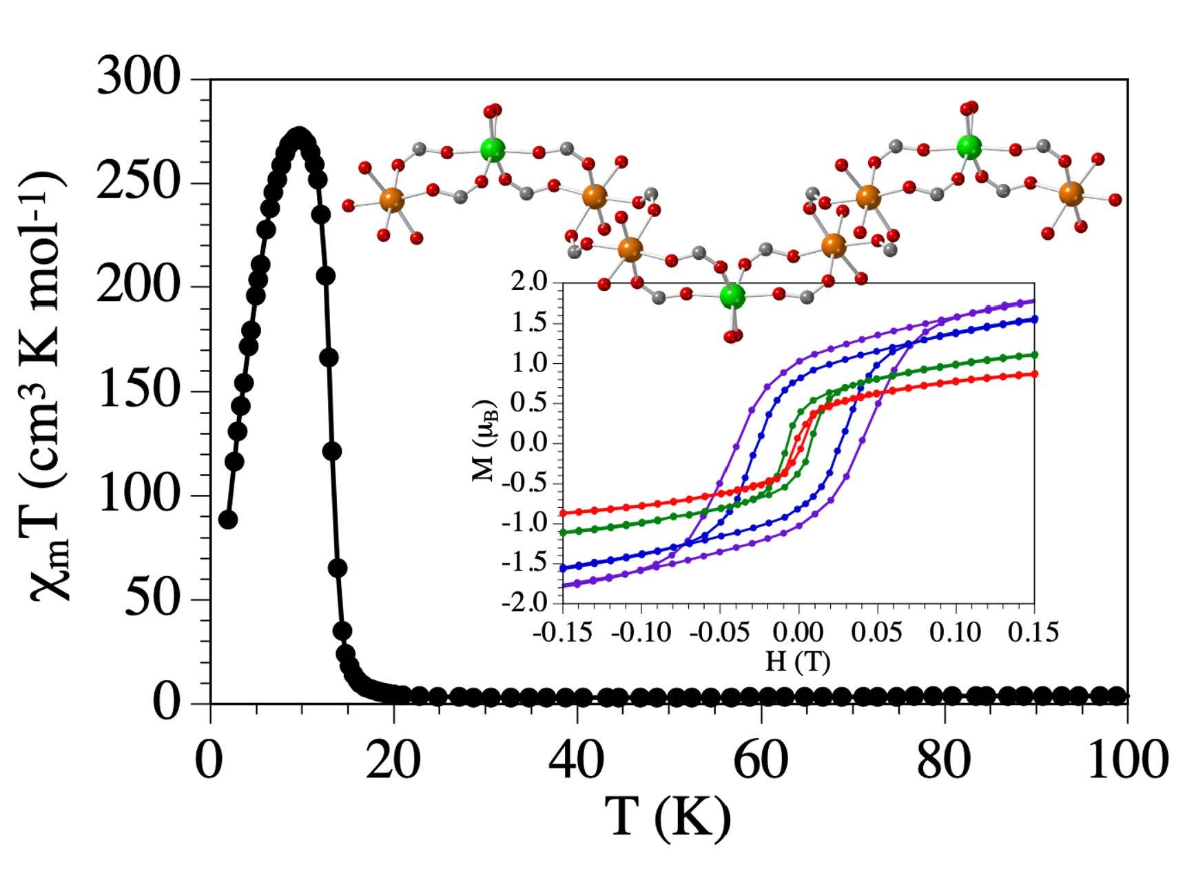
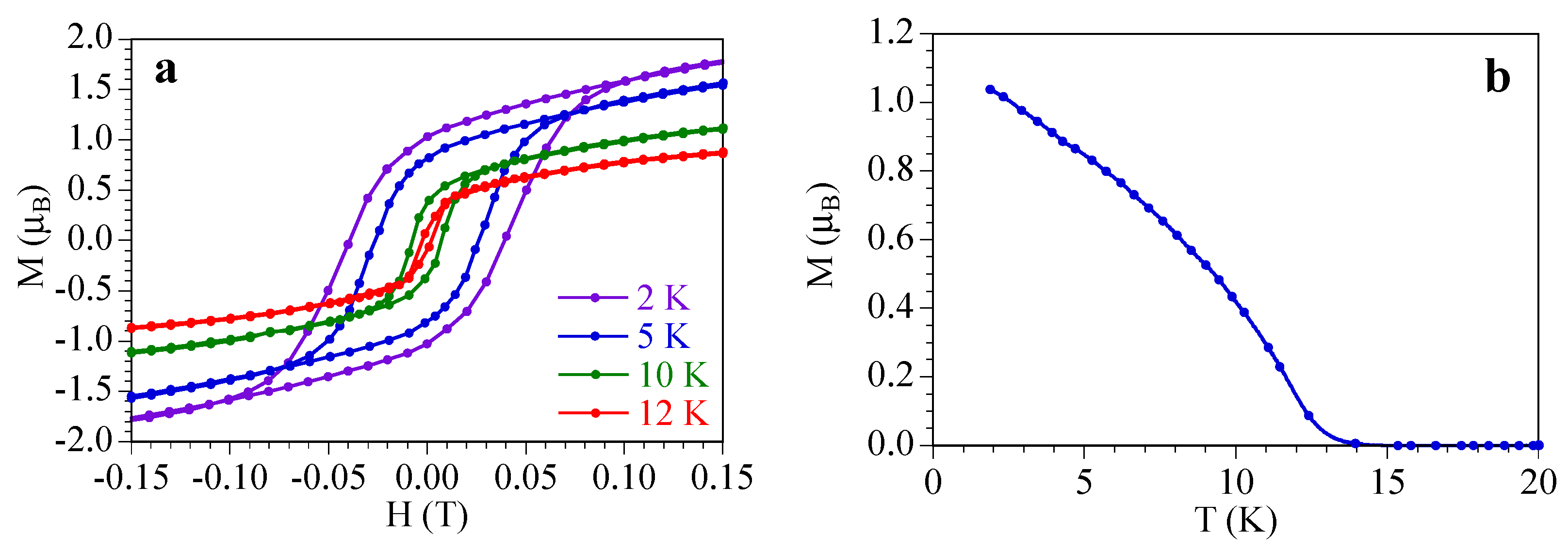
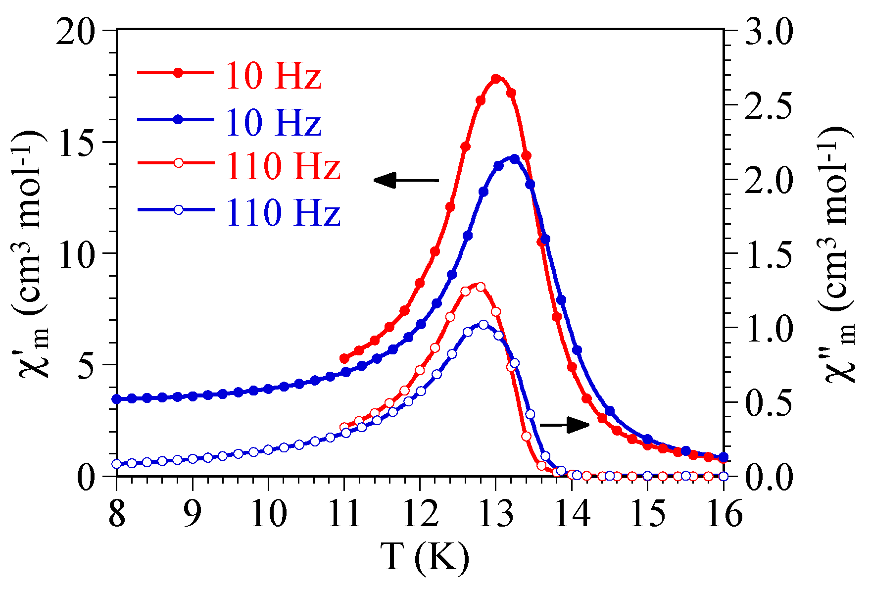
3.5. Magnetic Properties

The product of the molar magnetic susceptibility times the temperature (χmT) per Mn(II) ion for compound 1 shows at room temperature a value of ca. 4.4 cm3 K mol−1, very close to the expected value for an isolated Mn(II) ion with a ground spin state S = 5/2 and a g value close to 2. When the sample is cooled, χmT slowly decreases, reaching a minimum value of ca. 2.9 cm3 K mol−1 at around 42 K, indicating the presence of a weak antiferromagnetic coupling (Figure 6). Below this temperature, χmT shows a sharp increase and reaches a value of ca. 273 cm3 K mol−1 at 10 K. This abrupt increase indicates the presence of a long-range magnetic ordering and suggests the presence of a spin-canting since the Mn–Mn coupling is antiferromagnetic. Below 10 K, χmT sharply decreases due to saturation effects in χm to reach a value of ca. 90 cm3 K mol−1 at 2 K. The thermal variation of χm shows a sharp divergence with a maximum slope at around 14 K, corresponding to the approximate temperature of the canted antiferromagnetic long-range order (Figure S8).
Further confirmation of the canted ordering in 1 is provided by the isothermal magnetization at low temperatures that show an initial saturation value of ca. 2 μB (Figure S9) well below the expected one for an isolated MnII ion with g ≈ 2 (ca. 5 μB). Additionally, the isothermal magnetization shows hysteresis cycles of 40 mT at 2 K, 26 mT at 5 K, 7.5 mT at 10 K, and 2 mT at 12 K, indicating that the ordering temperature is above 12 K (Figure 7a). In fact, the thermal variation of the remnant magnetization obtained after cooling the sample under a magnetic field of 5 T shows a value of ca. 1.05 μB that vanishes above 14 K, confirming the presence of a canted antiferromagnetic order below ca. 14 K (Figure 7b).
To further confirm the presence of a long-range ordering, we have performed AC measurements of compound 1. These measurements show a frequency-independent peak at around 13 K with an out-of-phase signal (χ″m) that becomes non-zero below 14 K (Figure 8). This behaviour confirms the presence of a long-range order with an ordering temperature of 14 K.
Although not very common, the presence of a canted antiferromagnetic coupling between manganese ions through double and single syn-anti carboxylate bridges has already been reported in other manganese(II) [51] and manganese(III) [52,53] compounds and has been attributed to the lack of a symmetry centre between the manganese ions, that favours the Dzyaloshinsky–Moriya (D-M) interaction along the syn-anti carboxylate bridge [54].

4. Conclusions

In summary, we have prepared and structurally characterized a new 3D manganese(II) compound formulated as [Mn3(HL)6] (1), with H2L = 6-hydroxypicolinic acid, that represents the first coordination polymer with this ligand and Mn(II) ions. The 3D structure of 1 can be described as zigzag Mn chains with double syn-anti carboxylate bridges that are further connected to four other chains by four HL ligands through their keto groups to generate a 3D coordination polymer. The 6-hydroxypicolinic acid-based ligand remains in its keto form in the structure with the proton located on the pyridyl N atom. Hirshfeld surface analysis indicates the presence of strong hydrogen bonding interactions between the protonated pyridyl N atoms and the oxygen atoms of the ligand within compound 1. The DC and AC magnetic measurements show that compound 1 presents a weak antiferromagnetic Mn–Mn coupling through the double syn-anti carboxylate bridges but shows a long-range canted antiferromagnetic long-range order below 14 K with a coercive field of 40 mT at 2 K. This ligand demonstrates, thus, its capacity to construct 3D coordination polymers with interesting magnetic properties.

Supplementary Materials

The following supporting information can be downloaded at: https://www.mdpi.com/article/10.3390/magnetochemistry9020055/s1, Table S1: Structural data of all the reported structures with 6-hydroxypicolininc acid, Table S2: Coordination bond lengths (Å) and angles (°) in compound 1, Table S3: Hydrogen bond dimensions for compound 1; Figure S1: Simulated and experimental X-ray powder diffractograms for compound 1, Figure S2: IR spectra of compound [Mn(HL)2] (1), Figure S3: µ3-bridging mode of the ligand through carboxylate and hydroxy groups. Colour code: C = gray, O = red, N = blue, and Mn = purple), Figure S4: Hydrogen bonding interactions present in compound 1. Colour code: C = black, O = red, N = blue, and Mn = green. Pink lines indicate the H-bonds, Figure S5: Two-dimensional fingerprint plots for (a) O···H, (b) H···O, (c) N···H, (d) H···N, (e) N···O and (f) O···O interactions, Figure S6: Solid-state absorption spectra of compound 1, Figure S7: Solid-state emission spectra of compound 1, Figure S8: Thermal variation of χmT for compound 1. Inset shows the low-temperature region, Figure S9: Isothermal magnetization at different temperatures for compound 1. CCDC number for compound 1 is 2232982. References [36,37,39,49,55,56,57,58,59,60,61,62,63,64,65,66,67,68] have been cited in Supplementary Materials.

Author Contributions

S.K.D., M.P., S.B. and R.S. have synthesized and characterized the material. K.G. has performed the Hirshfeld surface analysis. C.J.G.-G. have collected and analyzed the magnetic data. All the authors have written and revised the manuscript. All authors have read and agreed to the published version of the manuscript.

Funding

R.S. is beneficiary of the grant (Z21-070) for the requalification of the Spanish University system from the Ministry of Universities of the Government of Spain, modality “Maria Zambrano”, financed by the European Union. S.B. thankfully acknowledges DST Inspire Faculty Research Grant (DST/INSPIRE/04/2018/000329, Faculty Registration No.: IFA18-CH304). S.K.D. thanks DST Inspire Faculty Research Grant to S.K.D. for financial assistance. R.S. also acknowledges UGC for their STIDE GRANT to Kazi Nazrul University (Ref No: KNU/R/Letter/913/21). This research was funded by the Advanced Materials program and was supported by MCIN with funding from European Union NextGeneration EU (PRTR-C17.I1) and the Generalitat Valenciana (project MFA-2022-057). We also thank the Generalidad Valenciana (Prometeo/2019/076) and the project PID2021-125907NB-I00, financed by MCIN/AEI/10.13039/501100011033/FEDER, UE, for financial support.

Institutional Review Board Statement

Not applicable.

Informed Consent Statement

Not applicable.

Data Availability Statement

Data are available upon request to the corresponding authors.

Conflicts of Interest

The authors declare no conflict of interest.

References

  1. Sakamaki, Y.; Tsuji, M.; Heidrick, Z.; Watson, O.; Durchman, J.; Salmon, C.; Burgin, S.R.; Beyzavi, H. Preparation and Applications of Metal−Organic Frameworks (MOFs): A Laboratory Activity and Demonstration for High School and/or Undergraduate Students. J. Chem. Educ. 2020, 97, 1109–1116. [Google Scholar] [CrossRef]
  2. Furukawa, H.; Cordova, K.E.; O’Keeffe, M.; Yaghi, O.M. The Chemistry and Applications of Metal-Organic Frameworks. Science 2013, 341, 1230444. [Google Scholar] [CrossRef]
  3. Kuppler, R.J.; Timmons, D.J.; Fang, Q.; Li, J.; Makal, T.A.; Young, M.D.; Yuan, D.; Zhao, D.; Zhuang, W.; Zhou, H. Potential applications of metal-organic frameworks. Coord. Chem. Rev. 2009, 253, 3042–3066. [Google Scholar] [CrossRef]
  4. Chen, L.; Zhang, X.; Cheng, X.; Xie, Z.; Kuang, Q.; Zheng, L. The function of metal–organic frameworks in the application of MOF-based composites. Nanoscale Adv. 2020, 2, 2628. [Google Scholar] [CrossRef] [PubMed]
  5. Russo, V.; Hmoudah, M.; Broccoli, F.; Iesce, M.R.; Jung, O.; Serio1, M.D. Applications of Metal Organic Frameworks in Wastewater Treatment: A Review on Adsorption and Photodegradation. Front. Chem. Eng. 2020, 2, 581487. [Google Scholar] [CrossRef]
  6. Thorarinsdottir, A.E.; Harris, T.D. Metal-Organic Framework Magnets. Chem. Rev. 2020, 120, 8716–8789. [Google Scholar] [CrossRef] [PubMed]
  7. Yadav, S.; Dixit, R.; Sharma, S.; Dutta, S.; Solanki, K.; Sharma, R.K. Magnetic metal–organic framework composites: Structurally advanced catalytic materials for organic transformations. Mater. Adv. 2021, 2, 2153–2187. [Google Scholar] [CrossRef]
  8. Gao, Y.; Liu, G.; Gao, M.; Huang, X.; Xu, D. Recent Advances and Applications of Magnetic Metal-Organic Frameworks in Adsorption and Enrichment Removal of Food and Environmental Pollutants. Crit. Rev. Anal. Chem. 2020, 50, 472–484. [Google Scholar] [CrossRef] [PubMed]
  9. Kurmoo, M. Magnetic metal–organic frameworks. Chem. Soc. Rev. 2009, 38, 1353–1379. [Google Scholar] [CrossRef] [PubMed]
  10. Coronado, E.; Espallargas, G.M. Dynamic magnetic MOFs. Chem. Soc. Rev. 2013, 42, 1525. [Google Scholar] [CrossRef]
  11. Ricco, R.; Malfatti, L.; Takahashi, M.; Hill, A.J.; Falcaro, P. Applications of magnetic metal–organic framework composites. J. Mater. Chem. A 2013, 1, 13033. [Google Scholar] [CrossRef]
  12. Ammari, Y.; Baaalla, N.; Hlil, E.K.; Abid, S. Structure, optical and magnetic properties of a novel homometallic coordination polymers: Experimental and computational studies. Nat. Sci. Rep. 2020, 10, 1316. [Google Scholar] [CrossRef] [PubMed]
  13. Wang, H.; Wu, Y.; Leong, C.F.; D’Alessandro, D.M.; Zuo, J. Crystal Structures, Magnetic Properties, and Electrochemical Properties of Coordination Polymers Based on the Tetra(4-pyridyl)- tetrathiafulvalene Ligand. Inorg. Chem. 2015, 54, 10766–10775. [Google Scholar] [CrossRef]
  14. Shao, D.; Moorthy, S.; Yang, X.; Yang, J.; Shi, L.; Singh, S.K.; Tian, Z. Tuning the structure and magnetic properties via distinct pyridine derivatives in cobalt(II) coordination polymers. Dalton Trans. 2022, 51, 695. [Google Scholar] [CrossRef] [PubMed]
  15. Zhang, Y.; Gao, L.; Zhou, W.; Wei, X.; Hu, T. Synthesis and magnetic properties of two Mn based coordination polymers constructed by a mixed-ligand strategy. CrystEngComm 2020, 22, 7123. [Google Scholar] [CrossRef]
  16. Pajuelo-Corral, O.; García, J.A.; Castillo, O.; Luque, A.; Rodríguez-Diéguez, A.; Cepeda, J. Single-Ion Magnet and Photoluminescence Properties of Lanthanide(III) Coordination Polymers Based on Pyrimidine-4,6-Dicarboxylate. Magnetochemistry 2021, 7, 8. [Google Scholar] [CrossRef]
  17. Zhou, Y.; Hong, M.; Wu, X. Lanthanide–transition metal coordination polymers based on multiple N- and O-donor ligands. Chem. Commun. 2006, 2, 135–143. [Google Scholar] [CrossRef] [PubMed]
  18. Feng, X.; Liu, L.; Wang, L.; Song, H.; Qiang, Z.; Wu, S.X.; Ng, S. Lanthanide coordination polymers based on multi-donor ligand containing pyridine and phthalate moieties: Structures, luminescence and magnetic properties. J. Solid State Chem. 2013, 206, 277–285. [Google Scholar] [CrossRef]
  19. Robin, A.Y.; Fromm, K.M. Coordination polymer networks with O- and N-donors: What they are, why and how they are made. Coord. Chem. Rev. 2006, 250, 2127–2157. [Google Scholar] [CrossRef]
  20. Galán-Mascarós, J.R.; Dunbar, K.R. A Self-Assembled 2D Molecule-Based Magnet: The Honeycomb Layered Material {Co3Cl4(H2O)2[Co(Hbbiz)3]2]. Angew. Chem. Int. Ed. 2003, 42, 2289. [Google Scholar] [CrossRef]
  21. Maspoch, D.; Ruiz-Molina, D.; Veciana, J. Magnetic nanoporous coordination polymers. J. Mater. Chem. 2004, 14, 2713. [Google Scholar] [CrossRef]
  22. Poulsen, R.D.; Bentien, A.; Chevalier, M.; Iversen, B.B. Synthesis, Physical Properties, Multitemperature Crystal Structure, and 20 K Synchrotron X-ray Charge Density of a Magnetic Metal Organic Framework Structure, Mn3(C8O4H4)3(C5H11ON)2. J. Am. Chem. Soc. 2005, 127, 9156. [Google Scholar] [CrossRef] [PubMed]
  23. Miyasaka, H.; Clérac, R.; Mizushima, K.; Sugiura, K.I.; Yamashita, M.; Wernsdorfer, W.; Coulon, C. [Mn2(saltmen)2Ni(pao)2(L)2](A)2 with L = Pyridine, 4-Picoline, 4-tert-Butylpyridine, N-Methylimidazole and A = ClO4, BF4, PF6, ReO4:  A Family of Single-Chain Magnets. Inorg. Chem. 2003, 42, 8203–8213. [Google Scholar] [CrossRef] [PubMed]
  24. Clérac, R.; Miyasaka, H.; Yamashita, M.; Coulon, C. Evidence for Single-Chain Magnet Behavior in a MnIII−NiII Chain Designed with High Spin Magnetic Units:  A Route to High Temperature Metastable Magnets. J. Am. Chem. Soc. 2002, 124, 12837–12844. [Google Scholar] [CrossRef]
  25. Kimura, S.; Matsuoka, R.; Kimura, S.; Nishihara, H.; Kusamoto, T. Radical-Based Coordination Polymers as a Platform for Magnetoluminescence. J. Am. Chem. Soc. 2021, 143, 5610–5615. [Google Scholar] [CrossRef]
  26. Yong, G.; Qiao, S.; Wang, Z. A One-Dimensional Coordination Polymer Based on Novel Radical Anion Ligand Generated In Situ: Notable Magnetic and Luminescence Properties. Cryst. Growth Des. 2008, 8, 1465–1467. [Google Scholar] [CrossRef]
  27. Benmansour, S.; Abherve, A.; Gómez-Claramunt, P.; Vallés-García, C.; Gómez-García, C.J. Nanosheets of Two-Dimensional Magnetic and Conducting Fe(II)/Fe(III) Mixed-Valence Metal−Organic Frameworks. ACS Appl. Mater. Interfaces 2017, 9, 26210–26218. [Google Scholar] [CrossRef]
  28. Aulakh, D.; Liu, L.; Varghese, J.R.; Xie, H.; Islamoglu, T.; Duell, K.; Kung, C.; Hsiung, C.; Zhang, Y.; Drout, R.J.; et al. Direct Imaging of Isolated Single-Molecule Magnets in Metal-Organic Frameworks. J. Am. Chem. Soc. 2019, 141, 2997–3005. [Google Scholar] [CrossRef]
  29. Palacios-Corella, M.; García-López, V.; Sánchez-Sánchez, C.; Clemente-Juan, J.M.; Clemente-León, M.; Coronado, E. Insertion of single-ion magnets based on mononuclear Co(II) complexes into ferromagnetic oxalate-based networks. Dalton Trans. 2021, 50, 5931. [Google Scholar] [CrossRef]
  30. Kusumoto, S.; Umeno, H.; Kim, Y.; Sekine, Y.; Nakamura, M.; Hayami, S. Structural and Magnetic Characterization of Homo- and Heterometallic Trinuclear Ni(II) and Cu(II) Clusters with N2O6 Acyclic Polydentate Ligand. Chem. Lett. 2021, 50, 1945–1948. [Google Scholar] [CrossRef]
  31. Andrews, P.C.; Deacon, G.B.; Frank, R.; Fraser, B.H.; Junk, P.C.; MacLellan, J.G.; Massi, M.; Moubaraki, B.; Murray, K.S.; Silberstein, M. Formation of HoIII Trinuclear Clusters and GdIII Monodimensional Polymers Induced by ortho and para Regioisomers of Pyridyl-Functionalised β-Diketones: Synthesis, Structure, and Magnetic Properties. Eur. J. Inorg. Chem. 2009, 2009, 744–751. [Google Scholar] [CrossRef]
  32. Gao, E.; Liu, N.; Cheng, A.; Gao, S. Novel frustrated magnetic lattice based on triangular [Mn33-F)] clusters with tetrazole ligands. Chem. Commun. 2007, 24, 2470–2472. [Google Scholar] [CrossRef] [PubMed]
  33. Sawada, K.; Ohashi, Y. 2-Oxo- 1,2-dihydropyridine-6-carboxylic Acid. Acta Cryst. 1998, C54, 1491–1493. [Google Scholar] [CrossRef]
  34. Kazemi, S.H.; Eshtiagh-Hosseini, H.; Izadyar, M.; Mirzaei, M. Computational Study of the Intramolecular Proton Transfer between 6- Hydroxypicolinic Acid Tautomeric Forms and Intermolecular Hydrogen Bonding in their Dimers. Phys. Chem. Res. 2013, 1, 117–125. [Google Scholar]
  35. Liu, C.; Song, Y. An Uneven Chain-like Ferromagnetic Copper(II) Coordination Polymer Displaying Metamagnetic Behavior and Long-Range Magnetic Ordering. Magnetochemistry 2022, 8, 2. [Google Scholar] [CrossRef]
  36. Sun, C.; Zheng, X.; Li, W.; Wang, M.; Fang, C. Assembly of Supramolecular Networks with the Inclusion of Water Chains, Cyclic Hepta and Octa Water Clusters. Z. Anorg. Allg. Chem. 2008, 634, 26632669. [Google Scholar] [CrossRef]
  37. Bian, G.Q.; Kuroda-Sowa, T.; Konaka, H.; Maekawa, M.; Munakata, M. Bis(μ-6-hydroxypicolinato)-μ-oxobis[dipyridinemanganese (III)] monohydrate. Acta Crystallogr. Sect. C 2004, 60, m338–m340. [Google Scholar] [CrossRef]
  38. Sun, C.; Zheng, X.; Jin, L.Z. Syntheses and Structures of the First Examples of Lanthanide Complexes with 6-Hydroxypicolinic Acid. Z. Anorg. Allg. Chem. 2004, 630, 13421347. [Google Scholar] [CrossRef]
  39. Kukovec, B.; Vaz, P.D.; Calhorda, M.J.; Popovic, Z. Disappearing and Concomitant Polymorphism of Nickel(II) Complexes with 6-Hydroxypicolinic Acid. Structural and Density Functional Theory Studies. Cryst. Growth Des. 2010, 10, 3685–3693. [Google Scholar] [CrossRef]
  40. Bruker, APEX2, SAINT and SADABS, BRUKER AXS, Inc.: Madison, WI, USA, 2008.
  41. Sheldrick, G.M. A short history of SHELX. Acta Cryst. 2008, A64, 112. [Google Scholar] [CrossRef]
  42. Farrugia, L.J. WinGX and ORTEP for Windows, an update. J. Appl. Crystallogr. 2012, 45, 849. [Google Scholar] [CrossRef]
  43. Sheldrick, G.M. Crystal structure refinement with SHELX. Acta Cryst. 2015, C71, 3. [Google Scholar]
  44. Spek, A.L. Structure validation in chemical crystallography. Acta Cryst. 2009, D65, 148. [Google Scholar] [CrossRef] [PubMed]
  45. Farrugia, L.J. ORTEP-3 for Windows—A version of ORTEP-III with a Graphical User Interface (GUI). J. Appl. Crystallogr. 1997, 30, 565. [Google Scholar] [CrossRef]
  46. Bain, G.A.; Berry, J.F. Diamagnetic corrections and Pascal’s constants. J. Chem. Educ. 2008, 85, 532–536. [Google Scholar] [CrossRef]
  47. McKinnon, J.J.; Jayatilaka, D.; Spackman, M.A. Towards quantitative analysis of intermolecular interactions with Hirshfeld surfaces. Chem. Commun. 2007, 37, 3814–3816. [Google Scholar] [CrossRef]
  48. Spackman, M.A.; McKinnon, J.J. Fingerprinting intermolecular interactions in molecular crystals. CrystEngComm 2002, 4, 378–392. [Google Scholar] [CrossRef]
  49. Kukovec, B.; Popovića, Z.; Pavlović, G.; Linarić, M.R. Synthesis and structure of cobalt(II) complexes with hydroxyl derivatives of pyridinecarboxylic acids: Conformation analysis of ligands in the solid state. J. Mol. Struct. 2008, 882, 47–55. [Google Scholar] [CrossRef]
  50. Blatov, V.A.; Shevchenko, A.P.; Proserpio, D.M. Applied Topological Analysis of Crystal Structures with the Program Package ToposPro. Cryst. Growth Des. 2014, 14, 3576–3586. [Google Scholar] [CrossRef]
  51. Wang, X.; Wei, H.; Wang, Z.; Chen, Z.; Gao, S. Formate—The Analogue of Azide:  Structural and Magnetic Properties of M(HCOO)2(4,4‘-Bpy)·nH2O (M = Mn, Co, Ni; n = 0, 5). Inorg. Chem. 2005, 44, 572–583. [Google Scholar] [CrossRef]
  52. Kar, P.; Guha, P.M.; Drew, M.G.B.; Ishida, T.; Ghosh, A. Spin-Canted Antiferromagnetic Phase Transitions in Alternating Phenoxo- and Carboxylato-Bridged MnIII-Salen Complexes. Eur. J. Inorg. Chem. 2011, 2011, 2075–2085. [Google Scholar] [CrossRef]
  53. Mossin, S.; Weihe, H.; Osholm Sørensen, H.; Lima, N.; Sessoli, R. Rationalisation of Weak Ferromagnetism in Manganese(iii) Chains: The Relation between Structure and Ordering Phenomena. Dalton Trans. 2004, 4, 632–639. [Google Scholar] [CrossRef] [PubMed]
  54. Kahn, O. Molecular Magnetism; VCH Publishers: New York, NY, USA, 1993. [Google Scholar]
  55. Kang, S.K.; Shim, Y.S. Poly[(m6-6-Oxidopyridinium-2-Carboxylato)Caesium]. Acta Cryst. E 2011, 67, m1237. [Google Scholar] [CrossRef]
  56. Małecki, J.G.; Krompiec, S.; Maroń, A.; Penkala, M. Synthesis, Molecular, Spectroscopic and Catalytic Characterization of Ruthenium(II) Complexes with Pyridine-2-Carboxylic Acid Derivatives Ligands. Polyhedron 2012, 48, 21–30. [Google Scholar] [CrossRef]
  57. Kukovec, B.-M.; Popovic, Z.; Pavlovic, G. Copper(II) Complexes with 3- and 6-hydroxypicolinic Acid. Preparation, Structural, Spectroscopic and Thermal Study. Acta Chim. Slov. 2008, 55, 779–787. [Google Scholar]
  58. Kukovec, B.M.; Kaksa, M.; Popovic, Z. Synthesis and Characterization of a Copper(II) Complex with 6-Hydroxypicolinic Acid and 3-Picoline Croatica. Chem. Acta 2012, 85, 479–483. [Google Scholar]
  59. Sengül, A.; Büyükgüngör, O. Trans-Di-Aqua-Bis(6-Hydroxy-Picolinato-k2-N,O2)Copper(II). Acta Cryst. E 2005, 61, m119–m121. [Google Scholar]
  60. You-Zhu Yu, Song-Yang Chang, Xi Han, Guang-Xin Chen, Ya-Wei Xuan, Xian-Li Wu, Fang Wang, Hydrothermal Syntheses, Crystal Structures and Luminescence Properties of Cu(II) and Cd(II) Complexes Assembled by 6-Hydroxypicolinic Acid and 1,10-Phenanthroline. Jiegou Huaxue 2019, 38, 651–659.
  61. Sun, C.; Zhou, J.; Li, W.; Jin, L. Supramolecular Stuctures from Mononuclear, Binuclear and 2D Net of Copper(II) Complexes with 6-Hydroxypicolinic Acid. Z. Anorg. Allg. Chem. 2008, 634, 549–554. [Google Scholar] [CrossRef]
  62. Su, W.; Guo, Y.; Yu, Y.; Wang, Y. Synthesis, Structure and Magnetic Property of a Linear Trinuclear Dy(III) Single Molecular Magnet. Inorg. Chem. Commun. 2020, 120, 108161. [Google Scholar] [CrossRef]
  63. Nakasone, T.; Nishioka, T.; Asato, E.; Kinoshita, I.; Takara, S. Synthesis and Structural Characterization of Novel Ruthenium(II) Complexes Bearing Hydroxypicolinato Ligands. Polyhedron 2012, 45, 152–157. [Google Scholar] [CrossRef]
  64. Casas, J.S.; Castellano, E.E.; Ellena, J.; García-Tasende, M.S.; Sánchez, A.; Sordo, J.; Toma, M. Dimethylthallium(III) Complexes with Picolinic Acid and its Hydroxyl Derivatives. Polyhedron 2008, 27, 1296–1302. [Google Scholar] [CrossRef]
  65. Chattopadhyay, S.; Fanwick, P.E.; Walton, R.A. Reactions of the Triply-Bonded Complex cis-Re2(μ-O2CCH3)2Cl2(μ-Dppm)2 with Pyridine Carboxylic Acids. the Isolation and Structural Characterization of a Third Structural Isomer of Re2(Dipic)Cl2(μ-Dppm)2 (Dipic = Pyridine-2,6-Dicarboxylate). Dalton Trans. 2003, 18, 3617–3621. [Google Scholar] [CrossRef]
  66. Mukiza, J.; Hosten, E.C.; Gerber, T.I.A. Rhenium(III), (IV) and (V) Complexes with 6-Hydroxypicolinic Acid. Polyhedron 2016, 110, 106–113. [Google Scholar] [CrossRef]
  67. Nakama, Y.; Nishioka, T.; Nakasone, T.; Asato, E.; Kinoshita, I.; Takara, S. Synthesis And Crystal Structure of [RuCl(6-hydroxypicolinato)(2,2’;6’,2”-terpyridine)]·(N,N-dimethylformamide). X-ray Struct. Anal. Online 2010, 26, 33–34. [Google Scholar] [CrossRef]
  68. Sun, C.; Jin, L. Supramolecular Architectures from the Self-Assembly of Lanthanide Ions with 6-Hydroxypicolinic Acid and 1,10-Phenanthroline. J. Mol. Struct. 2005, 741, 241–247. [Google Scholar] [CrossRef]
Scheme 1. Synthetic scheme of compound 1.
Magnetochemistry 09 00055 sch001
Figure 1. ORTEP diagram of the asymmetric unit with labelling scheme of compound 1. Ellipsoids are drawn at 50% probability (except the H atoms).
Magnetochemistry 09 00055 g001
Figure 2. View of the -Mn-(OC(R)O)2-Mn- chains formed by double syn-anti carboxylate bridges in 1 showing the A-type and B-type Mn dimers.
Magnetochemistry 09 00055 g002
Figure 3. (a) Projection of the structure of compound 1 down the [101] direction (perpendicular to the chains). (b) View of one isolated chain along the same direction. Colour code: C = gray, O = red, N = blue, Mn1 = green, and Mn2 = orange.
Magnetochemistry 09 00055 g003
Figure 4. Hirshfeld surfaces mapped with (a) dnorm, (b) shape index, (c) curvedness, and (d) fragment patch for compound 1.
Magnetochemistry 09 00055 g004
Figure 5. (a) Two-dimensional fingerprint plots for all contacts. (b) Two-dimensional fingerprint plot only for the O···H interactions.
Magnetochemistry 09 00055 g005
Figure 6. Thermal variation of χmT for compound 1. Inset shows the high-temperature region.
Magnetochemistry 09 00055 g006
Figure 7. (a) Hysteresis cycles at different temperatures for compound 1. (b) Thermal variation of the remnant magnetization of compound 1 after cooling the sample under 5 T.
Magnetochemistry 09 00055 g007
Figure 8. Thermal variation of the in-phase (χ′m, left scale, filled circles) and out-of-phase (χ″m, right scale, empty circles) signals of compound 1 at two different frequencies.
Magnetochemistry 09 00055 g008
Table 1. Crystallographic data collection and refinement parameters.
FormulaC36H24Mn3N6O18
Formula weight993.43
Crystal systemMonoclinic
Space groupC2/c (No. 15)
a (Å)12.3872 (13)
b (Å)15.4999 (13)
c (Å)20.7172 (17)
α (°)90
β (°)105.713 (4)
γ (°)90
V (Å3)3829.1 (6)
Z4
ρcalc (g/cm3)1.723
μ (Mo Kα) (mm)1.065
F(000)2004
Crystal size (mm3)0.10 × 0.12 × 0.16
Temperature, T (K)127
θmin-max (deg)2.6, 27.2
Total data21,514
Unique data4249
Rint0.079
Observed data [I > 2.0 σ(I)]3104
Nref4249
Npar294
R0.0479
wR20.1177
S1.06
w = [(Fo2) + (0.0287 P)2 + 13.3720 P] where P = (Fo2 + 2Fc2)/3.
Disclaimer/Publisher’s Note: The statements, opinions and data contained in all publications are solely those of the individual author(s) and contributor(s) and not of MDPI and/or the editor(s). MDPI and/or the editor(s) disclaim responsibility for any injury to people or property resulting from any ideas, methods, instructions or products referred to in the content.

