Nostoc edaphicum CCNP1411 from the Baltic Sea—A New Producer of Nostocyclopeptides
by
 , , and
Anna Fidor
1 ,
,
Michał Grabski
2,
Jan Gawor
3,
Robert Gromadka
3,
Grzegorz Węgrzyn
2 and
Hanna Mazur-Marzec
1,*
1
Division of Marine Biotechnology, Faculty of Oceanography and Geography, University of Gdańsk, Marszałka J. Piłsudskiego 46, PL-81378 Gdynia, Poland
2
Department of Molecular Biology, University of Gdansk, Wita Stwosza 59, 80-308 Gdansk, Poland
3
DNA Sequencing and Oligonucleotide Synthesis Laboratory, Polish Academy of Sciences, Institute of Biochemistry and Biophysics, 02-106 Warsaw, Poland
*
Author to whom correspondence should be addressed.
Mar. Drugs 2020, 18(9), 442; https://doi.org/10.3390/md18090442
Submission received: 31 July 2020
/
Revised: 21 August 2020
/
Accepted: 25 August 2020
/
Published: 26 August 2020
(This article belongs to the Special Issue Bioactive Molecules from Marine Microorganisms)
Abstract
:Nostocyclopeptides (Ncps) constitute a small class of nonribosomal peptides, exclusively produced by cyanobacteria of the genus Nostoc. The peptides inhibit the organic anion transporters, OATP1B3 and OATP1B1, and prevent the transport of the toxic microcystins and nodularin into hepatocytes. So far, only three structural analogues, Ncp-A1, Ncp-A2 and Ncp-M1, and their linear forms were identified in Nostoc strains as naturally produced cyanometabolites. In the current work, the whole genome sequence of the new Ncps producer, N. edaphicum CCNP1411 from the Baltic Sea, has been determined. The genome consists of the circular chromosome (7,733,505 bps) and five circular plasmids (from 44.5 kb to 264.8 kb). The nostocyclopeptide biosynthetic gene cluster (located between positions 7,609,981–7,643,289 bps of the chromosome) has been identified and characterized in silico. The LC-MS/MS analyzes of N. edaphicum CCNP1411 cell extracts prepared in aqueous methanol revealed several products of the genes. Besides the known peptides, Ncp-A1 and Ncp-A2, six other compounds putatively characterized as new noctocyclopeptide analogues were detected. This includes Ncp-E1 and E2 and their linear forms (Ncp-E1-L and E2-L), a cyclic Ncp-E3 and a linear Ncp-E4-L. Regardless of the extraction conditions, the cell contents of the linear nostocyclopeptides were found to be higher than the cyclic ones, suggesting a slow rate of the macrocyclization process.
1. Introduction
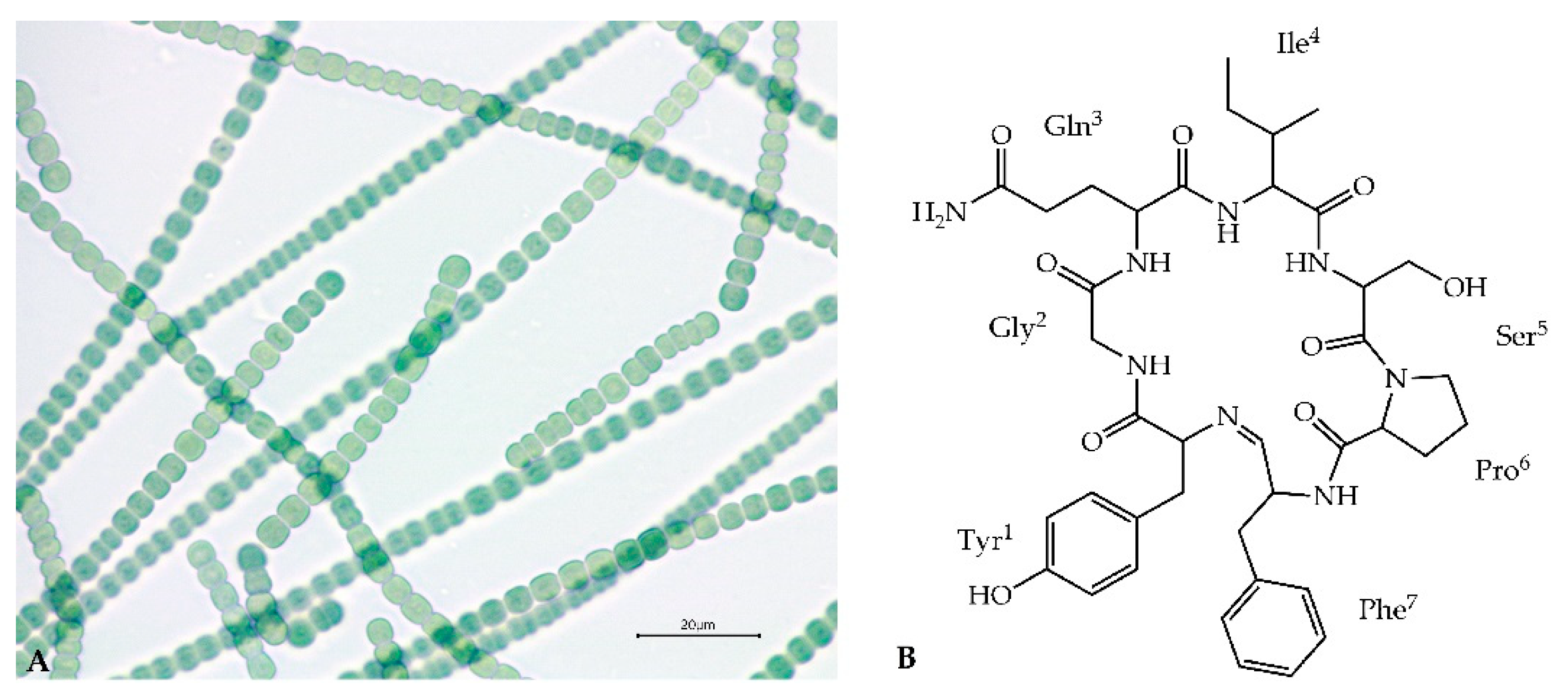
Secondary metabolites produced by cyanobacteria of the genus Nostoc (Nostocales) are characterized by a high variety of structures and biological activities [1,2,3,4,5,6]. On the basis of chemical structure, these compounds are mainly classified to peptides, polyketides, lipids, polysaccharides and alkaloids [7]. Abundantly produced cyanopeptides with anticancer, antimicrobial, antiviral and enzyme-inhibiting activity, have attracted attention of many research groups [6,8,9,10,11,12]. Some of the metabolites, such as nostocyclopeptides (Ncps) or cryptophycins are exclusively produced by the cyanobacteria of the genus Nostoc (Figure 1A). Ncps constitute a small class of nonribosomal peptides. Thus far, only three analogues of the compounds and their linear forms have been discovered. This includes Ncp-A1 and Ncp-A2 detected in Nostoc sp. ATCC53789 isolated from a lichen collected at Arron Island in Scotland [13]. The same peptides were detected in Nostoc sp. ASN_M, isolated from soil samples of paddy fields in the Golestan province in Iran [14] and in Nostoc strains from liverwort Blasia pusilla L. collected in Northern Norway [15]. A different analogue, Ncp-M1, was found in Nostoc sp. XSPORK 13A, the cyanobacterium living in symbiosis with gastropod from shallow seawaters at the Cape of Porkkala (Baltic Sea) [16].
Ncps are composed of seven residues and a unique imino linkage formed between C-terminal aldehyde and an N-terminal amine group of the conserved Tyr1 (Figure 1B) [13,16]. The presence of modified amino acid residues, e.g., 4-methylproline, homoserine and D-configured glutamine, indicated the nonribosomal biosynthetic pathway of the molecules. Genetic analysis of Nostoc sp. ATCC53789 revealed the presence of the 33-kb Ncp gene cluster composed of two genes, ncpA and ncpB, encoding NcpA1-A3 and NcpB1-B4 modules. These proteins catalyze the activation and incorporation of Tyr, Gly, Gln, Ile and Ser into the Ncp structure [17]. They show high similarity to NosE and NosF which take part in the biosynthesis of nostopeptolides in Nostoc sp. GSV224 [18]. The ncpFGCDE fragment of the Ncp gene cluster is involved in the synthesis of MePro (ncpCDE), transport (ncpF) and proteolysis (ncpG) of the peptides. The characteristic features of the Ncp enzymatic complex in Nostoc sp. ATCC53789 is the presence of the epimerase domain (NcpA3) responsible for D-configuration of glutamine, and the unique reductase domain at C-terminal end of NcpB which catalyze the reductive release of a linear peptide aldehyde [19,20].
The activity of Ncps have been explored [13] and their potential as antitoxins, inhibiting the transport of hepatotoxic microcystin-LR and nodularin into the rat hepatocytes through the organic anion transporter polypeptides OATP1B1/1B3 was revealed [21]. As OATP1B3 is overexpressed in some malignant tumors (e.g., colon carcinomas) [22], Ncps, as inhibitors of this transporter protein, are suggested to be promising lead compounds for new drug development.
In our previous studies, Nostoc edaphicum CCNP1411 (Figure 1A) from the Baltic Sea was found to be a rich source of cyanopeptolins, the nonribosomal peptides with potent inhibitory activity against serine proteases [6]. In the current work, the potential of the strain to produce other bioactive metabolites was explored. The whole-genome sequence of N. edaphicum CCNP1411 has been determined, and the nostocyclopeptide biosynthetic gene cluster has been identified in the strain and characterized in silico for the first time. Furthermore, the new products of the Ncp gene cluster have been detected and their structures have been characterized by LC-MS/MS.
2. Results and Discussion
2.1. Analysis of N. edaphicum CCNP1411 Genome
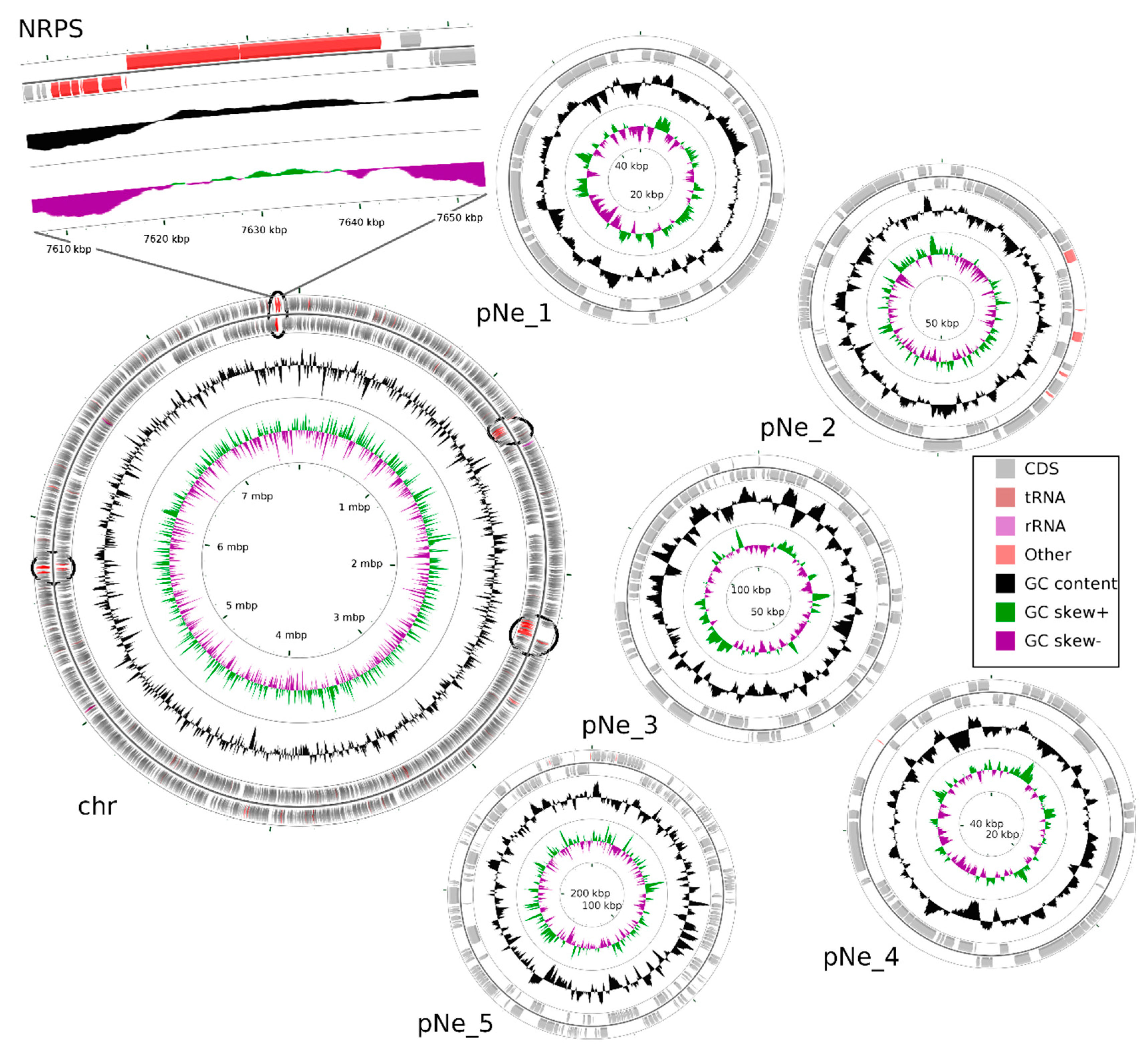
Total DNA has been isolated from N. edaphicum CCNP1411, and the whole genome sequence has been determined. Identified replicons of N. edaphicum CCNP1411 genome consist of the circular chromosome of 7,733,505 bps, and five circular plasmids (Table 1). Within the total size of 8,316,316 bps genome (chromosome and plasmids, Figure 2), we have distinguished, according to annotation, the total number of 6957 genes from which 6458 potentially code for proteins (CDSs), 415 are classified as pseudo-genes and 84 are coding for non-translatable RNA molecules. Pseudo-genes can be divided into subcategories due to the shift in the coding frame (180), internal stop codons (77), incomplete sequence (228), or occurrence of multiple problems (63). Genes coding for functional RNAs consist of those encoding ribosomal (rRNA) (9), transporting (tRNA) (71) and regulatory noncoding (ncRNA) (4), all embedded on the chromosome. Out of total coding and pseudo-genes sequences (6873), the vast majority (5846) initiates with the ATG start codon, while GTG and TTG occur less frequently (561 and 217 times, respectively). The frequencies of stop codons were set out as follows: TAA (3455), TAG (1750), TGA (1526). Coding and pseudo-genes sequences are distributed almost equally on the leading and complementary strand, including 3408 and 3465 sequences, respectively.
2.2. Non-Ribosomal Peptide Synthetase (NRPS) Gene Cluster of Nostocyclopeptides
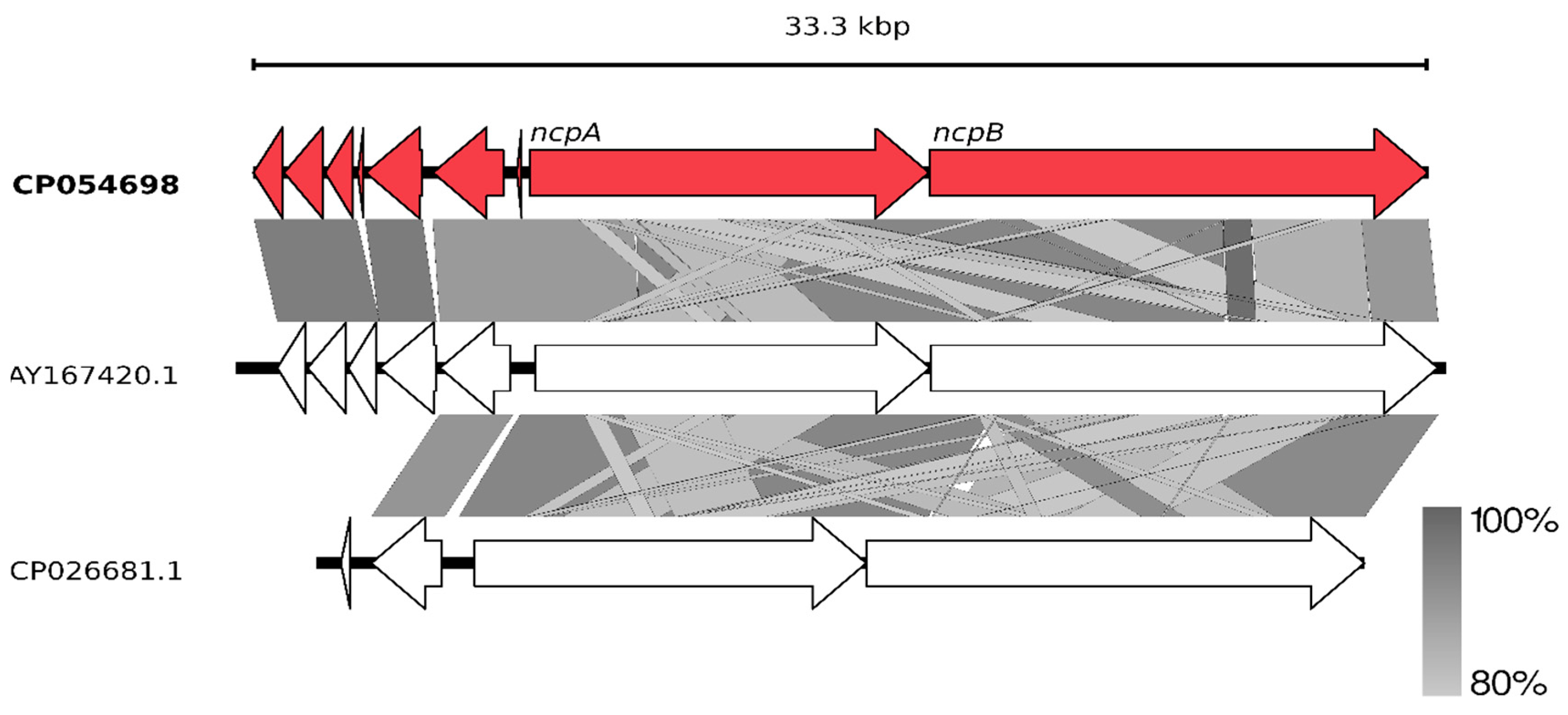
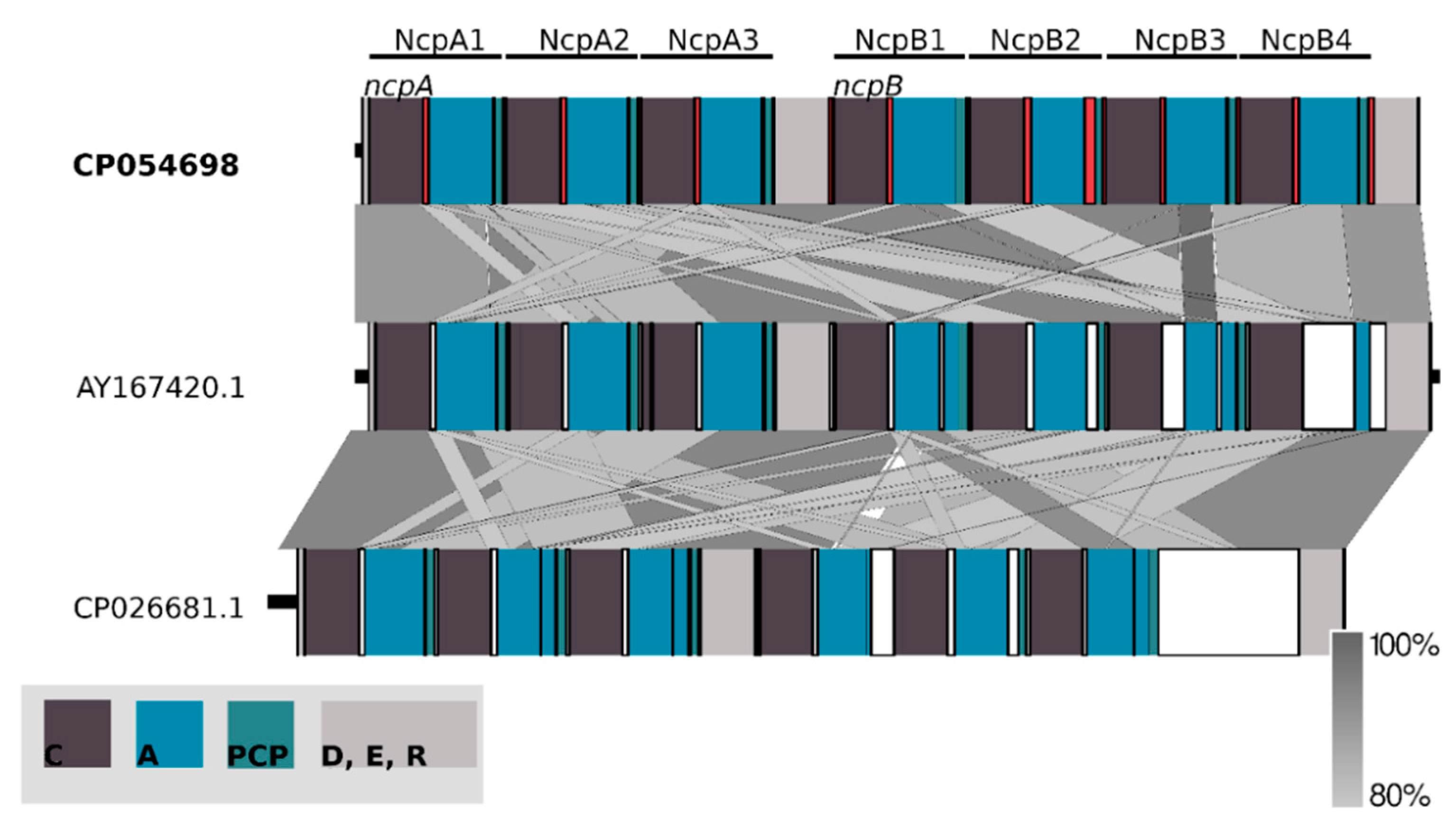
Having the whole genome sequence of N. edaphicum CCNP1411, we have analyzed in detail the non-ribosomal peptide synthetase (NRPS) cluster, containing potential genes coding for enzymes involved in the synthesis of nostocyclopeptides. To establish correct spans for non-ribosomal peptide synthetases, 35 complete nucleotide sequence clusters derived from Cyanobacteria phylum were aligned resulting in hits scattered around positions 2,287,143–2,323,617 and 7,609,981–7,643,289 within the N. edaphicum CCNP1411 chromosome (7.7 Mbp) (Figure 2). This method of characterization presented the overall similarity of selected spans to micropeptin (cyanopeptolin) biosynthetic gene cluster [23] and nostocyclopeptide biosynthetic gene cluster [17], respectively. To confirm these results, the antiSMASH analysis was employed resulting in confirmation of previously defined NRPS spans and adding two more regions 1,213,069–1,258,319 and 5,735,625–5,780,238, to small extent (12% and 30%, respectively) similar to anabaenopeptin gene cluster [24]. For the purpose of this study, we focused on putative nostocyclopeptide producing non-ribosomal peptide synthetase. Annotation of the selected region revealed nine putative open reading frames (ORFs), transcribed in reverse (7) and forward (2) direction. The identified cluster was arranged in a similar fashion to AY167420.1 (nostocyclopeptide biosynthetic gene cluster from Nostoc sp. ATCC 53789), with the exception of two ORFs (>170 bp), intersecting operon (ncpFGCDE) putatively encoding proteins involved in MePro assembly, efflux and hydrolysis of products of the second putative operon ncpAB (Figure 3).
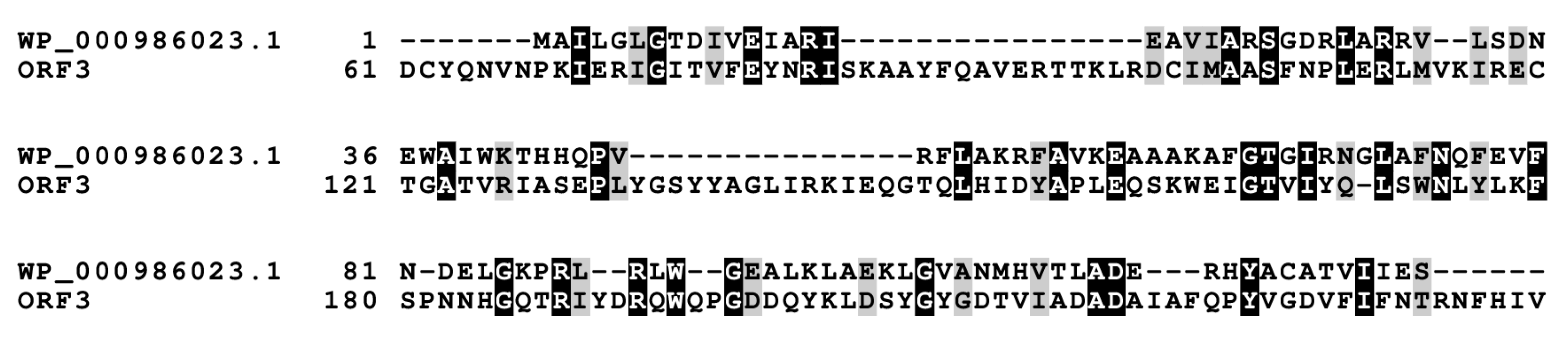
Two sequences ORF1 (HUN01_34350) (837 bp) and ORF2 (HUN01_34355) (1107 bp) embedded on 3′ end of nostocyclopeptide gene cluster resemble nosF and nosE genes, found in the nostopeptolide (nos) gene cluster [18] with 96% nucleotide sequence identities in both instances, putatively encoding for zinc-dependent long-chain dehydrogenase and a Δ1-pyrroline-5-carboxylic acid reductase. Further upstream, there is an ORF3 (HUN01_34360) (798 bp) of 98% homology to unknown gene from AF204805.2 gene cluster, suggested previously to be involved in 4-methylproline biosynthesis [17,25], due to close proximity of downstream genes encompassing this reaction, but no experimental evidence was presented. Alignment of the sequence of this putative protein have shown a sequence homology, to some extent, to 4′-phosphopantetheinyl transferase, crucial for PCP aminoacyl substrate binding (Figure 4) [26]. Moreover, partially present adenylate-forming domain within ORF4 (HUN01_34365) (165 bp) belongs to the acyl- and aryl- CoA ligases family, and may putatively engage substrate for post-translational modification of the PCP domain. Facing the same direction, an ORF5 (HUN01_34370) (1605 bp)-bearing putative domain classified as transpeptidase superfamily DD-carboxypeptidase and ORF6 (HUN01_34375) (2010 bp) homologous to ABC transporter ATP-binding protein/permease may be engaged in ncpAB peptide product transport [27]. Neither the ORF7 (HUN01_34780) Shine–Delgarno (SD) sequence upfront translation start codon could be assigned nor the TA-like signal ~12 nucleotides upstream could be found.
The main part of the Ncp biosynthetic gene cluster is located on the forward strand comprising two large genes which nucleotide sequences are homologous over 80% to ncpA and ncpB subunits of the ncp cluster in Nostoc sp. ATCC53789 [17]. Both these genes code for proteins consisting of repetitive modules incorporating single residue into elongating peptide. ORF 8 (HUN01_34785) (11,334 bp) encompasses three of these modules, whereas ORF 9 (HUN01_34380) (14,157 bp) encodes four modules. The core of one NRPS module consists of three succeeding domains: condensation (C), adenylation (A) and peptidyl carrier protein (PCP). Moreover, adjacent to coding spans of extreme modules, two tailoring domains were found within ORF8 and ORF9 genes (Figure 5).
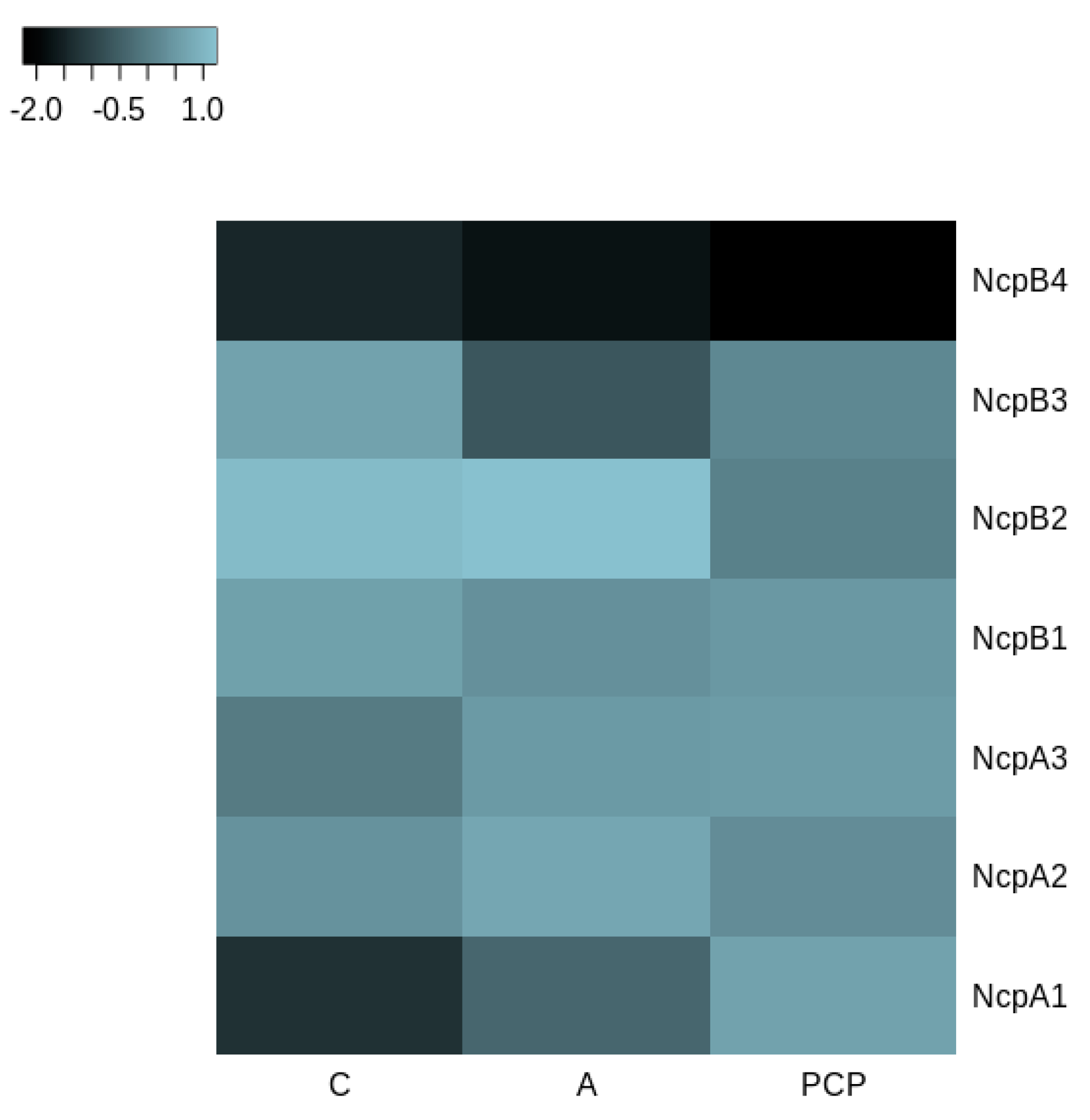
Alignment of nucleotide sequences to the ncpAB operon revealed major differences in consecutive NcpB3 and NcpB4 modules. Utilizing the selected spans conjoined with conserved domain search allowed us to distinguish and compare C, A and PCP amino-acid sequences (Figure 6). Intrinsic modules of NRPS, with an exception of NcpB3 adenylation domain sequence, were found homologous above 91%, whereas extremes have shown the biggest composition differences ranging from 13–15% to 24% in the NcpB4 adenylation domain (Figure 6).
The determination of the whole genome sequence of N. edaphicum CCNP1411 allowed us to perform analyses of genes coding for enzymes involved in the synthesis of nostocyclopeptides. The general analysis demonstrated homology of the NRPS/PKS clusters of N. edaphicum CCNP1411 to systems occurring in other cyanobacteria, however, with some differences. The non-ribosomal consensus code [28,29] allowed to recognize and predict the substrate specificities of NRPS adenylation domains: tyrosine (NcpA1), glycine (NcpA2), glutamine (NcpA3) for NcpA and isoleucine/valine (NcpB1) serine (NcpB2) 4-methylproline/proline (NcpB3) phenyloalanine/leucin/tyrosine (NcpB4) for NcpB (Table 2). This prediction was found to be in line with the structures of the Ncps detected in N. edaphicum CCNP1411.
To devolve elongating product onto subsequent condensation domain, the studied synthetase utilizes PCP domains, subunits responsible for thiolation of nascent peptide intermediates, where post-transcriptional modification of conserved serine residue shifts the state of the domain from inactive holo to active apo. Modification of this residue is related to PPTase which transfers a covalently-bound 4′-phosphopantetheine arm of CoA onto the PCP active site, enabling peptide intermediates to bind as reactive thioesters. Case residue which undergoes a nucleophilic attack by the hydroxyl group was conserved in every module within the PCP domain predicted at the front of the second helix [30].
The stand-alone docking domain (D) (7,617,812–7,617,964 bp) found on N-terminus of NcpA may be an essential component mediating interactions, recognition and specific association within NRPS subunits. The potential acceptor domain, based on sequence homology of conserved residues to C-terminal communication-mediating donor domains (COM), was found at the NcpB4 PCP domain second helix, encompassing conserved serine residue within potential binding sequence [31]. Moreover, this communication-mediating domain may putatively bind to C-terminus of NcpB3 and NcpB4 condensation (C) domains based on conserved motif LLEGIV, found by sequence homology to last five amino-acids of C-terminal docking domains residues, key for their interactions [32]. Within the same β-hairpin, a group of charged residues (ExxxxxKxR) putatively determines the binding affinity of the N-terminal domain [33].
Two tailoring domains encoded at the 5′ ends of ncpA and ncpB genes were classified as epimerization (E) (7,627,742–7,629,043 bp) domain and reductase (R) (7,642,183–7,643,238 bp) domain, accordingly. Epimerization domain catalyzes the conversion of L-amino acids to D-amino acids, a reaction coherent with D-stereochemistry of the peptide glutamine residue, where His of the conserved HHxxxDG motif and Glu from the upstream EGHGRE motif raceB comprise an epimerisation reaction active site [34]. Homologous HHxxxDG conserved motif sequence is found in condensation domains (C), where a similar reaction is catalyzed within peptide bond formation, putatively by the second His residue [35]. As in ncp cluster [17], module NcpA1 motif includes degenerate sequences in two positions HQIVGDL with leucine instead of phenylalanine residue at the start of the helix. The second histidine site-directed mutagenesis abolished enzymatic activity which might suggest that NcpA1 condensation domain is inactive [36].
Reductase domain (R) found at the C-terminus of NRPS was classified as oxidoreductase. Despite 15% discrepancy in domain composition compared to NcpB core catalytic triad Thr-Tyr-Lys and Rossmann-fold, a NAD (P) H nucleotide-binding motif GxxGxxG positions were not affected. The mechanism driving this chain release utilizes NAD (P) H cofactor for redox reaction of the final moiety of the nascent peptide to aldehyde or alcohol [37,38].
2.3. Structure Characterization of Ncps Produced by N. edaphicum CCNP1411
Thus far, only three Ncps, Ncp-A1, A2 and M1, and their linear aldehydes were isolated as pure natural products of Nostoc strains [13,16]. Ncp-A3, with MePhe in the C-terminal position, was obtained through aberrant biosynthesis in the Nostoc sp. ATCC53789 culture supplemented with MePhe [13]. The linear aldehydes of Ncp-A1 and Ncp-A2, with Pro instead of MePro, were chemically synthesized and used to study the Ncps epimerization and macrocyclization equilibria [19,20]. In our work, ten Ncps, differing mainly in position 4 and 7, were detected by LC-MS/MS in the N. edaphicum CCNP1411 cell extract (Table 3, Figure 1, Figure 7, Figure 8 and Figure S1–S7). These include five cyclic structures, four linear Ncp aldehydes, and one linear hexapeptide Ncp. The putative structures of the six peptides, which were found to be naturally produced by Nostoc for the first time, are marked in Table 3 in bold (Ncp-E1, Ncp-E1-L, Ncp-E2, Ncp-E2-L, Ncp-E3 and Ncp-E4-L).
The process of de novo structure elucidation was performed manually, based mainly on a series of b and y fragment ions produced by a cleavage of the peptide bonds (Figure 7, Figure 8 and Figure 9, Figures S1–S7), and on the presence of immonium ions (e.g., m/z 70 for Pro, 84 for MePro, 136 for Tyr) in the product ion mass spectra of the peptides. The process of structure characterization was additionally supported by the previously published MS/MS spectra of Ncps [14]. The fragment ions that derived from the two amino acids in C-terminus usually belonged to the most intensive ions in the spectra and in this study they facilitated the structure characterization. For example, in the product ion mass spectrum of Ncp-A1 (Figure S1) and Ncp-E3 (Figure S7), ions at m/z 209 [MePro+Leu+H] and m/z 181 [MePro+Leu+H−CO] were present, while in the spectrum of Ncp-E2 (Figure S5) with Pro (instead MePro), the corresponding ions at 14 unit lower m/z values, i.e., 195 and 167 were observed. The spectra of the linear Ncps contained the intensive Tyr immonium ion at m/z 136. Based on the previously determined structures of Ncp-A1 and Ncp-A2 [13], we assumed that in Ncp-E2, the amino acids in position 4 and 7, are Ile and Leu, respectively (Table 3; Figure S5). These two amino acids are difficult to distinguish by MS. Definitely, the NMR analyses are required to confirm the structures of the Ncps. The presence of Val in position 4, instead of Ile, distinguishes the Ncp-E3 from other Ncps produced by N. edaphicum CCNP1411. As it was previously reported [17], and also confirmed in this study, the predicted substrates of the NcpB1 protein encoded by ncpB and involved in the incorporation of the residue in position 4 are Ile/Leu and Val. However, the domain preferentially activates Ile, which explains why only traces of Val-containing Ncps were detected in N. edaphicum CCNP1411 (Table 3).
Methylated Pro (MePro) in position 6 is quite conserved. MePro is a rare non-proteinogenic amino acid biosynthesized from Leu through the activity of the zinc-dependent long chain dehydrogenases and Δ1-pyrroline-5-carboxylic acid (P5C) reductase homologue encoded by the gene cassette ncpCDE [17,18,25]. The genes involved in the biosynthesis of MePro were found in 30 of the 116 tested cyanobacterial strains, majority (80%) of which belonged to the genus Nostoc [39]. The new Ncp-E1 and Ncp-E2, detected at trace amounts, are the only Ncps produced by N. edaphicum CCNP1411 which contain Pro (Table 3). The presence of m/z 84 ion in the fragmentation spectra of the two Ncps complicated the process of de novo structure elucidation. This ion corresponds to the immonium ion of MePro and could indicate the presence of this residue. However, the two ions m/z 101 and 129, which together with ion at m/z 84, are characteristic of Gln, suggested the presence of this amino acid in Ncp-E1 and Ncp-E2. The detailed characterization of Ncp fragmentation pathways is presented in Figure 7, Figure 8 and Figure 9 and in Supplementary Materials (Figures S1–S7).
In addition to the heptapeptide Ncps, N. edaphicum CCNP1411 produces a small amount of the linear hexapeptide, Ncp-E4-L, whose putative structure is Tyr+Gly+Gln+Ile+Ser+MePro (Table 3, Figure 9). This Ncp was detected only when higher biomass of Nostoc was extracted. As the proposed amino acids sequence in this molecule is the same as the sequence of the first six residues in Ncp-A1 and Ncp-A2, the hexapeptide can be a precursor of the two Ncps. The other option is that the cell concentration of the Ncps is self-regulated and the Ncp-E4-L is released through proteolytic cleavage of the final products. This hypothesis could be verified when the role of the Ncps for the producer is discovered. In the ncp gene cluster, the presence of ncpG encoding the NcpG peptidase, with high homology to enzymes hydrolyzing D-amino acid-containing peptides was revealed by Becker et al. [17] and also confirmed in this study. Therefore, the in-cell degradation of Ncps by the NcpG peptidase is possible, but it probably proceeds at D-Gln and gives other products than Ncp-E4-L.
2.4. Production of Ncps by N. edaphicum CCNP1411
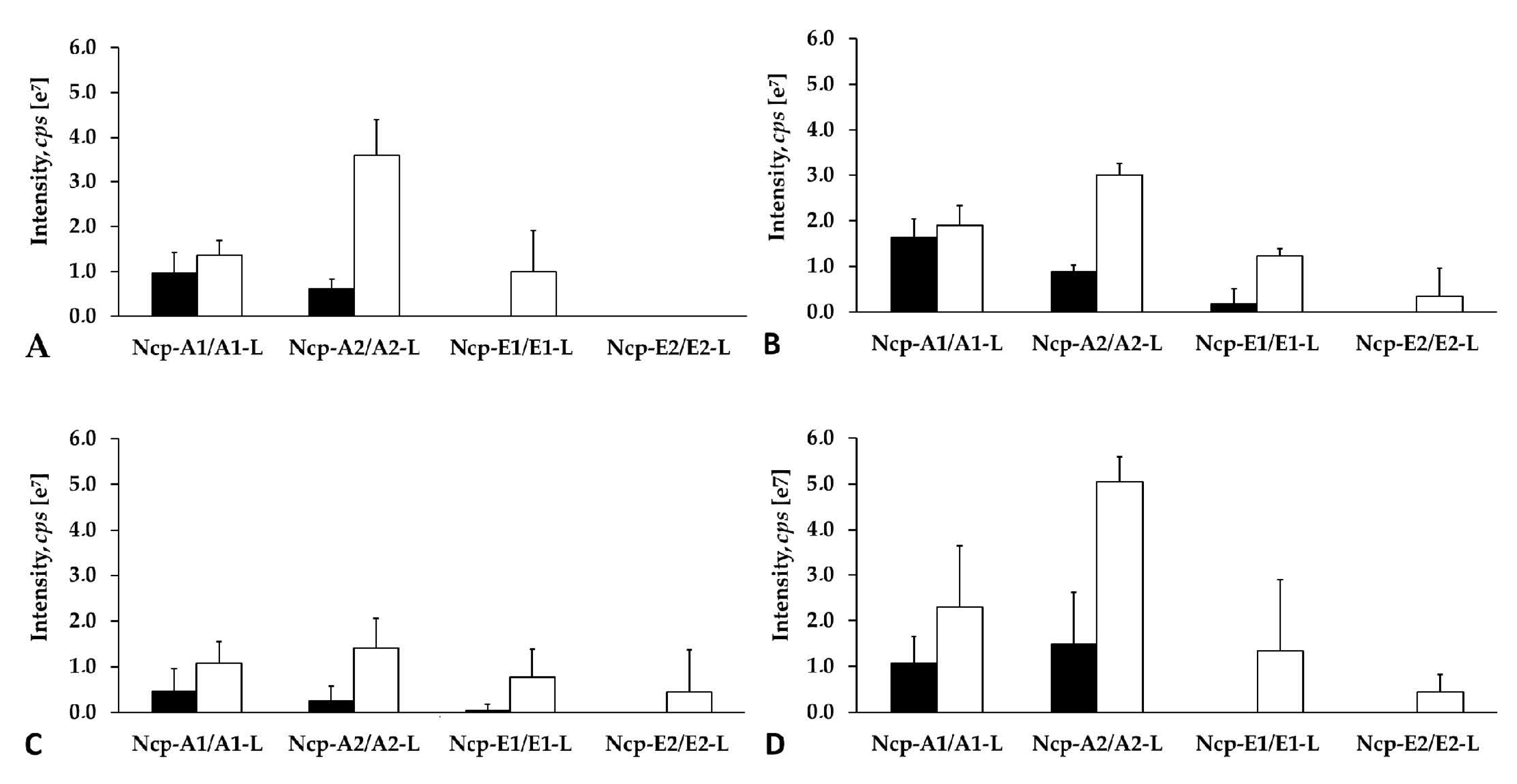
Apart from the structural analysis, we also made attempts to determine the relative amounts of the individual Ncps produced by N. edaphicum CCNP1411. To exclude the effect of the extraction procedure on the amounts of the detected peptides, different solvents and pH were applied. As the process of Ncp linearization during long storage of the freeze-dried material was suggested [16], both the fresh and lyophilized biomasses were analyzed. Regardless of the extraction procedure, Ncp-A2-L with Phe in C-terminus was always found to be the main Ncp analogue (Figure 10A–D). In addition, when MePro and Pro-containing peptide were compared separately, the peak intensity of the linear Ncps with Phe in C-terminus (i.e., Ncp-A2-L and Ncp-E1-L) was higher than the Ncps with Leu. These results might indicate preferential incorporation of Phe into the synthesized peptide chain.
The study also showed that the cell contents of the linear Ncps are higher than the cyclic ones. (Figure 10A–D and Figure S8). The release of Ncps from the synthetase as linear aldehydes is catalyzed by a reductase domain, located in the C-terminal part of the NcpB [17]. This reductive release triggers the spontaneous, and enzyme independent, macrocyclization of the linear peptide [19,20]. The reaction leads to the formation of a stable imino bond between the C-terminal aldehyde and the N-terminal amine group of Tyr [19,20]. In N. edaphicum CCNP1411 cells, depending on the Ncp analogue, the analyzed material (fresh or lyophilized) and extraction solvent, the cyclic Ncps constituted from even less than 10% (Ncp-A2, fresh biomass) to over 90% of the linear peptide (Figure 10A–D and Figure S8). In case of Ncp-A1, with MePro-Leu in C-terminus, the contribution of the cyclic form was always most significant, and at pH 8 it reached up to 91.7% of the linear peptide aldehyde (Ncp-A1-L) (Figure 10A–D and Figure S8). The cyclic analogues, Ncp-E1 and Ncp-E3 were produced in trace amounts and their spectra were sporadically recorded. It was proven that the macrocyclization process of Ncps is determined by the geometry of the linear peptide aldehyde and the conformation of D-Gln and Gly is crucial for the folding and formation of the imino bond [19]. As these two residues are present in all detected Ncps, then, probably other elements of the structure affect the cyclization process, as well. We hypothesize that due to the steric hindrances, the cyclization of Ncp-A1 with Leu in C-terminal position is easier than the cyclization of Ncp-A2 with Phe. As a consequence, the proportion of the cyclic Leu-containing Ncp-A1 to the linear form of the peptide is higher.
Thus far, Ncps synthesis was reported in few Nostoc strains, and the structural diversity of the peptides was found to be low. Other classes of NRPs were detected in cyanobacteria representing different orders and genera, and within one class of the peptides numerous analogues were detected. For example, the number of naturally produced cyclic heptapeptide microcystins (MCs), is over 270 [40,41] and in one cyanobacterial strains even 47 MCs analogues were detected [40]. Cyanopeptolins are produced by many cyanobacterial taxa and so far more than 190 structural analogues of the peptides have been discovered [41]. In this work, cyanopeptolin gene cluster was identified in N. edaphicum CCNP1411 and thirteen products of the genes were previously reported [6]. These peptides contain seven amino acids and a short fatty acid chain, and only one element of the structure, 3-amino-6-hydroxy-2-piperidone (Ahp), is conserved [6]. The structural diversity of NRPs is generated by frequent genetic recombination events and point mutations in the NRP gene cluster. The changes in gene sequences affect the structure and substrate specificity of the encoded enzymes. The tailoring enzymes can further modify the product, leading to even higher diversity of the synthetized peptides [42]. In case of Ncps, both the number of the producing organisms and the structural diversity of the peptides are limited. Ncp-M1 from Nostoc living in symbiosis with gastropod [16] is the only Ncp with structure distinctly different from Ncp-A1, Ncp-A2 and other Ncps described in this work.
The diversity within one class of bioactive metabolites offers a good opportunity for structure-activity relationship studies, without the need to synthesize the variants. The studies are of paramount importance when the efficacy and safety of a drug candidate are optimized. Therefore, in our future work, when sufficient quantities of pure Ncps are isolated, the activity of individual analogues against different cellular targets will be tested and compared, in order to select the lead compound for further studies.
3. Materials and Methods
3.1. Isolation, Purification and Culturing of Nostoc CCNP1411
Nostoc strain CCNP1411 was isolated in 2010 from the Gulf of Gdańsk, southern Baltic Sea, by Dr. Justyna Kobos. Based on the 16S rRNA sequence (GenBank accession number KJ161445) and morphological features, such as the shape of trichomes, cell size (4.56 ± 0.30 µm wide and 4.12 ± 0.72 µm long) and lack of akinetes [43,44], the strain was classified to N. edaphicum species. Purification of the strain was carried out by multiple transfers to a liquid and solid (1% bacterial agar) Z8 medium supplemented with NaCl to obtain the salinity of 7.3 [45]. To establish the strain as a monoculture, free from accompanying heterotrophic bacteria, a third-generation cephalosporin, ceftriaxone (100 µg/mL) (Pol-Aura, Olsztyn, Poland) was used. In addition, the purity of the culture was regularly tested by inoculation on LA agar (solid LB medium with 1.5% agar) [46] and on agar Columbia +5% sheep blood (BTL Ltd. Łódź, Poland), a highly nutritious medium, recommended for fastidious bacteria. Cyanobacteria cultures were grown in liquid Z8 medium (100 mL) at 22 ± 1 °C, continuous light of 5–10 µmol photons m−2 s−1. After three weeks of growth, the cyanobacterial biomass was harvested by passing the culture through a nylon net (mesh size 25 µm) and then freeze-dried before further processing.
3.2. Isolation and Sequencing of Genomic DNA
Genomic DNA of N. edaphicum CCNP1411 was isolated using SDS/Phenol method as described previously [47,48]. DNA quality control was performed by measuring the absorbance at 260/230 nm, template concentration was determined using Qubit fluorimeter (Thermo Fisher Scientific, Waltham, MA, USA), and DNA integrity was analyzed by 0.8% agarose gel electrophoresis and by PFGE using Biorad CHEF-III instrument (BioRad, Hercules, CA, USA).
Paired-end sequencing library was constructed using the NEB Ultra II FS Preparation Kit (New England Biolabs, Beverly, CA, USA) according to the manufacturer’s instructions. The library was sequenced using an Illumina MiSeq platform (Illumina, San Diego, CA, USA) with 2 × 300 paired-end reads. Sequence quality metrics were assessed using FASTQC (http://www.bioinformatics.babraham.ac.uk/projects/fastqc/) [49].
The long reads were obtained using the GridION sequencer (Oxford Nanopore Technologies, Oxford, UK). Prior to long-read library preparation, genomic DNA was sheared into 30 kb fragments using 26 G needle followed by size selection on Bluepippin instrument (Sage Science, Beverly, MA, USA). DNA fragments above 20 kb were recovered using PAC30 kb cassette. 5 µg of recovered DNA was taken for 1D library construction using SQK-LSK109 kit and 0.5 µg of the final library was loaded into R9.4.1 flowcell and sequenced on MinION sequencer.
3.3. Genome Assembling
Raw nanopore data was basecalled using Guppy v3.2.2 (Oxford Nanopore Technologies, Oxford, UK). After quality filtering using NanoFilt [50] and residual adapter removal using Porechop (https://github.com/rrwick/Porechop), the obtained dataset was quality checked using NanoPlot [50]. Long nanopore reads were then assembled using Flye v2.6 [51]. Flye assembled contigs were further polished using Illumina sequencing reads and Unicycler_polish pipeline (https://github.com/rrwick/Unicycler).
3.4. Genome and NRPS Alignment
Genome assembly was annotated using the NCBI Prokaryotic Genome Annotation Pipeline [52] with the assistance of prokka [53] refine annotation, with additionally curated database comprised of sequences selected by Nostocales order from NCBI non-redundant and refseq_genomes (280 positions) databases, enriched by 35 NRPS/PKS clusters selected by cyanobacteria phylum. To create circular maps of N. edaphicum CCNP1411 genome, the CGView Comparison Tool [54] was engaged with additional GC skew and GC content analyses.
Selected span for potential NRPS cluster was confirmed with BLASTn, BLASTp [55] and antiSMASH [56]. ORFs start codons within a putative cluster were verified by the presence of ribosome binding sites, 4–12 nucleotides upstream of the start codon. Schematic comparison of ORF BLASTn from relative synthetases, AY167420.1 and CP026681.1, was visualized by EasyFig program (http://mjsull.github.io/Easyfig/files.html). Annotated regions of NRPS span were subjected for NCBI Conserved Domain Database search [57] with a set e-value threshold (10−3), determining evolutionarily-conserved protein domains and motifs against CDD v3.18 database. Recognized motifs were selected using samtools v.1.9 and were subjected for protein structure and function prediction by I-TASSER [58], and results were confirmed with literature reports, PKS/NRPS Analysis Web-site prediction [59] and reevaluated using MEGAX suite [60]. Amino-acid sequence was visualized by BOXSHADE 3.2 program (https://embnet.vital-it.ch/software/BOX_form.html). Determination of domain ligand binding and active sites was achieved using COFACTOR and COACH part of I-TASSER analyses confirmed by MUSCLE amino acid alignment from MEGA X.
3.5. Data Deposition
Genomic sequences generated and analyzed in this study were deposited in the GenBank database under BioProject number: PRJNA638531.
3.6. Extraction and LC-MS/MS Analysis
For LC-MS/MS analyses of Ncps, the lyophilized (10 mg DW) biomass of N. edaphicum CCNP1411 was homogenized by grinding with mortar and pestle, and extracted in 1 mL of milliQ water, 20% methanol (pH 3.5, 6.0 and 8.0) and 50% methanol in water. The pH was adjusted with 0.5 M HCl and 1.0 M NaOH. In addition, the fresh material (500 mg FW) was extracted in 20% methanol in water. The samples were vortexed for 15 min and centrifuged at 14,000 rpm for 10 min, at 4 °C. The collected supernatants were directly analyzed by LC-MS/MS system.
The LC-MS/MS was carried out on an Agilent 1200 HPLC (Agilent Technologies, Waldbronn, Germany) coupled to a hybrid triple quadrupole/linear ion trap mass spectrometer QTRAP5500 (Applied Biosystems MDS Sciex, Concord, ON, Canada). The separation was achieved on a Zorbax Eclipse XDB-C18 column (4.6 mm ID × 150 mm, 5 µm; Agilent Technologies, Santa Clara, CA, USA). The extract components were separated by gradient elution from 10% to 100% B (acetonitrile with 0.1% formic acid) over 25 min, at a flow rate of 0.6 mL/min. As solvent A, 5% acetonitrile in MilliQ water with 0.1% formic acid was used. The mass spectrometer was operated in positive mode, with turbo ion source (5.5 kV; 550 °C). An information-dependent acquisition method at the following settings was used: for ions within the m/z range 500–1100 and signal intensity above the threshold of 500,000 cps the MS/MS spectra were acquired within the m/z range 50–1000, at a collision energy of 60 eV and declustering potential of 80 eV. Data were acquired with the Analyst ® Sofware (version 1.7 Applied Biosystems, Concord, ON, Canada).
4. Conclusions
Genes coding for subunits of the non-ribosomal peptide synthetase, in nostocyclopeptide-producing N. edaphicum CCNP1411, revealed differences in nucleotide compositions, compared to the previously described ncp cluster of Nostoc sp. ATCC53789. Although the analysis of fragments of genes coding for active sites and ligand binding sites of the conserved protein domains derived from N. edaphicum CCNP1411 and Nostoc sp. ATCC53789 indicated identical amino-acid compositions, residues within adenylation domains and substrate binding sites were different between compared sequences. This finding may highlight sites prone to mutations within regions accounted for structure and substrate stability. Analysis of ncp gene products in N. edaphicum CCNP1411 led to the detection of new nostocyclopeptide analogues. However, modifications in their structure were minor and limited to three positions of the heptapeptides. Although the naturally produced nostocyclopeptides were previously described as cyclic structures, in N. edaphicum CCNP1411 they are mainly present as linear peptide aldehydes, indicating a slow cyclization process.
Supplementary Materials
The following are available online at https://www.mdpi.com/1660-3397/18/9/442/s1. Figure S1: Structure and enhanced product ion mass spectrum of the cyclic nostocyclopeptide Ncp-A1 cyclo [Tyr+Gly+Gln+Ile+Ser+MePro+Leu] identified based on the following fragment ions: m/z 757 [M+H]; 739 [M+H−H2O]; 729 [M+H−CO]; 721 [M+H−2H2O]; 628 [M+H−MePro−H2O], 626 [M+H−Ile−H2O]; 594 [M+H−Tyr]; 549 [Tyr+Gly+Gln+Ile+Ser+H]; 541 [M+H−(Ser+MePro)−H2O], 446 [M+H−(Ile+Ser+MePro)]; 428 [M+H−(Ile+Ser+MePro)−H2O]; 386 [Gly+Gln+Ile+Ser+H]; 372 [MePro+Leu+Tyr+H]; 300 [Leu+Tyr+Gly+H−H2O]; 209 [MePro+Leu+H]; 181 [MePro+Leu+H−CO]; 86−Ile/Leu immonium; 84 MePro immonium, Figure S2: Structure and enhanced product ion mass spectrum of the linear peptide aldehyde Ncp-A1-L (linear aldehyde of Ncp-A1) Tyr+Gly+Gln+Ile+Ser+MePro+Leu identified based on the following fragment ions: m/z 775 [M+H]; 757 [M+H – H2O]; 739 [M+H−2H2O]; 660 [M+H−Leu]; 549 [Tyr+Gly+Gln+Ile+Ser+H]; 531 [Tyr+Gly+Gln+Ile+Ser+H−H2O]; 521 [Tyr+Gly+Gln+Ile+Ser+H−CO]; 532 [M+H−(MePro+Leu)−H2O]; 462 [Tyr+Gly+Gln+Ile+H]; 434 [Tyr+Gly+Gln+Ile+H−CO]; 386 [Gly+Gln+Ile+Ser+H]; 349 [Tyr+Gly+Gln+H]; 301 [Gln+Ile+Ser+H−CO]; 227 [MePro+Leu+H]; 221 [Tyr+Gly+H]; 209 [MePro+Leu+H−H2O]; 181 [MePro+Ile+H−H2O−CO]; 148 [Tyr−NH2]; 136 Tyr immonium; 86−Ile/Leu immonium; 84, 101 (immonium),129 Gln; 84 MePro immonium, Figure S3: Structure and enhanced product ion mass spectrum of the cyclic nostocyclopeptide Ncp-A2 cyclo[Tyr+Gly+Gln+Ile+Ser+MePro+Phe] identified based on the following fragment ions: m/z 791 [M+H]; 773 [M+H−H2O]; 763 [M+H−CO]; 755 [M+H−2H2O]; 745 [M+H−CO−H2O]; 678 [M+H−Ile]; 628 [M+H−Tyr]; 593 [M+H−(Ser+MePro)]; 549 [Tyr+Gly+Gln+Ile+Ser+H]; 531 [Tyr+Gly+Gln+Ile+Ser+H−H2O]; 480 [M+H−(Ile+Ser+MePro)]; 462 [Tyr+Gly+Gln+Ile+H]; 406 [MePro+Phe+Tyr+H]; 379 [MePro+Phe+Tyr+H−CO]; 349 [Tyr+Gly+Gln+H]; 335 [Phe+Tyr+Gly+H−H2O]; 312 [Ile+Ser+MePro+H]; 307 [Phe+Tyr+Gly+H−H2O−CO]; 243 [MePro+Phe+H]; 215 [MePro+Phe+H−CO]; 158 [Gly+Gln+H−CO]; 132 Phe; 84 MePro immonium, Figure S4: Structure and enhanced product ion mass spectrum of the linear nostocyclopeptide aldehyde Ncp-A2-L (linear aldehyde of Ncp-A2) Tyr+Gly+Gln+Ile+Ser+MePro+Phe identified based on the following fragment ions: m/z 809 [M+H]; 791 [M+H−H2O]; 773 [M+H−2H2O]; 763 [M+H−CO−H2O]; 660 [M+H−Phe]; 628 [M+H−Tyr−H2O]; 549 [Tyr+Gly+Gln+Ile+Ser+H]; 531 [M+H−(MePro+Phe)−H2O]; 462 [Tyr+Gly+Gln+Ile+H]; 434 [Tyr+Gly+Gln+Ile+H−CO]; 312 [Ile+Ser+MePro+H]; 261 [MePro+Phe+H]; 243 [MePro+Phe+H – H2O]; 221 [Tyr+Gly+H], 193 [Tyr+Gly+ H−CO]; 148 [Tyr−NH2]; 136 Tyr immonium; 84, 101 (immonium), 129 Gln; 84 MePro immonium, Figure S5: Proposed structure and enhanced product ion mass spectrum of cyclic nostocyclopeptide Ncp-E2 cyclo[Tyr+Gly+Gln+Ile+Ser+Pro+Leu] characterized based on the following fragment ions: m/z 743 [M+H]; 725 [M+H−H2O]; 715 [M+H−CO]; 707 [M+H−2H2O]; 697 [M+H – H2O−CO]; 656 [M+H−Ser]; 638 [M+H−Ser−H2O]; 628 [M+H−Ser−CO]; 612 [M+H−Ile−H2O]; 549 [Tyr+Gly+Gln+Ile+Ser+H]; 541 [M+H−(Ser+Pro)−H2O]; 531 [Tyr+Gly+Gln+Ile+Ser+H−H2O]; 428 [M+H−(Ile+Ser+Pro)−H2O]; 349 [Tyr+Gly+Gln+H]; 300 [Leu+Tyr+Gly+H−H2O]; 195 [Pro+Leu+H]; 167 [Pro+Leu+H−CO]; 84 Gln; 70 Pro immonium, Figure S6: Proposed structure and enhanced product ion mass spectrum of the linear nostocyclopeptide aldehyde Ncp-E2-L (linear aldehyde of Ncp-E2) with general structure Tyr+Gly+Gln+Ile+Ser+Pro+Leu characterized based on the following fragment ions: m/z 761 [M+H]; 743 [M+H−H2O]; 725 [M+H−2H2O]; 549 [Tyr+Gly+Gln+Ile+Ser+H]; 532 [Tyr+Gly+Gln+Ile+Ser+H−H2O]; 462 [Tyr+Gly+Gln+Ile+H]; 349 [Tyr+Gly+Gln+H]; 434 [Tyr+Gly+Gln+Ile+H−CO]; 300 [Ser+Pro+Leu+H]; 221 [Tyr+Gly+H]; 213 [Pro+Leu+H]; 195 [Pro+Leu+H−H2O]; 148 [Tyr−NH2]; 136 Tyr immonium; 84, 101 (immonium), 129 Gln; 70 Pro immonium, Figure S7: Proposed structure and enhanced product ion mass spectrum of cyclic nostocyclopeptide Ncp-E3 cyclo[Tyr+Gly+Gln+Val+Ser+MePro+Leu] characterized based on the following fragment ions: m/z 743 [M+H]; 725 [M+H−H2O]; 715 [M+H−CO]; 707 [M+H – 2H2O]; 697 [M+H−H2O−CO]; 645 [M+H−Val]; 580 [M+H−Tyr]; 527 [M+H−(Ser+MePro)−H2O]; 428 [M+H−(Val+Ser+MePro)−H2O]; 410 [M+H−(Val+Ser+MePro)−2H2O]; 372 [Gly+Gln+Val+Ser+H]; 344 [Gly+Gln+Val+Ser+H−CO]; 300 [Leu+Tyr+Gly+H−H2O]; 233 [Leu+Tyr+H−CO]; 209 [MePro+Leu+H]; 181 [MePro−Leu+H−CO]; 84 MePro immonium; 72 Val immonium, Figure S8: Relative contents of nostocyclopeptides extracted from 10 mg of lyophilized biomass of N. edaphicum CCNP1411 with 20% MeOH of different pH (3.5, 6 and 8)
Author Contributions
Conceptualization, H.M.-M. and G.W.; methodology, H.M.-M. and G.W.; software, M.G., J.G. and R.G.; validation, M.G., J.G. and R.G.; formal analysis, A.F. and M.G.; investigation, A.F. and M.G.; resources, data curation, M.G., H.M.-M., J.G. and R.G.; writing—original draft preparation, A.F. and M.G.; writing—review and editing, A.F., M.G.; H.M.-M. and G.W.; visualization, A.F. and M.G.; supervision, H.M.-M. and G.W.; project administration, H.M.-M.; funding acquisition, H.M.-M. All authors have read and agreed to the published version of the manuscript.
Funding
This research was funded by the National Science Centre in Poland 2016/21/B/NZ9/02304.
Conflicts of Interest
The authors declare no conflict of interest.
References
- Moore, R. Cyclic peptides and depsipeptides from cyanobacteria: A review. J. Ind. Microbiol. 1996, 16, 134–143. [Google Scholar] [CrossRef] [PubMed]
- Golakoti, T.; Ogino, J.; Heltzel, C.; Le Husebo, T.; Jensen, C.; Larsen, L.; Patterson, G.; Moore, R.; Mooberry, S.; Corbett, T.; et al. Structure determination, conformational analysis, chemical stability studies, and antitumor evaluation of the cryptophycins. Isolation of new 18 analogs from Nostoc sp. strain GSV 224. J. Am. Chem. Soc. 1995, 117, 12030–12049. [Google Scholar] [CrossRef]
- Boyd, M.; Gustafson, K.; McMahon, J.; Shoemaker, R.; O’Keefe, B.; Mori, T.; Gulakowski, R.; Wu, L.; Rivera, M.; Laurencot, C.; et al. Discovery of cyanovirin-N, a novel human immunodeficiency virus-inactivating protein that binds viral surface envelope glycoprotein gp120: Potential applications to microbicide development. Antimicrob. Agents Chemother. 1997, 41, 1521–1530. [Google Scholar] [CrossRef] [Green Version]
- Ploutno, A.; Carmeli, S. Nostocyclyne A, a novel antimicrobial cyclophane from the cyanobacterium Nostoc sp. J. Nat. Prod. 2000, 63, 1524–1526. [Google Scholar] [CrossRef]
- El-Sheekh, M.; Osman, M.; Dyan, M.; Amer, M. Production and characterization of antimicrobial active substance from the cyanobacterium Nostoc muscorum. Environ. Toxicol. Pharmacol. 2006, 21, 42–50. [Google Scholar] [CrossRef] [PubMed]
- Mazur-Marzec, H.; Fidor, A.; Cegłowska, M.; Wieczerzak, E.; Kropidłowska, M.; Goua, M.; Macaskill, J.; Edwards, C. Cyanopeptolins with trypsin and chymotrypsin inhibitory activity from the cyanobacterium Nostoc edaphicum CCNP1411. Mar. Drugs 2018, 16, 220. [Google Scholar] [CrossRef] [PubMed] [Green Version]
- Řezanka, T.; Dor, I.; Dembitsky, V. Fatty acid composition of six freshwater wild cyanobacterial species. Folia Microbiol. 2003, 48, 71–75. [Google Scholar] [CrossRef]
- Schwartz, R.; Hirsch, C.; Sesin, D.; Flor, J.; Chartrain, M.; Fromtling, R.; Harris, G.; Salvatore, M.; Liesch, J.; Yudin, K. Pharmaceuticals from cultured algae. J. Ind. Microbiol. 1990, 5, 113–124. [Google Scholar] [CrossRef]
- Gustafson, K.; Sowder, R.; Henderson, L.; Cardellina, J.; McMahon, J.; Rajamani, U.; Pannell, L.; Boyd, M. Isolation, primary sequence determination, and disulfide bond structure of cyanovirin-N, an anti-HIV (Human Immunodeficiency Virus) protein from the cyanobacterium Nostoc ellipsosporum. Biochem. Biophys. Res. Commun. 1997, 238, 223–228. [Google Scholar] [CrossRef]
- Okino, T.; Qi, S.; Matsuda, H.; Murakami, M.; Yamaguchi, K. Nostopeptins A and B, elastase inhibitors from the cyanobacterium Nostoc minutum. J. Nat. Prod. 1997, 60, 158–161. [Google Scholar] [CrossRef]
- Kaya, K.; Sano, T.; Beattie, K.; Codd, G. Nostocyclin, a novel 3-amino-6-hydroxy-2-piperidone containing cyclic depsipeptide from the cyanobacterium Nostoc sp. Tetrahedron Lett. 1996, 37, 6725–6728. [Google Scholar] [CrossRef]
- Mehner, C.; Müller, D.; Kehraus, S.; Hautmann, S.; Gütschow, M.; König, G. New peptolides from the cyanobacterium Nostoc insulare as selective and potent inhibitors of human leukocyte elastase. ChemBioChem 2008, 9, 2692–2703. [Google Scholar] [CrossRef] [PubMed]
- Golakoti, T.; Yoshida, W.; Chaganty, S.; Moore, R. Isolation and structure determination of nostocyclopeptides A1 and A2 from the terrestrial cyanobacterium Nostoc sp. ATCC53789. J. Nat. Prod. 2001, 64, 54–59. [Google Scholar] [CrossRef] [PubMed]
- Nowruzi, B.; Khavari-Nejad, R.; Sivonen, K.; Kazemi, B.; Najafi, F.; Nejadsattari, T. Indentification and toxigenic potential of Nostoc sp. Algae 2012, 27, 303–313. [Google Scholar] [CrossRef]
- Liaimer, A.; Jensen, J.; Dittmann, E. A genetic and chemical perspective on symbiotic recruitment of cyanobacteria of the genus Nostoc into the host plant Blasia pusilla L. Front. Microbiol. 2016, 7, 1963. [Google Scholar] [CrossRef] [PubMed]
- Jokela, J.; Herfindal, L.; Wahlsten, M.; Permi, P.; Selheim, F.; Vasconçelos, V.; Døskeland, S.; Sivonen, K. A novel cyanobacterial nostocyklopeptide is a potent antitoxin against Microcystis. ChemBioChem 2010, 11, 1594–1599. [Google Scholar] [CrossRef]
- Becker, J.; Moore, R.; Moore, B. Cloning, sequencing, and biochemical characterization of the nostocycyclopeptide biosynthetic gene cluster: Molecular basis for imine macrocyclization. Gene 2004, 325, 35–42. [Google Scholar] [CrossRef]
- Hoffmann, D.; Hevel, J.; Moore, R.; Moore, B. Sequence analysis and biochemical characterization of the nostopeptolide A biosynthetic gene cluster from Nostoc sp. GSV224. Gene 2003, 311, 171–180. [Google Scholar] [CrossRef]
- Kopp, F.; Mahlet, C.; Grünewald, J.; Marahiel, M. Peptide macrocyclization: The reductase of the nostocyclopeptide synthetase triggers the self-assembly of a macrocyclic imine. J. Am. Chem. Soc. 2006, 128, 16478–16479. [Google Scholar] [CrossRef]
- Enck, S.; Kopp, F.; Marahiel, M.; Geyer, A. The entropy balance of nostocyclopeptide macrocyclization analysed by NMR spectroscopy. ChemBioChem 2008, 9, 2597–2601. [Google Scholar] [CrossRef]
- Herfindal, L.; Myhren, L.; Kleppe, R.; Krakstad, C.; Selheim, F.; Jokela, J.; Sivonen, K.; Døskeland, S. Nostocyclopeptide-M1: A potent, nontoxic inhibitor of the hepatocyte drug trasporters OATP1B3 and OATP1B1. Mol. Pharm. 2011, 8, 360–367. [Google Scholar] [CrossRef]
- Lee, W.; Belkhiri, A.; Lockhart, A.; Merchant, N.; Glaeser, H.; Harris, E.; Washington, M.; Brunt, E.; Zaika, A.; Kim, R.; et al. Overexpression of OATP1B3 confers apoptotic resistance in colon cancer. Cancer Res. 2008, 68, 10315–10323. [Google Scholar] [CrossRef] [PubMed] [Green Version]
- Nishizawa, T.; Ueda, A.; Nakano, T.; Nishizawa, A.; Miura, T.; Asayama, M.; Fuji, K.; Harada, K.; Shirai, M. Characterization of the locus of genes encoding enzymes producing heptadepsipeptide micropeptin in the unicellular cyanobacterium Microcystis. J. Biochem. 2011, 149, 475–485. [Google Scholar] [CrossRef] [PubMed]
- Rouhiainen, L.; Jokela, J.; Fewer, D.; Urmann, M.; Sivonen, K. Two alternative starter modules for the non-ribosomal biosynthesis of specific anabaenopeptin variants in Anabaena (cyanobacteria). Chem. Biol. 2010, 17, 265–273. [Google Scholar] [CrossRef] [PubMed] [Green Version]
- Luesch, H.; Hoffmann, D.; Hevel, J.; Becker, J.; Golakoti, T.; Moore, R. Biosynthesis of 4-Methylproline in Cyanobacteria: Cloning of nosE and nosF and biochemical characterization of the encoded dehydrogenase and reductase activities. J. Org. Chem. 2003, 68, 83–91. [Google Scholar] [CrossRef] [PubMed]
- Lambalot, R.; Walsh, C. Cloning, Overproduction, and Characterization of the Escherichia coli Holo-acyl Carrier Protein Synthase. J. Biol. Chem. 1995, 270, 24658–24661. [Google Scholar] [CrossRef] [Green Version]
- Marchler-Bauer, A.; Derbyshire, M.; Gonzales, N.; Lu, S.; Chitsaz, F.; Geer, L.; Geer, R.; He, J.; Gwadz, M.; Hurwitz, D.; et al. CDD: NCBI’s conserved domain database. Nucleic Acids Res. 2015, 43, d222–d226. [Google Scholar] [CrossRef] [Green Version]
- Stachelhaus, T.; Mootz, H.; Marahiel, M. The specificity-conferring code of adenylation domains in nonribosomal peptide synthetases. Chem. Biol. 1999, 6, 493–505. [Google Scholar] [CrossRef] [Green Version]
- Challis, G.; Ravel, J.; Townsend, C. Predictive, structure-based model of amino acid recognition by nonribosomal peptide synthetase adenylation domains. Chem. Biol. 2000, 7, 211–224. [Google Scholar] [CrossRef] [Green Version]
- Stein, T.; Vater, J.; Kruft, V.; Otto, A.; Wittmann-Liebold, B.; Franke, P.; Panico, M.; McDowell, R.; Morris, H. The multiple carrier model of nonribosomal peptide biosynthesis at modular multienzymatic templates. J. Biol. Chem. 1996, 271, 15428–15435. [Google Scholar] [CrossRef] [Green Version]
- Dehling, E.; Volkmann, G.; Matern, J.; Dörner, W.; Alfermann, J.; Diecker, J.; Mootz, H. Mapping of the Communication-Mediating Interface in Nonribosomal Peptide Synthetases Using a Genetically Encoded Photocrosslinker Supports an Upside-Down Helix-Hand Motif. J. Mol. Biol. 2016, 428, 4345–4360. [Google Scholar] [CrossRef]
- Hacker, C.; Cai, X.; Kegler, C.; Zhao, L.; Weickhmann, K.; Wurm, J.; Bode, H.; Wöhnert, J. Structure-based redesign of docking domain interactions modulates the product spectrum of a rhabdopeptide-synthesizing NRPS. Nat. Commun. 2018, 9, 4366. [Google Scholar] [CrossRef] [Green Version]
- Richter, C.; Nietlispach, D.; Broadhurst, R.; Weissman, K. Multienzyme docking in hybrid megasynthetases. Nat. Chem. Biol. 2008, 4, 75–81. [Google Scholar] [CrossRef] [PubMed]
- Haese, A.; Schubert, M.; Herrmann, M.; Zocher, R. Molecular characterization of the enniatin synthetase gene encoding a multifunctional enzyme catalysing N-methyldepsipeptide formation in Fusarium scirpi. Mol. Biol. 1993, 7, 905–914. [Google Scholar] [CrossRef]
- Marahiel, M.; Stachelhaus, T.; Mootz, H. Modular peptide synthetases involved in nonribosomal peptide synthesis. Chem. Rev. 1997, 97, 2651–2674. [Google Scholar] [CrossRef]
- Chang, C.; Lohman, J.; Huang, T.; Michalska, K.; Bigelow, L.; Rudolf, J.; Jędrzejczak, R.; Yan, X.; Ma, M.; Babnigg, G.; et al. Structural Insights into the Free-Standing Condensation Enzyme SgcC5 Catalyzing Ester-Bond Formation in the Biosynthesis of the Enediyne Antitumor Antibiotic C-1027. Biochemistry 2018, 57, 3278–3288. [Google Scholar] [CrossRef]
- Du, L.; Lou, L. PKS and NRPS release mechanisms. Nat. Prod. Rep. 2010, 27, 255–278. [Google Scholar] [CrossRef] [PubMed]
- Koketsu, K.; Minami, A.; Watanabe, K.; Oguri, H.; Oikawa, H. Pictet-Spenglerase involved in tetrahydroisoquinoline antibiotic biosynthesis. Curr. Opin. Chem. Biol. 2012, 16, 142–149. [Google Scholar] [CrossRef] [PubMed]
- Liu, L.; Jokela, J.; Herfindal, L.; Wahlsten, M.; Sinkkonen, J.; Permi, P.; Fewer, D.; Døskeland, S.; Sivonen, K. 4-Methylproline guided natural product discovery: Co-occurrence of a 4-hydroxy- and 4-methtylprolines in nostoweipeptins and nostopeptolides. ACS Chem. Biol. 2014, 9, 2646–2655. [Google Scholar] [CrossRef]
- Bouaïcha, N.; Miles, C.; Beach, D.; Labidi, Z.; Djabri, A.; Benayache, N.; Nguyen-Quang, T. Structural diversity, characterization and toxicology of microcystins. Toxins 2019, 11, 714. [Google Scholar] [CrossRef] [Green Version]
- Jones, M.; Pinto, E.; Torres, M.; Dörr, F.; Mazur-Marzec, H.; Szubert, K.; Tartaglione, L.; Dell’Aversano, C.; Miles, C.; Beach, D.; et al. Comprehensive database of secondary metabolites from cyanobacteria. BioRxiv 2020. [Google Scholar] [CrossRef] [Green Version]
- Meyer, S.; Kehr, J.; Mainz, A.; Dehm, D.; Petras, D.; Süssmuth, R.; Dittmann, E. Biochemical dissection of the natural diversification of microcystin provides lessons for synthetic biology of NRPS. Cell. Chem. Biol. 2016, 23, 462–471. [Google Scholar] [CrossRef] [PubMed] [Green Version]
- Kondratyeva, N.V. Novyi vyd synio-zelenykh vodorostey—Nostoc edaphicum sp. n. [A new species of blue-green algae—Nostoc edapicum sp. n. Ukr. Bot. J. 1962, 19, 58–65. [Google Scholar]
- Komárek, J. Süsswasserflora von Mitteleuropa. Cyanoprokaryota: 3rd Part: Heterocystous Genera; Springer Spectrum: Berlin/Heidelberg, Germany, 2013; Volume 19, pp. 1–1130. [Google Scholar]
- Kotai, J. Instructions for Preparation of Modified Nutrient Solution Z8 for Algae; Publication: B-11/69; Norwegian Institute for Water Research: Oslo, Norway, 1972; p. 5. [Google Scholar]
- Bertani, G. Studies on lysogenesis. I. The mode of phage liberation by lysogenic Escherichia Coli. J. Bacteriol. 1951, 62, 293–300. [Google Scholar] [CrossRef] [PubMed] [Green Version]
- Nowak, R.; Jastrzębski, J.; Kuśmirek, W.; Sałamatin, R.; Rydzanicz, M.; Sobczyk-Kopcioł, A.; Sulima-Celińska, A.; Paukszto, Ł.; Makowaczenko, K.; Płoski, R.; et al. Hybrid de novo whole genome assembly and annotation of the model tapeworm Hymenolepis diminuta. Sci. Data 2019, 6, 302. [Google Scholar] [CrossRef] [Green Version]
- Wilson, K. Preparation of genomic DNA from bacteria. In Current Protocols in Molecular Biology; Ausubel, R., Bent, R.E., Eds.; Kingston: Fountain Valley, CA, USA; Wiley & Sons: New York, NY, USA, 1987; pp. 2.10–2.12. [Google Scholar]
- Andrews, S. Babraham Bioinformatics, FastQC—A Quality Control Application for High Throughput Sequence Data. 2010. Available online: http://www.bioinformatics.babraham.ac.uk/projects/fastqc/ (accessed on 11 February 2020).
- De Coster, W.; D’Hert, S.; Schultz, D.; Cruts, M.; Van Broeckhoven, C. NanoPack: Visualizing and processing long–read sequencing data. Bioinformatics 2018, 34, 2666–2669. [Google Scholar] [CrossRef]
- Kolmogorov, M.; Yuan, J.; Lin, Y.; Pevzner, P. Assembly of long, error–prone reads using repeat graphs. Nat. Biotechnol. 2019, 37, 540–546. [Google Scholar] [CrossRef]
- Tatusova, T.; DiCuccio, M.; Badretdin, A.; Chetvernin, V.; Nawrocki, E.; Zaslavsky, L.; Lomsadze, A.; Pruitt, K.; Borodovsky, M.; Ostell, J. NCBI prokaryotic genome annotation pipeline. Nucleic Acids Res. 2016, 44, 6614–6624. [Google Scholar] [CrossRef]
- Seemann, T. Prokka: Rapid prokaryotic genome annotation. Bioinformatics 2014, 30, 2068–2069. [Google Scholar] [CrossRef]
- Grant, J.; Arantes, A.; Stothard, P. Comparing thousands of circular genomes using the CGView comparison tool. BMC Genom. 2012, 13, 202. [Google Scholar] [CrossRef] [Green Version]
- Altschul, S.; Madden, T.; Schäffer, A.; Zhang, J.; Zhang, Z.; Miller, W.; Lipman, D. Gapped BLAST and PSI–BLAST: A new generation of protein database search programs. Nucleic Acids Res. 1997, 25, 3389–3402. [Google Scholar] [CrossRef] [PubMed] [Green Version]
- Medema, M.; Blin, K.; Cimermanic, P.; de Jager, V.; Zakrzewski, P.; Fischbach, M.; Weber, T.; Takano, E.; Breitling, R. antiSMASH: Rapid identification, annotation and analysis of secondary metabolites biosynthesis gene clusters in bacterial and fungal genome sequences. Nucleic Acids Res. 2011, 39, w339–w346. [Google Scholar] [CrossRef]
- Marchler-Bauer, A.; Lu, S.; Anderson, J.; Chitsaz, F.; Derbyshire, M.; DeWesse-Scott, C.; Fong, J.; Geer, L.; Geer, R.; Gonzales, N.; et al. CDD: A conserved database for the functional annotation of proteins. Nucleic Acids Res. 2011, 39, d225–d229. [Google Scholar] [CrossRef] [PubMed] [Green Version]
- Yang, J.; Yan, R.; Roy, A.; Xu, D.; Poisson, J.; Zhang, Y. The I–TASSER Suite: Protein structure and function prediction. Nat. Methods 2015, 12, 7–8. [Google Scholar] [CrossRef] [PubMed] [Green Version]
- Bachmann, B.; Ravel, J. Methods for in silico prediction of microbial polyketide and nonribosomal peptide biosynthetic pathways from DNA sequence data. Methods Enzymol. 2009, 458, 181–217. [Google Scholar]
- Kumar, S.; Stecher, G.; Li, M.; Knyaz, C.; Tamura, K. MEGA X: Molecular Evolutionary Genetics Analysis across computing platforms. Mol. Biol. Evol. 2018, 35, 1547–1549. [Google Scholar] [CrossRef]
Figure 1.
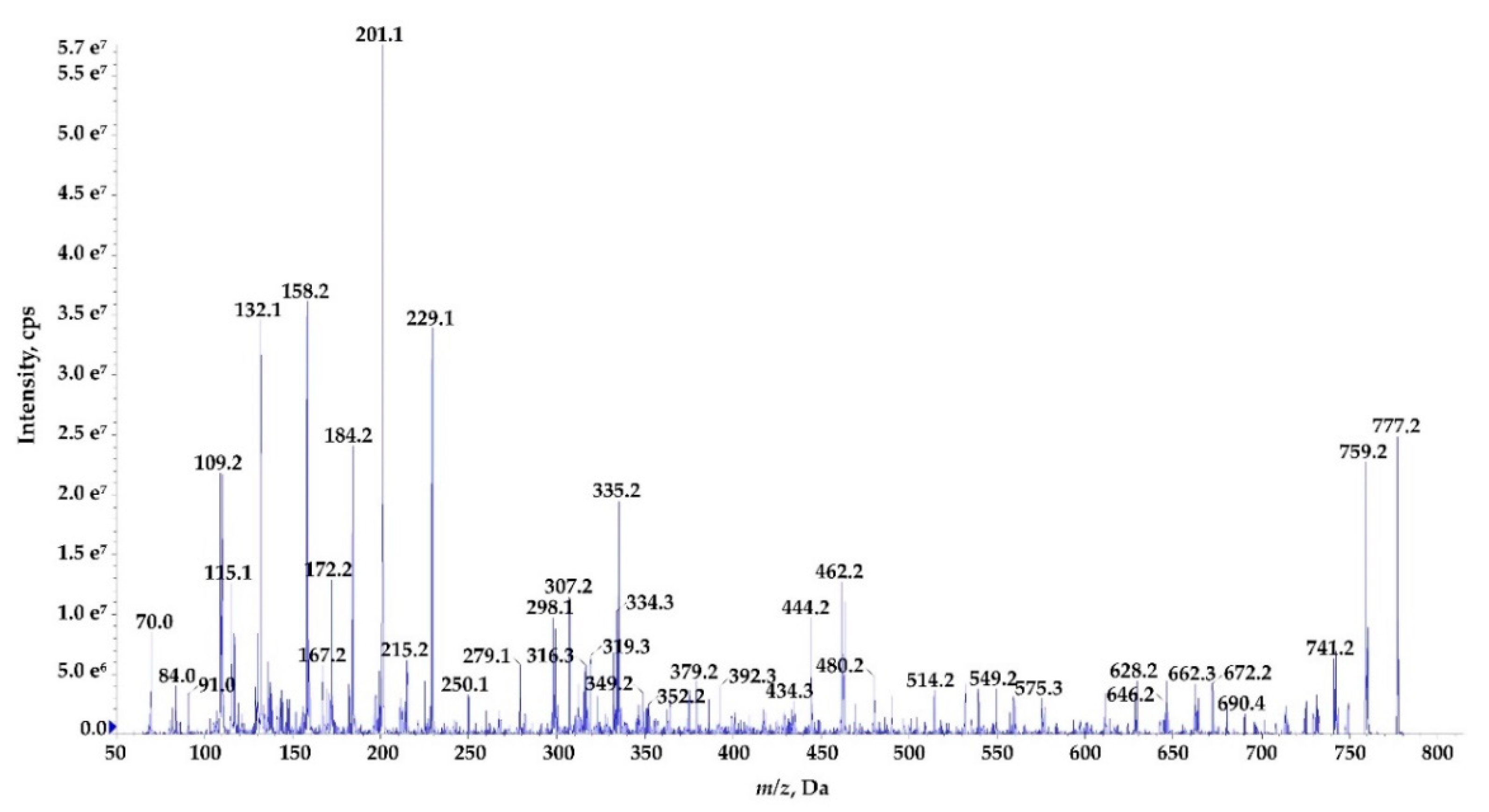
Nostoc edaphicum CCNP1411 (A) and the proposed chemical structure of Ncp-E1 (B).

Figure 2.
Map of the N. edaphicum CCNP1411 genome where chromosome (chr) and five plasmids (pNe_1–5) are presented. The ORFs are indicated with grey arrows split into two rings outermost showing ORFs on direct strand and inner showing complementary strand ORFs. The middle circle shows GC content (black) and the innermost circle shows GC skew (green and purple). Genes for putative NRPS/PKS are marked on the chromosome in their proper position (red within a black circle), with closeup on NRPS in position 7,609,981–7,643,289 putatively coding for Ncp biosynthetic gene cluster.

Figure 3.
Schematic alignment of genes coding for putative non-ribosomal peptide synthetase from N. edaphicum CCNP1411 (red) and two related Ncp-producing synthetases AY167420.1 and CP026681.1 (white). The grey bar in the lower right corner shows the identity percentage associated with color of the bars connecting homologous regions. The analysis was conducted at the nucleotide level.

Figure 4.
Structure-based sequence alignment of 4′-phosphopantetheinyl transferase and partial ORF3. Amino-acids highlighted in black color indicate conserved residues, whereas those in grey color indicate conservative mutations.
Figure 4.
Structure-based sequence alignment of 4′-phosphopantetheinyl transferase and partial ORF3. Amino-acids highlighted in black color indicate conserved residues, whereas those in grey color indicate conservative mutations.

Figure 5.
Schematic representation of conserved domains within ncpA and ncpB coding nucleotide sequences. They are composed of repetitive modules condensation (C), adenylation (A) and peptidyl carrier protein (PCP) domains adjacent to delineating docking, epimerization and reductase domains aligned with two related synthetases AY167420.1 and CP026681.1. The analysis was conducted at the nucleotide level.

Figure 6.
Heatmap of the highest (light blue) and lowest (black) percentage of similarities between NcpA and NcpB domains in N. edaphicum CCNP1411 and ATCC53789; values scaled by rows. The analysis was conducted at the amino acid level.
Figure 6.
Heatmap of the highest (light blue) and lowest (black) percentage of similarities between NcpA and NcpB domains in N. edaphicum CCNP1411 and ATCC53789; values scaled by rows. The analysis was conducted at the amino acid level.

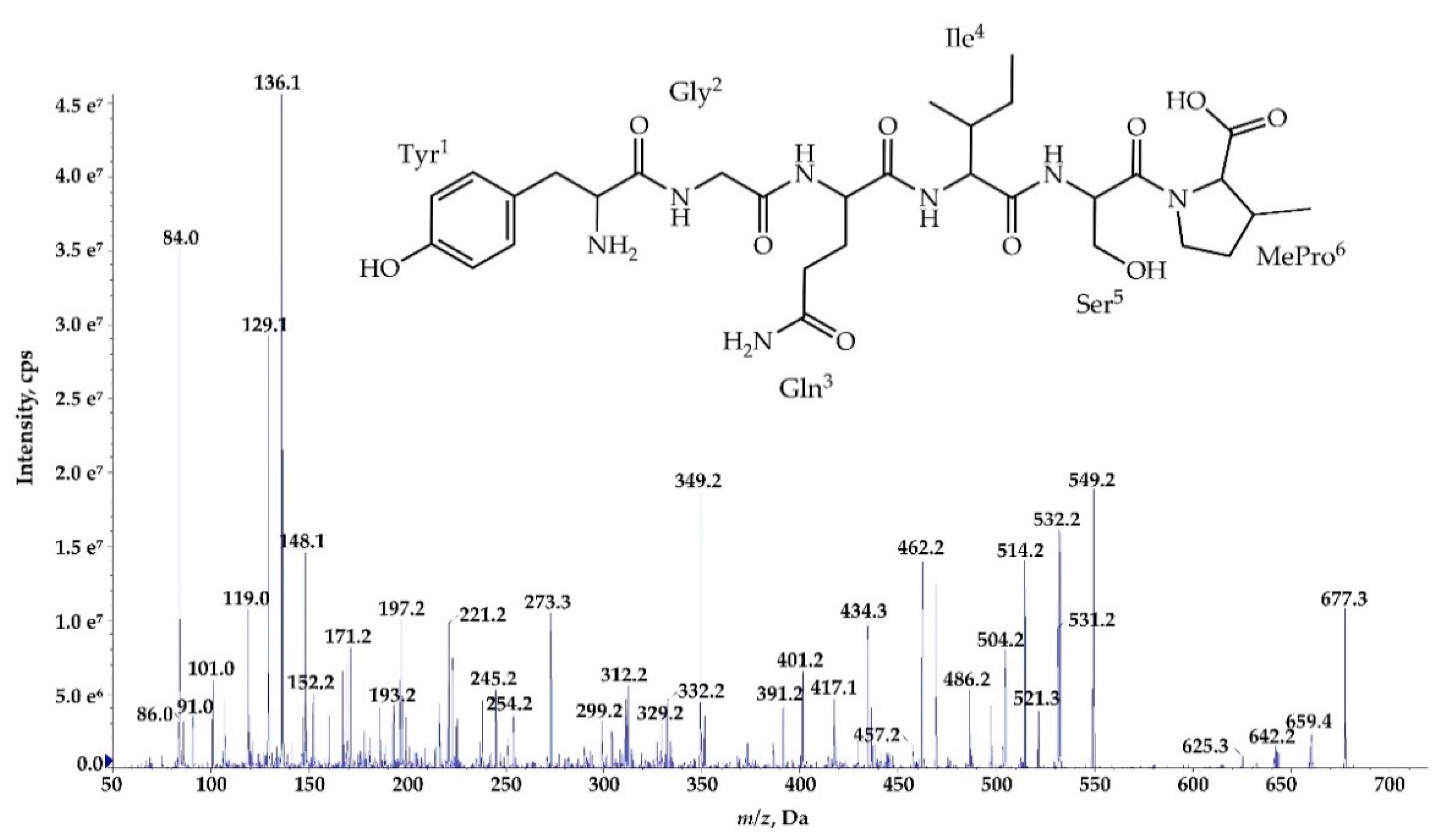
Figure 7.
Postulated structure and enhanced product ion mass spectrum of the linear aldehyde nostocyclopeptide Ncp-E1-L; Tyr+Gly+Gln+Ile+Ser+Pro+Phe characterized based on the following fragment ions: m/z 795 [M+H]; 777 [M+H−H2O]; 759 [M+H−2H2O]; 646 [M+H – Phe]; 614 [M+H – Tyr−H2O]; 575 [M+H−(Tyr+Gly)]; 549 [Tyr+Gly+Gln+Ile+Ser+H]; 531 [Tyr+Gly+Gln+Ile+Ser+H−H2O]; 462 [Tyr+Gly+Gln+Ile+H]; 349 [Tyr+Gly+Gln+H]; 334 [Ser+Pro+Phe+2H]; 247 [Phe+Pro+H]; 229 [Phe+Pro+H]; 201 [Phe+Pro+H – CO]; 148 [Tyr−NH2]; 136 Tyr immonium; 129, 101 (immonium), 84 Gln; 70 Pro immonium.

Figure 8.
Postulated tructure and enhanced product ion mass spectrum of a linear nostocyclopeptide Ncp-E4-L [Tyr+Gly+Gln+Ile+Ser+MePro] characterized based on the following fragment ions: m/z 677 [M+H]; 659 [M+H−H2O]; 642 [M+H−H2O−NH3]; 549 [Tyr+Gly+Gln+Ile+Ser+H]; 531 [Tyr+Gly+Gln+Ile+Ser+H−H2O]; 521 [Tyr+Gly+Gln+Ile+Ser+H−CO]; 462 [Tyr+Gly+Gln+Ile+H]; 434 [Tyr+Gly+Gln+Ile+H−CO]; 349 [Tyr+Gly+Gln+H]; 329 [Gln+Ile+Ser+H]; 312 [Ile+Ser+MePro+H]; 221 [Tyr+Gly+H]; 193 [Tyr+Gly+H−CO]; 148 [Tyr−NH2]; 136 Tyr immonium; 86 Ile immonium; 84, 101 (immonium), 129 Gln; 84 MePro immonium.

Figure 9.
Enhanced product ion mass spectrum of the cyclic nostocyclopeptide Ncp-E1 with putative structure cyclo[Tyr+Gly+Gln+Ile+Ser+Pro+Phe] characterised based on the following fragment ions: m/z 777 [M+H]; 759 [M+H−H2O]; 741 [M+H−2H2O]; 690 [M+H−Ser]; 672 [M+H−Ser−H2O]; 662 [M+H−Pro−H2O]; 646 [M+H−Phe]; 628 [M+H−Phe−H2O]; 575 [M+H−(Ser+Pro)−H2O]; 549 [Tyr+Gly+Glu+Ile+Ser+H]; 480 [Phe+Tyr+Gly+Gln+H]; 462 [Tyr+Gly+Gln+Ile+H]; 444 [Tyr+Gly+Gln+Ile+H−H2O]; 434 [Tyr+Gly+Gln+Ile+H−CO]; 392 [Pro+Phe+Tyr+H]; 352 [Phe+Tyr+Gly+H]; 335 [Phe+Tyr+Gly+H−H2O]; 316 [Ser+Pro+Phe+H]; 307 [Phe+Tyr+Gly+H−H2O -CO]; 298 [Ile+Ser+Pro+H]; 229 [Phe+Pro+H]; 201 [Phe+Pro+H−CO]; 158 [Gly+Gln+H−CO]; 132 Phe; 70 Pro immonium. Structure of the peptide is presented in Figure 1.

Figure 10.
Relative cell contents of nostocyclopeptides extracted from 10 mg of lyophilized (A,B,D) or 500 mg fresh (C) biomass of N. edaphicum CCNP1411 with MilliQ water (A), 50% methanol in water (B), and 20% methanol in water (C,D). The cell content was expressed as peak intensity in LC-MS/MS chromatogram.


Table 1.
Composition and coverage of N. edaphicum CCNP1411 genome.
| Replicon | Accession Number | Length (bp) | Topology | G+C Content (%) | Coverage (x) Nanopore Data | Coverage (x) Illumina Data |
|---|---|---|---|---|---|---|
| pNe_1 | CP054693.1 | 44,503 | Circular | 42.3 | 115.5 | 244.8 |
| pNe_2 | CP054694.1 | 99,098 | Circular | 40.2 | 168.4 | 135.3 |
| pNe_3 | CP054695.1 | 120,515 | Circular | 41.3 | 256.4 | 177.1 |
| pNe_4 | CP054696.1 | 53,840 | Circular | 41.6 | 102.4 | 211.5 |
| pNe_5 | CP054697.1 | 264,855 | Circular | 41.0 | 226.3 | 160.7 |
| chr | CP054698.1 | 7,733,505 | Circular | 41.6 | 160.7 | 116.9 |
Table 2.
Characterization of substrate binding pocket amino acid residues adenylation domains of NcpA and NcpB modules based on gramicidin S synthetase (GrsA) phenylalanine activating domain. Residues in brackets mark inconsistency with AY167420.1 residues.
Table 2.
Characterization of substrate binding pocket amino acid residues adenylation domains of NcpA and NcpB modules based on gramicidin S synthetase (GrsA) phenylalanine activating domain. Residues in brackets mark inconsistency with AY167420.1 residues.
| NRPS Module | Adenylation Domain Residue Position | Proposed Substrate | ||||||||
|---|---|---|---|---|---|---|---|---|---|---|
| 235 | 236 | 239 | 278 | 299 | 301 | 322 | 330 | 331 | ||
| NcpA1 | D | A | S | T | [I] | A | A | V | C | Tyr |
| NcpA2 | D | I | L | Q | L | G | L | I | W | Gly |
| NcpA3 | D | A | W | Q | F | G | L | I | D | Gln |
| NcpB1 | D | A | F | F | L | G | V | T | F | Ile/Val |
| NcpB2 | D | V | W | H | I | S | L | I | D | Ser |
| NcpB3 | D | V | Q | [F] | I | A | H | V | A | Pro/MePro |
| NcpB4 | D | A | W | [T] | I | G | [A] | V | C | Phe/Tyr/Leu |
Table 3.
The putative structures of nostocyclopeptides (Ncps) detected in the crude extract of N. edaphicum CCNP1411 and the structure of Ncp-M1 identified in Nostoc sp. XSPORK 13 A [16]. The new analogues are marked in bold and the peptides detected in trace amounts are marked with [T]. The variable residues in position 4 and 7 are marked in blue.
| Compound | Structure | m/z [M+H]+ | Retention Time [min] | |
|---|---|---|---|---|
| Cyclic | Linear–COH | |||
| Ncp-A1 | cyclo[Tyr+Gly+Gln+Ile+Ser+MePro+Leu] | 757 | 7.1 | |
| Ncp-A1-L | Tyr+Gly+Gln+Ile+Ser+MePro+Leu | 775 | 5.8 | |
| Ncp-A2 | cyclo[Tyr+Gly+Gln+Ile+Ser+MePro+Phe] | 791 | 6.0 | |
| Ncp-A2-L | Tyr+Gly+Gln+Ile+Ser+MePro+Phe | 809 | 5.6 | |
| Ncp-E1 | cyclo[Tyr+Gly+Gln+Ile+Ser+Pro+Phe] | 777 [T] | 7.2 | |
| Ncp-E1-L | Tyr+Gly+Gln+Ile+Ser+Pro+Phe | 795 | 5.7 | |
| Ncp-E2 | cyclo[Tyr+Gly+Gln+Ile+Ser+Pro+Leu] | 743 [T] | 6.3 | |
| Ncp-E2-L | Tyr+Gly+Gln+Ile+Ser+Pro+Leu | 761 | 5.1 | |
| Ncp-E3 | cyclo[Tyr+Gly+Gln+Val+Ser+MePro+Leu] | 743 [T] | 7.0 | |
| * Ncp-E4-L | [Tyr+Gly+Gln+Ile+Ser+MePro] | 677 [T] | 6.0 | |
| ** Ncp-M1 | cyclo[Tyr+Tyr+HSe+Pro+Val+MePro+Tyr] | 882 | 27.5 | |
* Ncp-E4-L is the only linear Ncps analogue with carboxyl group in C-terminus. ** Identified in Nostoc sp. XSPORK [16].
© 2020 by the authors. Licensee MDPI, Basel, Switzerland. This article is an open access article distributed under the terms and conditions of the Creative Commons Attribution (CC BY) license (http://creativecommons.org/licenses/by/4.0/).
Share and Cite
MDPI and ACS Style
Fidor, A.; Grabski, M.; Gawor, J.; Gromadka, R.; Węgrzyn, G.; Mazur-Marzec, H. Nostoc edaphicum CCNP1411 from the Baltic Sea—A New Producer of Nostocyclopeptides. Mar. Drugs 2020, 18, 442. https://doi.org/10.3390/md18090442
AMA Style
Fidor A, Grabski M, Gawor J, Gromadka R, Węgrzyn G, Mazur-Marzec H. Nostoc edaphicum CCNP1411 from the Baltic Sea—A New Producer of Nostocyclopeptides. Marine Drugs. 2020; 18(9):442. https://doi.org/10.3390/md18090442
Chicago/Turabian StyleFidor, Anna, Michał Grabski, Jan Gawor, Robert Gromadka, Grzegorz Węgrzyn, and Hanna Mazur-Marzec. 2020. "Nostoc edaphicum CCNP1411 from the Baltic Sea—A New Producer of Nostocyclopeptides" Marine Drugs 18, no. 9: 442. https://doi.org/10.3390/md18090442
Note that from the first issue of 2016, this journal uses article numbers instead of page numbers. See further details here.