Article

Anti-Inflammatory Polyketide Derivatives from the Sponge-Derived Fungus Pestalotiopsis sp. SWMU-WZ04-2

1
School of Pharmacy, Southwest Medical University, Luzhou 646000, China
2
Key Laboratory of Tropical Marine Bioresources and Ecology, Guangdong Key Laboratory of Marine Materia Medica, South China Sea Institute of Oceanology, Chinese Academy of Sciences, Guangzhou 510301, China
3
University of Chinese Academy of Sciences, 19 Yuquan Road, Beijing 100049, China
*
Authors to whom correspondence should be addressed.
Mar. Drugs 2022, 20(11), 711; https://doi.org/10.3390/md20110711
Submission received: 11 October 2022 / Revised: 5 November 2022 / Accepted: 9 November 2022 / Published: 13 November 2022
(This article belongs to the Special Issue Bioactive Compounds from the Deep-Sea-Derived Microorganisms)

Abstract

:
Five undescribed polyketide derivatives, pestaloketides A–E (15), along with eleven known analogues (616), were isolated from the sponge-derived fungus Pestalotiopsis sp. Their structures, including absolute configurations, were elucidated by analyses of NMR spectroscopic HRESIMS data and electronic circular dichroism (ECD) calculations. Compounds 5, 6, 9, and 14 exhibited weak cytotoxicities against four human cancer cell lines, with IC50 values ranging from 22.1 to 100 μM. Pestaloketide A (1) is an unusual polyketide, featuring a rare 5/10/5-fused ring system. Pestaloketides A (1) and B (2) exhibited moderately inhibited LPS-induced NO production activity, with IC50 values of 23.6 and 14.5 μM, respectively, without cytotoxicity observed. Preliminary bioactivity evaluations and molecular docking analysis indicated that pestaloketides A (1) and B (2) had the potential to be developed into anti-inflammatory activity drug leads.

1. Introduction

Marine sponge-derived fungi have been proven to be a large and promising source of novel drug leads [1]. Among them, the pestalotiopsis species isolated from specific habitats are especially recognized as important producers of structurally varied, biologically active metabolites [2,3,4]. Since the discovery of taxol from the fungal Pestalotiopsis microspora [5], many novel secondary metabolites with potential pharmaceutical properties have been reported from this genus, including anti-inflammatory, cytotoxic, antiviral, antioxidant, and antimicrobial activities [6,7,8]. Accordingly, these findings have inspired many researchers to investigate the bioactive metabolites produced by Pestalotiopsis species.
The pestalotiopsis species, mainly distributed in both terrestrial and marine habitats, can produce many secondary metabolites. Polyketides possessing a rearranged or a modified different carbon nucleus have been reported from Pestalotiopsis species [9,10]. However, a novel tricyclic 5/10/5 skeleton has not been declared.
As part of our ongoing excavation for new secondary metabolites from sponge-derived fungi [11], the fungus Pestalotiopsis sp. was investigated by the “epigenetic modification” strategy, including 5-aza-2-deoxycytidine [12]. Chemical exploration of ethyl acetate (EtOAc) extract of the fungus led to the isolation of five new polyketide derivatives (15) (Figure 1), along with eleven known compounds (616). Herein, details of the isolation, structural elucidation, and bioactivities of the isolated compounds are described.

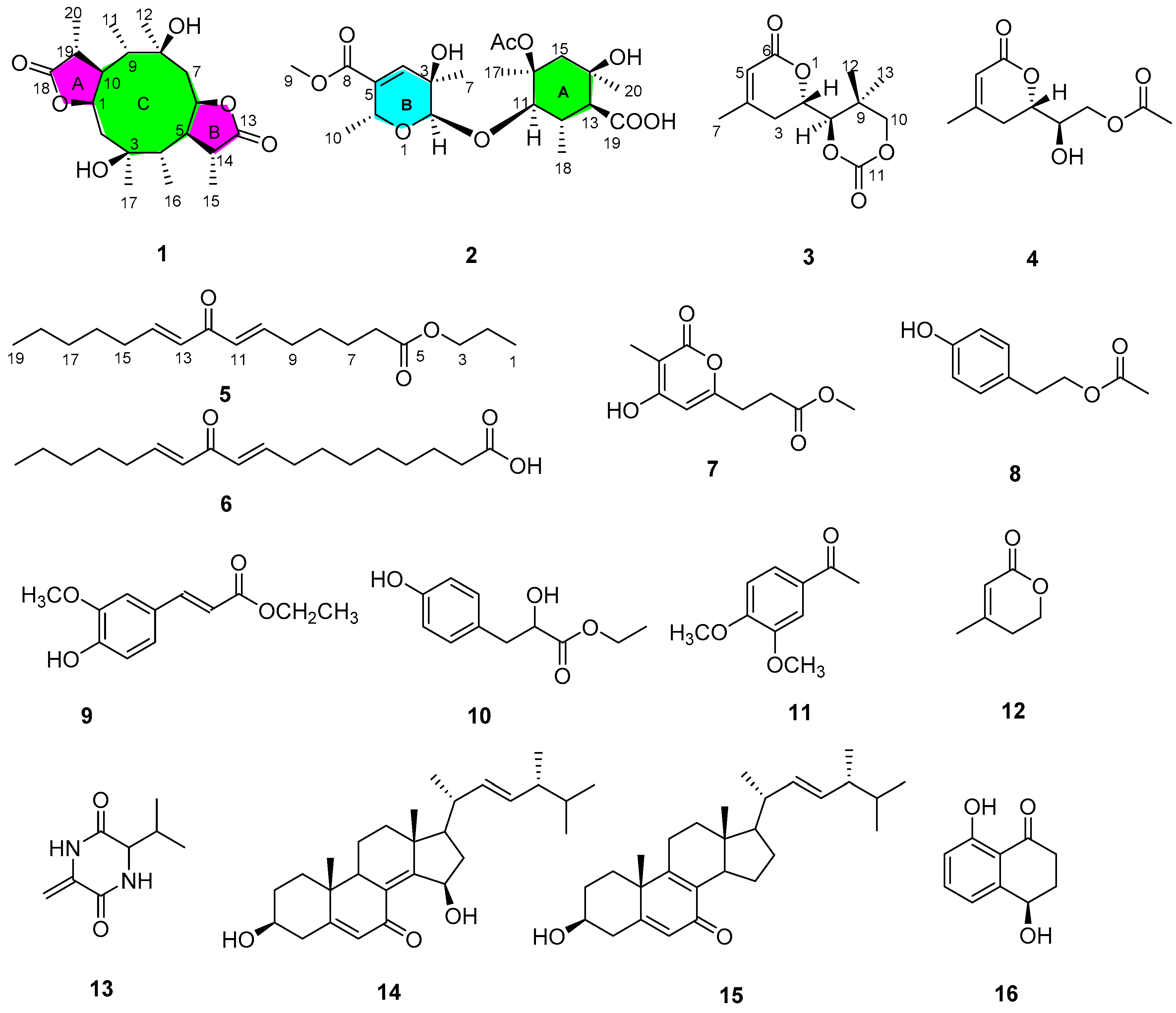
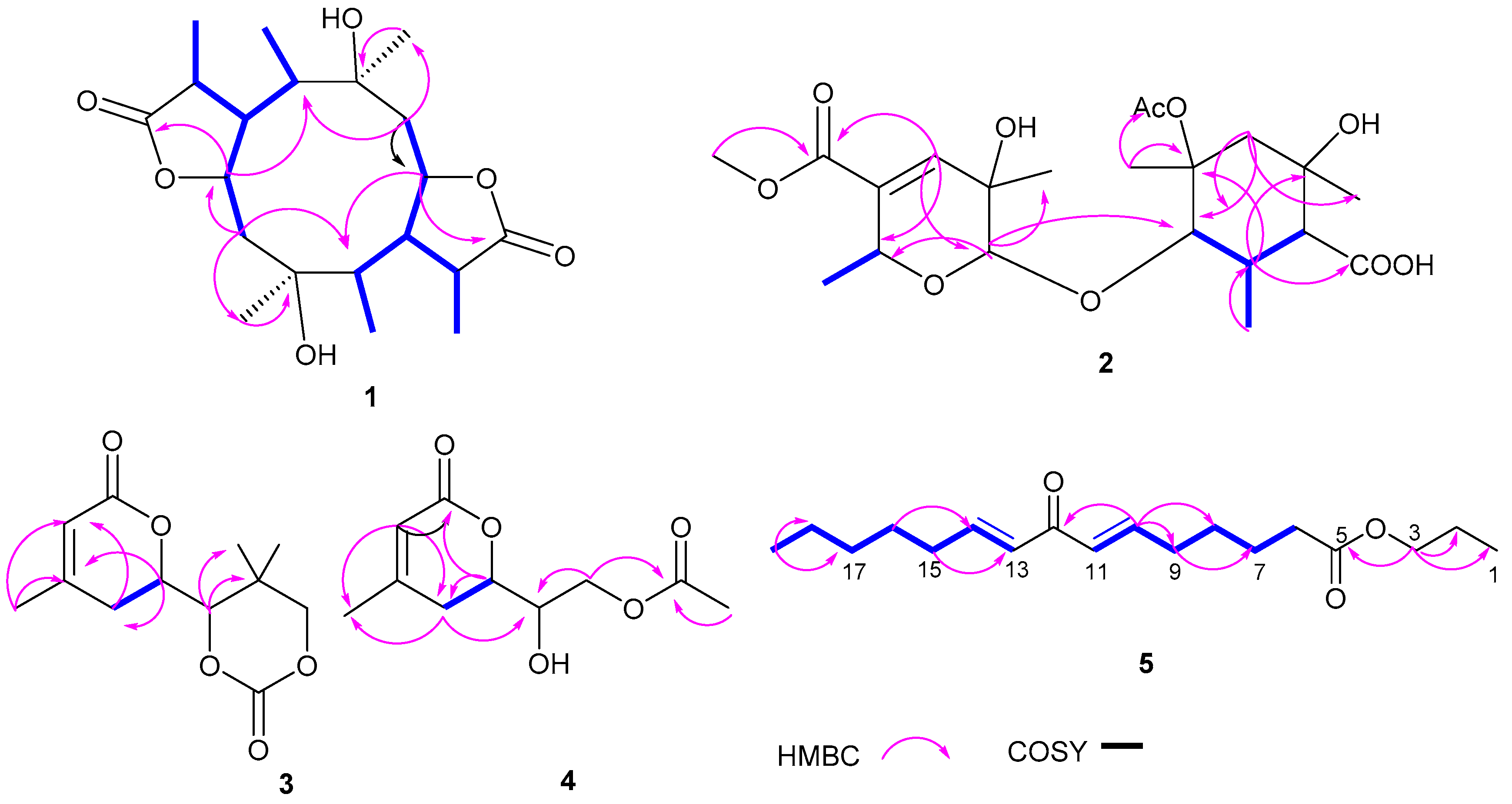
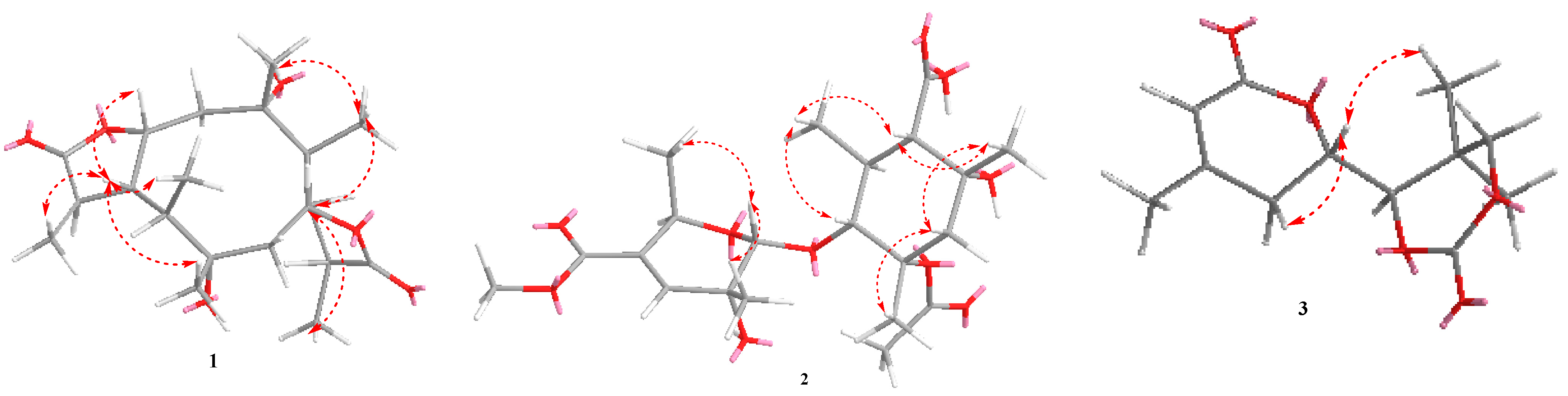
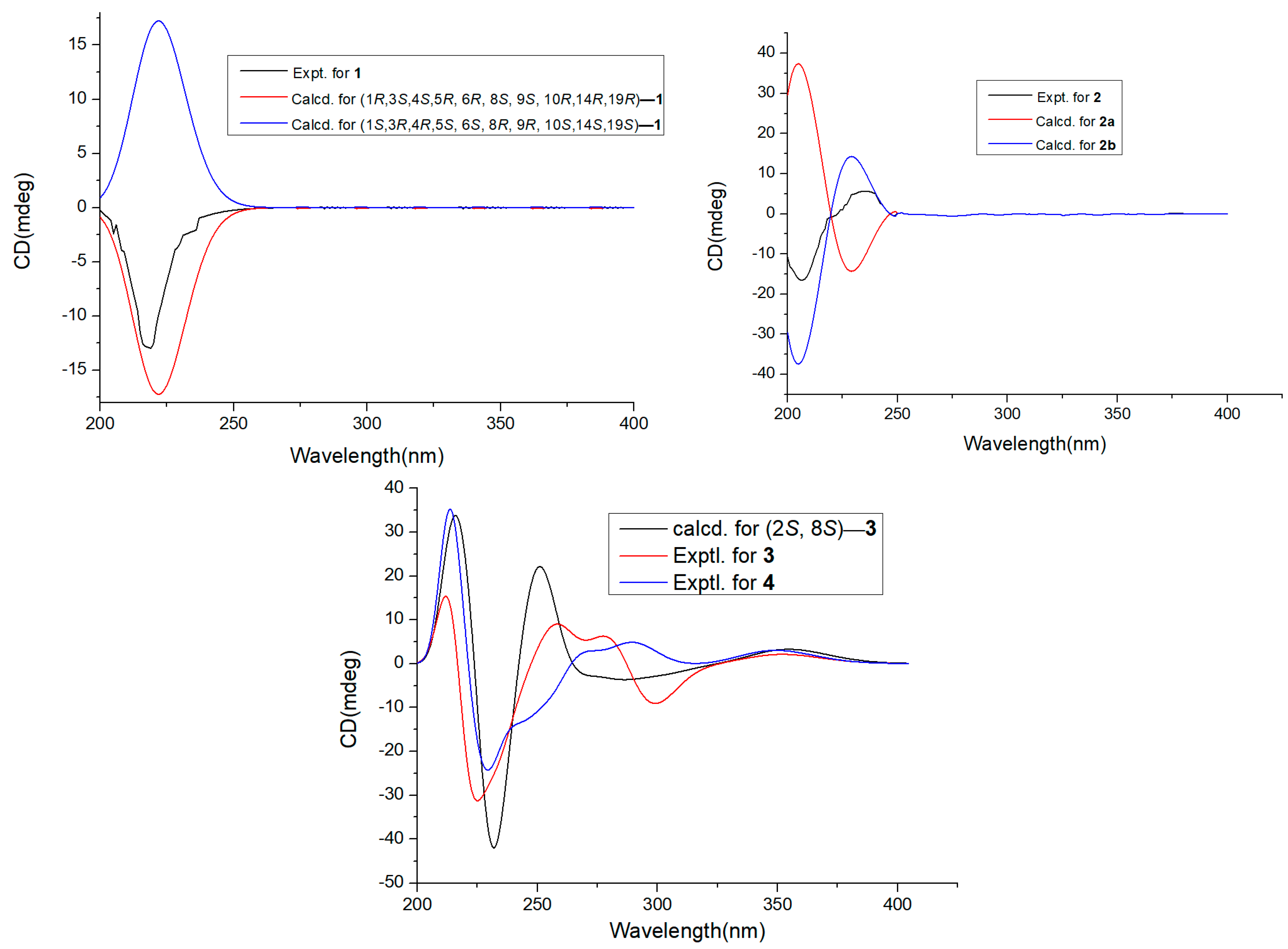
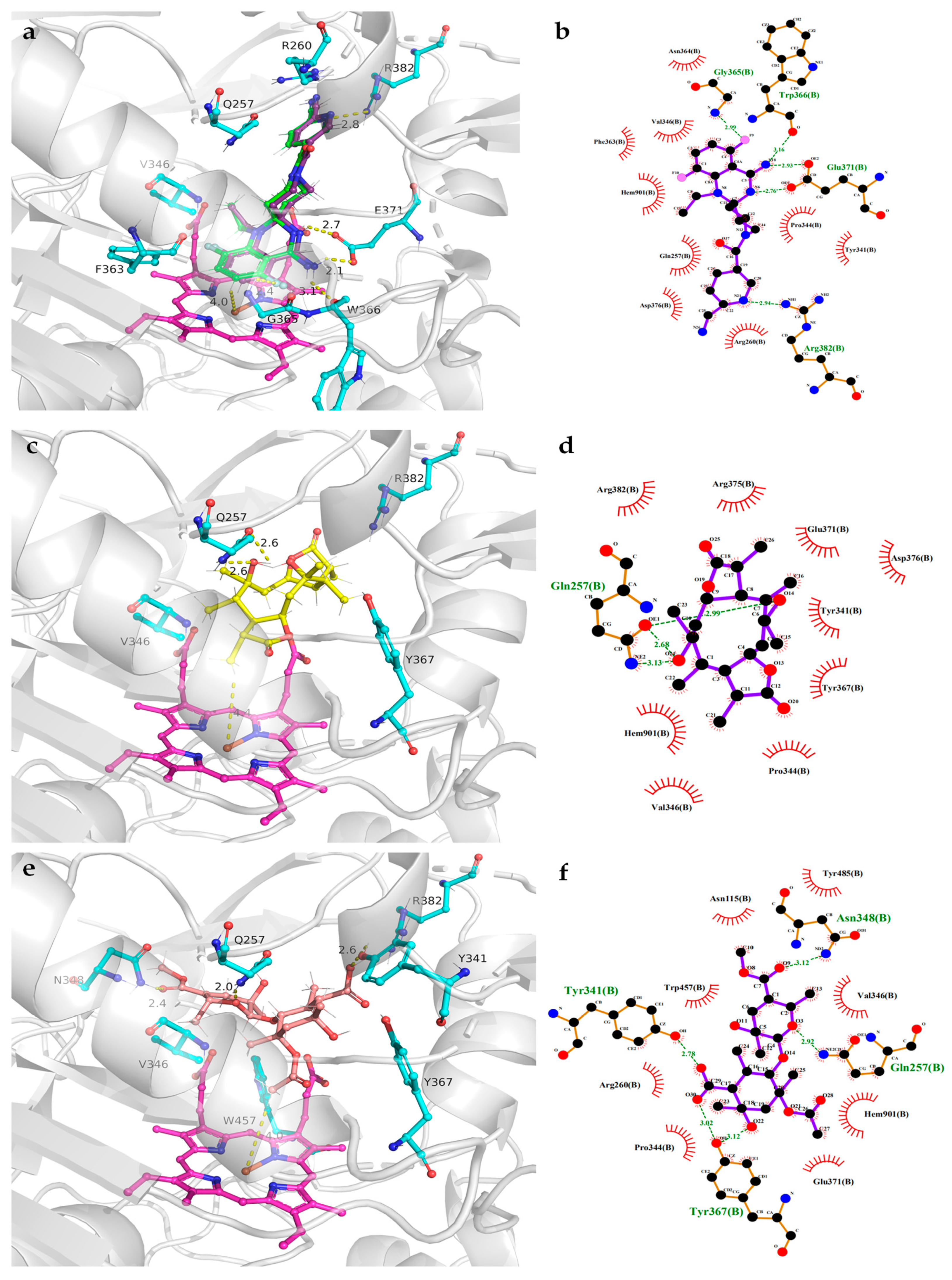
2. Results and Discussion

Compound 1 was detected as white oil. Its 1 H and 13C NMR data and HRESIMS spectrum data at m/z 391.2101 [M+Na]+ suggested that 1 had the molecular formula C20H32O6. Analysis of 1H NMR and HSQC spectrum (Table 1) of 1 showed six methyl groups (δH 0.95, d (J = 7.2 Hz, H3-11), 1.20, s, H3-12, 1.32, d (J = 7.6 Hz, H3-15), 0.97, d (J = 7.0 Hz, H3-16), 1.19, s, H3-17, and 1.29, d (J = 7.4 Hz, H3-20)), two oxygenated methines (δH 4.84, td (J = 2.4, 6.8 Hz, H-1), 4.93, td (J = 2.5, 7.3 Hz, H-6)), and two methylenes (δH 2.16, m; δH 2.21, m, 2.02, m). The 13C NMR and HSQC spectra of 1 exhibited 20 carbon signals, including six methyls, eight methines, two methylenes, and four oxygenated quaternary carbons. The above NMR data revealed the structure of 1 as a polyketide derivative, which was supported again by key HMBC correlations from H-19 (δH 2.88) to C-18 (δC 179.6), C-20 (δC 11.6), C-10 (δC 49.5), and C-9 (δC 44.0); and from H-14 (δH 2.70) to C-13 (δC 181.1), C-6 (δC 81.4), C-5 (δC 54.0), C-4 (δC 50.6), and C-15 (δC 18.3). The key HMBC correlations from H-2 (δH 2.16) to C-1 (δC 81.0), C-4 (δC 50.6), and C-17 (δC 23.8); and from H-7 (δH 2.02/2.21) to C-9 (δC 44.0), C-5 (δC 54.0), C-6 (δC 81.4), and C-12 (δC 23.8), the COSY correlations H-20/H3-19/H-10/H-9/H3-11/H-1/H-2 (fragment ) and H3-16/H-4/H-5/H-14/H3-15/H-6/H-7 (fragment ) (Figure 2) indicated that lactone rings (A and B) were connected by C-1 to C-10, C-5 to C-6 bond, which established a novel tricyclic 5/10/5 skeleton. The relative configuration of 1 was deduced from the key NOESY correlations (Figure 3). The cross-peaks from H-10 to H3-20, H3-11, and H-1; from H3-12 to H3-10 and H-6; and from H-5 to H3-15, H3-16, and H3-17, together with the correlation from H-1 to H-17, indicated that H-1, H-17, H3-16, H3-15, H-5, H-6, H3-12, H3-11, H3-20, and H-10 were on the same side. In order to discriminate between (1S, 3R, 4R, 5S, 6S, 8R, 9R, 10S, 14S, 19S)-1 and (1R, 3S, 4S, 5R, 6R, 8S, 9S, 10R, 14R, 19R)-1, calculations of the ECD spectrum of 1 were performed. As a result, the calculated spectrum of (1R, 3S, 4S, 5R, 6R, 8S, 9S, 10R, 14R, 19R)-1 coincided well with its experimental data (Figure 4), suggesting the absolute configuration of 1 to be 1R, 3S, 4S, 5R, 6R, 8S, 9S, 10R, 14R, 19R.
Compound 2 was yielded as a white solid. Its molecular formula C21H32O10 was established by 13C NMR data together with HRESIMS at m/z 443.1923, [M-H]-. Analysis of NMR spectra of 2 showed six methyl protons (δH 0.90, 1.04, 1.14, 1.30, 1.81, 1.35), one methylene protons (δH 2.12, 1.86), and one olefinic protons at δH 6.53 (1H, d, J = 2.0 Hz). The DEPT and 13C NMR spectra revealed 21 resonances including six methyl (δC 25.9, 18.0, 21.0, 14.2, 22.1, 22.6), one methylene (δC 45.7), six methines (δC 94.0, 139.5, 66.0, 80.2, 56.0, 43.9), seven nonprotonated carbons, one carbonyl group (δC 180.4), and two ester carbonyl groups (δC 179.5, 165.9). The key HMBC correlations from H-12 (δH 2.26) to C-13 (δC 43.9), C-18 (δC 14.2), C-11 (δC 80.2), C-16 (δC 74.4), and C-19 (δC 180.4); H-15 (δH 2.12, 1.85) to C-20 (δC 22.1), C-13 (δC 43.9), and C-14 (δC 80.0); H-17 (δH 1.35) to C-16 (δC 74.4), and C-1′ (δC 179.5), together with 1H-1H COSY correlation information between H-18/H-12/H-13/H-11 established the fragment A (Figure 2). Further, 1H-1H COSY correlation information between H-10 (δH 1.30) and H-6 (δH 4.54), along with the key HMBC correlations from H-4 (δH 6.53) to C-8 (δC 165.9), C-5 (δC 133.7), C-2 (δC 94.1), C-7 (δC 21.0), and C-6 (δC 66.0); and H-2 (δH 4.80) to C-4 (δC 139.5), C-6 (δC 66.0), C-7 (δC 21.0) and C-11 (δC 80.2) established the fragment B. Therefore, the structure of 2 was assigned (Figure 1). The relative configurations of 2 were investigated by key NOESY spectrum, as indicated in Figure 3. The key NOESY correlations of H-2/H3-10, H-2/H3-7, H3-20/H3-18, H3-20/H3-17, H-13/H3-18, and H-11/H3-17 in 2, suggested these groups were cofacial. In order to confirm the absolute configurations of 2, the ECD calculations were performed (2a and 2b) (Figure 4). As a result, the ECD calculations of 2b fitted well with the experimental curve. Thus, compound 2 was assigned and named as pestaloketide B.
Compound 3 was detected as yellow oil, giving the molecular formula of C12H16O5 from the analysis of their 13C NMR data and HRESIMS (Table 2). Carefully, analysis of the 1D NMR data, in combination with the HSQC spectrum, revealed characteristic signals corresponding to one olefinic proton (δH 5.83 (1H, s, H-5)) and three methyls (δH 1.08, s, δH 1.24, s, δH 2.03, s). The 13C NMR and HSQC spectroscopic data of 3 exhibited resonances for one lactone carbon (δC 164.8 (C-6)) and three oxygenated carbons (δC 65.9, 75.7, 76.4). Detailed analysis of these above data of 3 revealed that compound 3 was very similar to those of 12, 3-methyl-2-penten-5 [13], except for the presence of the lactonic ring groups at C-2 in 3. The aforementioned conclusion was supported again by the key correlations from H-7 to C-3/C-5/C-4, from H-8 to C-9/C-12/C-13, and from H-2 to C-3/C-4/C-9/C-6. The key NOESY correlations of H-2 with H3-12 and H-3β suggested that these protons were cofacial; thus, the relative configuration of compound 3 was deduced as 2S, 8S. The absolute configuration of C-2 and C-8 in 3 were elucidated by comparing the calculated ECD spectrum of the 2S, 8S-model and the experimental ECD curve of 3 (Figure 4). Thus, compound 3 was assigned (Figure 1).
Compound 4 was yielded as a yellow oil, gave the molecular formula of C10H14O5 from the HRESIMS ion at m/z 237.0747 [M+Na]+ and 13C NMR data. Analysis of the 1D NMR spectroscopic data between 4 and 3 indicated both compounds to be structurally similar (Table 2). The major difference was that the lactonic ring groups at C-2 in 3 were replaced by one acetate at C-2 in 4. The aforementioned results were supported by the key HMBC correlations from H-5 to C-6/C-3/C-7 (δC 22.9), from H-2 to C-3/C-4/C-6, and from H-3 to C-7/ C-2/C-5/C-8, and the COSY correlations H-2 and H-3. According to the above evidence, by the biosynthetic pathway, similar chemical shift, and specific rotation (4, [α ] D 25 -52 (c 0.2, MeOH, 3 ] D 25 -58 (c 0.20, MeOH)) data comparison, the relative configurations of 3 and 4 were concluded to be the same for C-2 and C-8. This assignment was proved by the ECD spectrum, the result of which showed good accordance with 3 (Figure 4). Thus, the structure of 4 was assigned and named pestaloketide C.
Compound 5 was a colorless oil. The 1H NMR and HSQC data revealed the presence for four olefinic methines (δH 6.97 (1H, dt, J = 15.6, 7.0 Hz), 6.94 (1H, dt, J = 15.6, 7.0 Hz), 6.4 (2H, d, J = 15.6 Hz)), eleven aliphatic methylenes (δH 1.34-2.26), and two methyls (δH 0.92 (3H, t, J = 6.0 Hz); δH 0.99 (3H, d, J = 6.7 Hz)). The 13C NMR and HSQC data (Table 2) of 5 showed 18 carbon resonances comprising two methyls, four olefinic methines, ten aliphatic methylenes, one oxygenated carbon, and two carbonyl carbons. The aforementioned data suggested that 5 was similar to compound 6, 11-keto-9(E), 12(E)-octadecadienoic acid [14]. The major difference was the absence of the carboxyl group and the presence of a butyl ester (δC 169.3 72.9, 29.1, 19.5) in 6. HMBC correlations from H-10 to C-12/C-11/C-9/C-8, from H-9 to C-12/C-10/C-9, and from H-3 to C-5/C-2/C-1, along with 1H-1H COSY correlations of H-13/H14/H-15/H-16/H17/H-18/H-19 and H-11/H10/H-9/H-8/H7/H-6 confirmed the planar structure of 5.
In order to further investigate the structure of compound 5, ESI-MS analysis was performed. It was found that compound 5 yielded the ion at m/z 294 in the mass spectrum, and further fragmentation of this ion gave an intense signal at m/z 179 [Frag 1] in Figure S35. Finally, the results of the ESI-MS analysis suggested that fragment at m/z 179 was attributed to [CH3(CH2)4(CH)2CO(CH)2(CH2)2] cations. These results assisted to reconfirm the configuration of compound 5.
Compounds 616 were determined to be the known 11-keto-9(E),12(E)-octadecadienoic acid (6) [14], scirpyrone K (7) [15], 4-hydroxy phenethyl acetate (8) [16], ethyl (2E)-3-(4-hydroxy-3-methoxyphenyl)prop-2-enoate (9) [17], ethylp-hydroxyphenyllactate (10) [18], 3,4-dimethoxyacetophenone (11) [19], 4-methyl-5,6-dihydropyren-2-one (12) [20], cyclo(L-Val-Dha) (13) [21], 3,15-dihydroxyl-(22E, 24 R)-ergosta-5,8(14),22-trien-7-one (14) [22], 3β-hydroxy-(22E,24R)-ergosta-5,8,22-trien-7-one (15) [23], and (-)-isosclerone (16) [24], by comparing their NMR data.
The cytotoxicity assay of compounds (116) was examined via MTT assay. Compounds 5, 6, 9, and 14 showed weak cytotoxicities against four human cancer cell lines, with IC50 values 22.1-100 μM (Table 3). Anti-inflammatory activities were performed for compounds 14, 78, 1013, and 1516 with NO production inhibitory activity. Pestalolactones A (1) and B (2) showed moderate inhibitory of NO production with IC50 values of 23.6 and 14.5 μM, respectively, without cytotoxicity observed. Others were inactive (100 μM). The result showed that pestalolactones A (1) and B (2) had the potential to be developed into anti-inflammatory activity drug leads (Table 4).
Interestingly, pestaloketide A (1) is reported to be the first tricyclic 5/10/5 skeleton polyketide from the sponge-derived fungus Pestalotiopsis sp. In addition, pestaloketides A (1) and B (2) exhibited moderately inhibited LPS-induced NO production activity. To further investigate the anti-inflammatory mechanism of pestaloketides A (1) and B (2), molecular docking of 1 and 2 with inducible NO oxidase (iNOS) as target was employed, and dexamethasone was used for redocking (Figure 5). Docking results display that the docking pose of dexamethasone (Figure 5a, green) fit well with its original pose (Figure 5a, purple) in cocrystal, and compounds 12 exhibited good interactions with the INOS target in its pocket. Pestaloketide A (1) had a hydrogen bond interaction with Q257, and had nonpolar interactions with residues V346, Y367, and R382 and the cofactor heme. For pestaloketide B (2), hydrogen bond interactions were formed with residues Q257, Y341, N348 and Y367, and nonpolar interactions were formed with residues V346, R382, and W457 and the cofactor heme. These results indicate that 2 has a stronger association with the INOS protein than 1, which is consistent with our in vitro biological activity experiment results. Therefore, both pestalolactones A (1) and B (2) had the potential to be developed into anti-inflammatory activity drug leads.

3. Materials and Methods

3.1. General Experimental Procedures

Optical rotations were measured using an (Anton) MCP500 polarimeter. HRESIMS were used for a Bruker maXis TOF-Q mass spectrometer, while infrared spectra were acquired on a Shimadzu IR spectrometer. NMR spectra were measured on Bruker spectrometer. The UV spectra were carried out on a Shimadzu UV-2600 PC spectrometer. Open chromatography column was performed on silica gel (100-300 mesh, Qingdao, China), YMC ODS-A (S-50 μm, 12 nm) (YMC Co., Ltd., Kyoto, Japan). HPLC was accomplished using ODS column (YMC-5μm, ODS-A) Sephadex LH-20 (GE, Sweden). The RAW 264.7 cells were obtained from the Chinese Academy of Sciences (Shanghai, China)

3.2. Fungal Material

Strain SWMU-WZ04-2 was obtained from the sponge collected in Weizhou Island, Guangxi province, China, in April 2018. It was identified as Pestalotiopsis sp. SWMU-WZ04-2 according to a molecular biological protocol by DNA amplification and sequencing of the ITS region. A voucher specimen (No. SWMU-WZ04-2) was deposited in the laboratory.

3.3. Fermentation, Extraction, and Isolation

The mass fermentation of the fungal strain SWMU-WZ04-2 was performed in 120 × 1 L Erlenmeyer flasks. The medium was grown (containing 200 g of natural rice, 3% sea salt; 200 mL of water, 10 μM of 5-aza-2-deoxycytidine) for 36 days at 28 °C. The fermented rice cultures were soaked and extracted with EtOAc to gain 59 g of residue.
The crude extract was chromatographed by silica gel cc (column chromatography), which was eluted with petroleum ether/EtOAc (50: 1 to 0: 1, v/v) and separated into 8 fractions (Fr-1–Fr-8). Fr-3 was applied to silica gel cc (petroleum ether/EtOAc, 10:1-5:1) to obtain five subfractions (Frs. 3.1-3.5). Fr. 3.3 was purified with Sephadex LH-20 (MeOH) and applied by C18 HPLC (80% H2O/MeOH) to obtain compounds 8 (6.0 mg) and 10 (4.0 mg). Fr. 3.4 was applied by ODS column chromatography eluting with MeOH/H2O (60%) and purified by Sephadex LH-20 column (MeOH) and HPLC (70%, MeOH/H2O) to give 9 (5.0 mg) and 11 (3.0 mg). Fr-4 was subjected by silica gel (petroleum ether-EtOAc, 6:1-0:1) and further separated by preparative TLC, (60%, H2O/MeOH), HPLC, and Sephadex LH-20 (MeOH), to yield 1 (7.0 mg), 3 (3.0 mg), and 4 (4.0 mg). Fr-5 was applied to silica gel cc (CH2Cl2/Acetone), followed by Sephadex LH-20 chromatography (MeOH) and HPLC (55% H2O/MeOH) to afford 2 (4.0 mg). Fr. 5.4 was applied by ODS (H2O/MeOH) column and further subjected by HPLC (H2O/MeOH 45:55) to obtain 7 (7.0 mg), 12 (3.0 mg), and 16 (5.0 mg). Fr-6 was divided into five subfractions (Frs. 6.1-6.5) by silica gel cc (CH2Cl2-MeOH,10:1- 0:1). Then, Fr. 6.3 was applied by HPLC (MeOH/H2O, 45%) to yield 5 (4.0 mg). Fr. 6.4 was applied by HPLC (MeOH/H2O, 40%) to afford 6 (8.0 mg). Fr. 6.5 was applied by Sephadex LH-20 (MeOH), preparative TLC and HPLC (H2O/MeOH) to yield 13 (6.0 mg), 14 (12.0 mg), and 15 (10.0 mg).
Pestaloketide A (1): white oil; [α ] D 25 −31 (c 0.45, MeOH); UV (MeOH) λmax(log ε) 210 (2.54) nm; IR (film)νmax 3329, 2952, 2851, 1680, 1589, 1440, 1348, 1024 cm−1; 1H NMR and 13C NMR data, Table 1; HRESIMS m/z 391.2101 [M+Na]+ (calcd for C20H32NaO6, 391.2109).
Pestaloketide B (2): white powder; [α ] D 25 +42 (c 0.1, MeOH); UV (MeOH) λmax(log ε) 205 (3.60), 233 (3.32) nm; IR (film)νmax 3368, 2925, 2859, 1702, 1646, 1604, 1380, 1349, 1258, 1226, 1121, 1053, 1034, 878 cm−1; 1H NMR and 13C NMR data, Table 1; HRESIMS at m/z 443.1923 [M-H] (calcd for C21H31O10, 443.1924).
Pestaloketide C (3): yellow oil; [α ] D 25 −58 (c 0.2, MeOH); UV (MeOH) λmax(log ε) 235 (4.26), 210 (3.38) nm; IR (film)νmax 3329, 2952, 2852, 1732, 1680, 1467, 1440, 1379, 1220, 1165, 1103 cm−1; 1H NMR and 13C NMR data, Table 2; HRESIMS m/z 263.1005 [M+Na]+ (calcd for C12H16NaO5, 263.0902).
Pestaloketide D (4): yellow oil; [α ] D 25 −52 (c 0.2, MeOH); UV (MeOH) λmax(log ε) 240 (4.23), 205 (3.26) nm; IR (film)νmax 3359, 3262, 2937, 1705, 1652, 1580, 1455, 1376, 1349, 1166, 1022 cm−1; 1H NMR and 13C NMR data, Table 2; HRESIMS m/z 237.0747 [M+Na]+ (calcd for C10H14NaO5, 237.0725).
Pestaloketide E (5): colorless oil; [α ] D 25 −26 (c 0.1, MeOH); UV (MeOH) λmax(log ε) 250 (4.16) nm; IR (film)νmax 2930, 2853, 1705, 1662, 1455, 1348, 1162, 950, 836, 710, 680 cm−1; 1H NMR (600 Hz) and 13C NMR(150 Hz) data, Table 2; HRESIMS m/z 293.2128 [M-H]- (calcd for C18H29O3, 293.2126).

3.4. Computational Section

The calculations were applied by the Spartan’14, Gaussian 09 software, and Merck Molecular Force Field (MMFF), respectively. The conformers of 14 were chosen at the B3LYP/6-311+G(d,p) level. The overall calculation of the ECD was performed using the TDDFT method for the stable conformers of new compounds. The spectra were obtained by SpecDis 1.6.

3.5. Cytotoxicity Assay

The method for the assay of cytotoxicity activity of 116 was conducted according to the one described previously [11]. Positive control (Adriamycin).

3.6. Inhibition of NO Production Assays

The activity of compounds 116 were examined by inhibited NO production in LPS-stimulated RAW. The detailed process of the assay is described in the previously published paper [9]. Positive control (dexamethasone).

3.7. Molecular Docking

The three-dimensional structure of INOS (PDB ID:3E6T) was acquired from the Protein Data Bank (http://www.rcsb.org, accessed on 30 October 2022) [25,26], for which the resolution was 2.5 Å. Using the Chain A of the INOS structure as the receptor, pestaloketides A (1) and B (2) were docked using Autodock vina [27] and AutoDockTools-1.5.6 [28]. The geometrical restraints for 1 and 2 were generated by Grade Web Server (http://grade.globalphasing.org, accessed on 29 October 2022). A grid box of a 48.02 Å × 42.58 Å × 33.75 Å size was centered on the catalytic site. All docking parameters were set to default values. The docking results were further analyzed and presented using PyMOL (http://www.pymol.org, accessed on 29 October 2022) and LigPlot+ [29].

4. Conclusions

In summary, five new compounds (15), together with other eleven known natural products (616), were isolated from the fungus Pestalotiopsis sp. SWMU-WZ04-2. Pestaloketide A (1) is an unusual polyketide featuring a rare 5/10/5-fused ring system. Compounds 5, 6, 9, and 14 showed weak cytotoxicities against four human cancer cell lines (IC50: 22.1–100 μM). Other compounds were inactive (100 μM). Anti-inflammatory activities were performed for compounds 14, 78, 1013, and 1516, and Pestalolactones A (1) and B (2) showed moderate inhibitory of NO production with IC50 values of 23.6 and 14.5 μM, respectively, without cytotoxicity observed.” Although the detailed mechanism of action is still undefined for pestaloketides A (1) and B (2), molecular docking analysis showed that both compounds had the potential to be developed into anti-inflammatory activity drug leads.

Supplementary Materials

The following supporting information can be downloaded at https://www.mdpi.com/article/10.3390/md20110711/s1, Figures S1–S35,S37,S38: 1D, 2D NMR, and HRESIMS spectra of compounds 15. S36: Computational details for compound 14 (ECD). Table SI–S3: Energy analysis for conformers of 1–3 at B3LYP/6-31G(d) level.

Author Contributions

P.J. and H.L. performed experiments and wrote the original draft; J.L. and B.T. isolated the fungal strain; Y.J. and L.J. performed the bioassay experiments; H.N. and D.Z. revised the manuscript; L.Z. conducted the molecular docking analysis. All authors have read and agreed to the published version of the manuscript.

Funding

This work was funded by Natural Science Foundation of Sichuan Province (2022NSFSC0109), the Project of Sichuan Industrial Institute of Antibiotics, Chengdu University (ARRLKF20-04), and “Student’s Platform for Innovation and Entrepreneurship Training Program” (S202210632194 and S202210632232).

Institutional Review Board Statement

Not applicable.

Data Availability Statement

The data presented in this study are available in the main text and the supplementary materials of this article.

Conflicts of Interest

The authors declare no conflict of interest.

References

  1. Zhang, B.; Zhang, T.; Xu, J.; Lu, J.; Qiu, P.; Wang, T.; Ding, L. Marine sponge-associated fungi as potential novel bioactive natural product sources for drug discovery. Mini Rev. Med. Chem. 2020, 20, 1966–2010. [Google Scholar] [CrossRef] [PubMed]
  2. Yang, X.L.; Zhang, J.Z.; Luo, D.Q. The taxonomy, biology and chemistry of the fungal Pestalotiopsis genus. Nat. Prod. Rep. 2012, 29, 622–641. [Google Scholar] [CrossRef] [PubMed]
  3. Xu, J.; Yang, X.B.; Lin, Q. Chemistry and biology of Pestalotiopsis-derived natural products. Fungal Divers. 2014, 66, 37–68. [Google Scholar] [CrossRef]
  4. Wu, B.; Wu, X.D.; Sun, M.; Li, M.H. Two novel tyrosinase inhibitory sesquiterpenes induced by CuCl2 from a marine-derived fungus Pestalotiopsis sp. Z233. Mar. Drugs 2013, 11, 2713–2721. [Google Scholar] [CrossRef] [PubMed] [Green Version]
  5. Strobel, G.; Yang, X.; Sears, J.; Kramer, R.; Sidhu, R.S.; Hess, W.M. Taxol from Pestalotiopsis microspora, an endophytic fungus of Taxus wallachiana. Microbiology 1996, 142, 435–440. [Google Scholar] [CrossRef] [Green Version]
  6. Xu, J.; Ebada, S.S.; Proksch, P. Pestalotiopsis a highly creative genus: Chemistry and bioactivity of secondary metabolites. Fungal Divers. 2010, 44, 15–31. [Google Scholar] [CrossRef]
  7. Zhang, Y.L.; Bai, J.; Yan, D.J.; Liu, B.Y.; Zhang, L.; Zhang, C.; Chen, M.H.; Mou, Y.H.; Hu, Y.C. Highly oxygenated caryophyllene-type sesquiterpenes from a plant-associated fungus, Pestalotiopsis hainanensis, and their biosynthetic gene cluster. J. Nat. Prod. 2020, 83, 3262–3269. [Google Scholar] [CrossRef]
  8. Feng, L.; Han, J.; Wang, J.; Zhang, A.X.; Miao, Y.Y.; Tan, N.H.; Wang, Z. Pestalopyrones A-D, four tricyclic pyrone derivatives from the endophytic fungus Pestalotiopsis neglecta S3. Phytochemistry 2020, 179, 112505. [Google Scholar] [CrossRef]
  9. Rivera-Chávez, J.; Zacatenco-Abarca, J.; Morales-Jiménez, J.; Martínez-Aviña, B.; Hernández-Ortega, S.; Hernández-Ortega, S.; Aguilar-Ramírez, E. Cuautepestalorin, a 7,8-dihydrochromene-oxoisochromane adduct bearing a hexacyclic scaffold from Pestalotiopsis sp. IQ-011. Org. Lett. 2019, 21, 3558–3562. [Google Scholar] [CrossRef]
  10. Liu, S.; Dai, H.F.; Makhloufi, G.; C Heering, C.; Janiak, C.; Hartmann, R.; Mándi, A.; Kurtán, T.; Müller, W.E.G.; Kassack, M.U.; et al. Cytotoxic 14-membered macrolides from a mangrove-derived endophytic fungus, Pestalotiopsis microspore. J. Nat. Prod. 2016, 79, 2332–2340. [Google Scholar] [CrossRef]
  11. Lei, H.; Lin, X.P.; Han, L.; Ma, J.; Ma, Q.J.; Zhong, J.L.; Liu, Y.H.; Sun, T.M.; Wang, J.H.; Huang, X.S. New metabolites and bioactive chlorinated benzophenone derivatives produced by a marine-derived fungus Pestalotiopsis heterocornis. Mar. Drugs 2017, 15, 69. [Google Scholar] [CrossRef] [PubMed] [Green Version]
  12. Rutledge, P.J.; Challis, G.L. Discovery of microbial natural products by activation of silent biosynthetic gene clusters. Nat. Rev. Microbiol. 2015, 13, 509–523. [Google Scholar] [CrossRef] [PubMed]
  13. He, Z.H.; Wu, J.; Xu, L.; Hu, M.Y.; Xie, M.M.; Hao, Y.J.; Li, S.J.; Shao, Z.Z.; Yang, X.W. Chemical constituents of the deep-sea-derived Penicillium Solitum. Mar. Drugs 2021, 19, 580. [Google Scholar] [CrossRef] [PubMed]
  14. Shinohara, C.; Hasumi, K.; Chikanishi, T.; Kikuchi, T.; Endo, A. 11-Keto-9 (E), 12 (E)-octadecadienoic acid, a novel fatty acid that enhances fibrinolytic activity of endothelial cells. J. Antibiot. 1999, 52, 171–174. [Google Scholar] [CrossRef] [PubMed] [Green Version]
  15. Zhang, Z.Z.; He, X.Q.; Che, Q.; Zhang, G.J.; Zhu, T.J.; Gu, Q.Q.; Li, D.H. Sorbicillasins A-B and scirpyrone K from a deep-sea-derived fungus, Phialocephala sp. FL30r. Mar. Drugs 2018, 16, 245. [Google Scholar] [CrossRef] [Green Version]
  16. Wu, H.H.; Tian, L.; Chen, G.; Xu, N.; Wang, Y.N.; Sun, S.; Pei, Y.H. Six compounds from marine fugus Y26-02. J. Asian Nat. Prod. REs 2009, 11, 748–751. [Google Scholar] [CrossRef]
  17. Yang, L.X.; Huang, K.X.; Li, H.B.; Gong, J.X.; Wang, F.; Feng, Y.B.; Tao, Q.F.; Wu, Y.H.; Li, X.K.; Wu, X.M.; et al. Design, synthesis, and examination of neuron protective properties of alkenylated and amidated dehydro-silybin derivatives. J. Med. Chem. 2009, 52, 7732–7752. [Google Scholar] [CrossRef]
  18. Takaya, Y.; Furukawa, T.; Miura, S.; Akutagawa, T.; Hotta, Y.; Ishikawa, N.; Niwa, M. Antioxidant constituents in distillation residue of awamori spirits. J. Agric. Food Chem. 2007, 55, 75–79. [Google Scholar] [CrossRef]
  19. Bose, P.; Banerji, J. Synthesis of 4-phenylcoumarins from Dalbergia volubilis and Exostema caribaeu. Phytochemistry 1991, 30, 2438–2439. [Google Scholar] [CrossRef]
  20. Reggelin, M.; Gerlach, M.; Vogt, M. Metallated 2-alkenyl sulfoximines in asymmetric synthesis: Regio-and stereoselective synthesis of highly substituted oxabicyclic ethers and studies towards the total syntheses of the euglobals G1 and G2 and arenaran A. Eur. J. Org. Chem. 1999, 5, 1011–1031. [Google Scholar] [CrossRef]
  21. Tian, S.Z.; Pu, X.; Luo, G.Y.; Zhao, L.X.; Xu, L.H.; Li, W.J.; Luo, Y.G. Isolation and characterization of new p-terphenyls with antifungal, antibacterial, and antioxidant activities from Halophilic actinomycete Nocardiopsis gilva YIM 90087. J. Agric. Food Chem. 2013, 61, 3006–3012. [Google Scholar] [CrossRef] [PubMed]
  22. Wang, F.Z.; Fang, Y.C.; Zhang, M.; Lin, A.Q.; Zhu, T.J.; Gu, Q.Q.; Zhu, W.M. Six new ergosterols from the marine-derived fungus Rhizopus sp. Steroids 2008, 73, 19–26. [Google Scholar] [CrossRef] [PubMed]
  23. Lei, H.M.; Ma, N.; Wang, T.; Zhao, P.J. Metabolites from the endophytic fungus colletotrichum sp. F168. Nat. Prod. Res. 2021, 35, 1077–1083. [Google Scholar] [CrossRef]
  24. Yang, Y.H.; Yang, D.S.; Lei, H.M.; Li, C.Y.; Li, G.H.; Zhao, P.J. Griseaketides A-D, new aromatic polyketides from the pathogenic fungus Magnaporthe grisea. Molecules 2020, 25, 72. [Google Scholar] [CrossRef] [PubMed] [Green Version]
  25. Garcin, E.D.; Arvai, A.S.; Rosenfeld, R.J.; Kroeger, M.D.; Crane, B.R.; Andersson, G.; Andrews, G.; Hamley, P.J.; Mallinder, P.R.; Nicholls, D.J.; et al. Anchored plasticity opens doors for selective inhibitor design in nitric oxide synthase. Nat. Chem. Biol. 2008, 4, 700–707. [Google Scholar] [CrossRef] [PubMed] [Green Version]
  26. Burley, S.K.; Bhikadiya, C.; Bi, C.; Bittrich, S.; Chen, L.; Crichlow, G.V.; Christie, C.H.; Dalenberg, K.; Di Costanzo, L.; Duarte, J.M.; et al. RCSB Protein Data Bank: Powerful new tools for exploring 3D structures of biological macromolecules for basic and applied research and education in fundamental biology, biomedicine, biotechnology, bioengineering and energy sciences. Nucleic Acids Res. 2021, 49, D437–D451. [Google Scholar] [CrossRef]
  27. Trott, O.; Olson, A.J. AutoDock Vina: Improving the speed and accuracy of docking with a new scoring function, efficient optimization, and multithreading. J. Comput. Chem. 2010, 31, 455–461. [Google Scholar] [CrossRef] [Green Version]
  28. Forli, S.; Huey, R.; Pique, M.E.; Sanner, M.F.; Goodsell, D.S.; Olson, A.J. Computational protein-ligand docking and virtual drug screening with the AutoDock suite. Nat. Protoc. 2016, 11, 905–919. [Google Scholar] [CrossRef] [Green Version]
  29. Laskowski, R.A. Swindells M.B. LigPlot+: Multiple ligand-protein interaction diagrams for drug discovery. J. Chem. Inf. Model. 2011, 51, 2778–2786. [Google Scholar] [CrossRef]
Figure 1. Structures of compounds 1–16.
Marinedrugs 20 00711 g001
Figure 2. COSY and key HMBC correlations of 1–5.
Marinedrugs 20 00711 g002
Figure 3. Key NOESY correlations of compounds 13.
Marinedrugs 20 00711 g003
Figure 4. ECD spectra of 13 as well as experimental ECD spectrum 4.
Marinedrugs 20 00711 g004
Figure 5. Representative docking poses of dexamethasone and pestaloketides A (1) and B (2) bound to the INOS protein (PDB ID:3E6T). Dexamethasone was used for redocking. The intermolecular interactions between INOS with dexamethasone and pestaloketides A (1) and B (2) are depicted as three-dimensional interaction maps ((a,c,e), respectively) and two-dimensional interaction maps ((b,d,f), respectively).
Marinedrugs 20 00711 g005
Table 1. 1 H (NMR) (500 MHz) and 13C NMR (125 MHz) data for 1 and 2.
1 a2 b
No.dC, TypedH (J in Hz)dC, TypedH (J in Hz)
181.0, CH4.84, td (2.4, 6.8)--
246.0, CH22.16, m94.1, CH4.80, s
381.2, C-67.2, C-
450.6, CH1.94, dt (7.0, 13.1)139.5, CH6.53, d (2.0)
554.0, CH2.14, m133.7, C-
681.4, CH4.93, td (2.5, 7.3)66.0, CH4.54, dd (6.7, 1.6)
746.4, CH22.21, m, 2.02, m21.0, CH31.14, s
880.9, C-165.9, C-
944.0, CH2.04, m50.8, OCH33.65, s
1049.5, CH2.54, dt (10.0, 7.1)18.0, CH31.30, d (6.7)
1115.9, CH30.95, d (7.2)80.2, CH4.78, s
1223.8, CH31.20, s56.0, CH2.26, d (7.2)
13181.1, C-43.9, CH2.09, m
1442.6, CH2.70, dd (7.6, 3.2)80.0, C-
1518.3, CH31.32, d (7.6)45.7, CH22.12, m, 1.85, m
1616.0, CH30.97, d (7.0)74.4, C-
1723.8, CH31.19, s25.9, CH31.35, s
18179.6, C-14.2, CH30.90, d (7.2)
1938.3, CH2.88, dq (10.0, 7.4)180.4, C-
2011.6, CH31.29, d (7.4)22.1, CH3 1.04, s
1′-OAc 179.5, C-
2′ 22.6, CH31.81, s
a Measured in CDCl3. b Measured in CD3OD.
Table 2. 1 H (NMR) (500 MHz) and 13C NMR (125 MHz) data for 3–5.
3 a4 b5 b
No.dC, TypedH (J in Hz)dC, TypedH (J in Hz)dC, TypedH (J in Hz)
1 19.5, CH30.99, d (6.7)
265.9, CH4.38,t(6.3)67.6, CH4.38, t (6.3)29.1, CH21.5, m
329.2, CH22.38, t (6.3)29.3, CH22.44, t (6.3)72.9, CH24.07, t (6.7)
4157.8, C 162.1, C
5116.8, CH5.83, s116.5, CH5.78, s169.3, C
6164.8, C 167.5, C 30.7, CH21.34, m
723.0, CH32.03, s22.9, C2.02, s29.4, CH21.5, m
875.7, CH4.12, d (6.3)68.3, CH4.02, m30.4, CH21.33, m
940.9, C 66.2, CH24.10, dd (11.3, 5.3)33.7, CH22.26, t (7.0)
1076.4, CH24.03, d (8.9), 3.95, d (8.9) 150.4, CH6.97, dt (15.6, 7.0)
11177.6, C 172.6, C 129.4, CH6.4, d (15.6)
1222.9, CH31.24, s20.7, CH32.06, s192.0, C
1318.8, CH31.08, s 129.4, CH6.4, d (15.6)
14 150.4, CH6.94, dt (15.6, 7.0)
15 33.7, CH2 2.26, t (7.0)
16 30.4, CH21.35, m
17 32.6, CH21.33, m
18 23.5, CH31.30, m
19 14.4, CH30.92, t (6.0)
a Measured in CDCl3. b Measured in CD3OD.
Table 3. Cytotoxicity of compounds 116 a (IC50 in μM).
CompoundSMMC-7721H460PC-3BGC-823
565.135.628.2>100
657.342.622.4>100
935.054.342.022.1
1473.564.3>10062.6
Adriamycin2.21.21.81.5
a Compounds that are not shown in this table did not exhibit activity (>100).
Table 4. Anti-inflammatory activities of the compounds 14, 78, 1013, and 1516 (IC50, μM).
Compound123–4, 7–8, 10–13, 15–16Positive a
IC5023.614.5-12.1
a Dexamethasone, - not exhibit activity.
Publisher’s Note: MDPI stays neutral with regard to jurisdictional claims in published maps and institutional affiliations.

