
Three-Dimensional Gait Analysis in Children Undergoing Gastrocsoleus Lengthening for Equinus Secondary to Cerebral Palsy


 , ,
, ,
Abstract
:1. Introduction
2. Materials and Methods
3. Results
3.1. Swing Phase Dorsiflexion
3.2. Gait Scores
3.3. Stability of Correction

| Study | CP Type/ No. of Subjects | Type of Surgery Eponym and Zone † | Stability of Correction | Follow Up in Months: Mean (Range) | Outcome Measures | MINORS |
|---|---|---|---|---|---|---|
| Baddar 2002 [14] | 34B | Isolated Zone 2 | - | 15 (9–23) | CE, 3DGA, EMG, gastroc-soleus length, SPK | 8 |
| Borton 2001 [15] | 45U, 89B | Isolated Zone 2/3 (Baker/ZTAL/Hoke TAL) | Recurrent equinus: U = 38%, B = 16% Calcaneus: U = 4%, B = 40% | (60–120) | CE, 3DGA, PRS | 11 |
| Cimolin 2011 [37] | 10U, 9B | Isolated Zone 2 (modified Vulpius) | - | 13 | 3DGA, GDI | 9 |
| Davids 2011 [4] | 33U, 20B | Isolated + SEMLS Zone 1/3 (Strayer/White) | - | 27 (20–65) | CE, 3DGA, SMC, SPK | 10 |
| Dreher 2012 [9] | 44B | SEMLS Zone 1 (Baumann) to 15–20° DF in KE/KF | Recurrent equinus—24% Calcaneus—9% early onset (<1 year), 11% late onset (>1 year) | 103 (12–156) | CE, 3DGA, SPK, GGI | 10 |
| Engsberg 2005 [27] | 32B | Isolated Zone 2/3 (Vulpius/White) | - | 14 | CE, 3DGA | 8 |
| Etnyre 1993 [28] | 11U, 13B | SEMLS Zone 1/2/3 (Strayer/Vulpius/Baker/Z-/step/sliding) | Recurrent equinus—12.5% (2U, 1B) | 8 | CE, EMG, 3DGA | 6 |
| Firth 2013 [16] | 44B | SEMLS Zone 1/3 (Strayer/modified Strayer/White slide) to 5° DF with KE | Recurrent equinus—35% Calcaneal gait—2.5% Revision calf surgery—13% | 48 | CE, 3DGA, GVS, GPS | 9 |
| Fujita 2020 [23] | 10U | SEMLS Zone 2 (Baker) + FHL transfer | Recurrent equinus—0% Calcaneus—30% | 35 (25–64) | CE, 3DGA, SPK, GVS, GPS | 7 |
| Galli 2009 [10] | 12B | Isolated Zone 2 (modified Vulpius) to neutral | Recurrent equinus—0% | (3–62) | CE, 3DGA, SPK | 7 |
| Galli 2005 [17] | 8U, 12B | Isolated Zone 2 (modified Vulpius) to neutral | - | 12 (+/−2) | CE, 3DGA, SMC | 7 |
| Jahn 2009 [42] | Not reported | Isolated Zone 2/3 (Vulpius/TAL) | - | 13 (8–20) | CE, 3DGA, muscle lengths | 7 |
| Kay 2004 [35] | 23U, 32B | SEMLS Zone 1/3 (GR/TAL) | Under-correction—3–28% Over-correction—19–22% | 19 (+/−10) | CE, 3DGA, SMC, PRS, SPK | 7 |
| Kim 2020 [29] | 14B | SEMLS Zone 2 (Endoscopic/open modified Vulpius) to 10° DF in KE | Surgical complications—none Recurrent equinus—7% Overcorrection—0% | 24 | CE, 3DGA, GDI, GPS | 13 ‡ |
| Klausler 2017 [30] | 12U, 8B | SEMLS Zone 3 (TATS + ZTAL) to 10° DF for U, 5–10° PF for B | Recurrent equinus—13% (3U) Overcorrection—0% | 70 | CE, 3DGA, GPS | 8 |
| Klotz 2016 [31] | 18B | SEMLS Zone 1/3 (Baumann/Strayer/Z/Hoke) | - | 13 | CE, 3DGA | 9 |
| Klotz 2013 [43] | 19B | SEMLS Zone 1/3 (Baumann/Strayer/Z/Hoke) | - | 14 | CE, 3DGA | 7 |
| Lofterod 2009 [12] | 16U, 18B | SEMLS Zone 2/3 (Vulpius/ZTAL) to 10° DF in KE | Persistent drop foot—48% Undercorrection—15% Overcorrection—2.5% | (11–21) | CE, 3DGA, FMS, SPK | 7 |
| Lofterod 2008 [18] | 6U, 9B | Isolated Zone 2/3 (Vulpius/ZTAL) to 10° DF in KE | Recurrent equinus—5% (at 3 years) Under-correction—20% Over-correction—5% | (13–55) | CE, 3DGA, SPK | 7 |
| Park 2006 [24] | 16U | Single level Zone 2/3 (?/TAL) | - | 15 (9–25) | CE, 3DGA, SPK | 7 |
| Patikas 2007 [39] | 16U, 18B | SEMLS | - | 30 | CE, EMG, 3DGA, GGI | 8 |
| Pilloni 2019 [19] | 18B | Isolated Zone 1 (bilateral modified Strayer) | Overcorrection—75% of true equinus | 12 | CE, 3DGA, GVS, GPS | 8 |
| Rajagopal 2020 [44] | Not reported | SEMLS | - | 14 | CE, 3DGA, ADI | 14 ‡ |
| Rose 1993 [36] | 5U, 15B | SEMLS Zone 2 (modified Baker) to 10° DF in KE | Crouch gait—0% | 13 | CE, 3DGA, SPK | 7 |
| Rutz 2011 [32] | 21U, 8B | SEMLS Zone 3 (TATS + ZTAL) to 10° DF for U, 5–10° PF for B | Surgical complications—none | 14 | CE, 3DGA, GPS, GDI, GGI | 9 |
| Rutz 2020 [45] | NA | - | - | - | Delphi consensus study | NA |
| Saraph 2000 [25] | 22B | SEMLS Zone 1 (Baumann) to neutral | Recurrent equinus—0% Under-correction—5% Over-correction—0% | (25–48) | CE, 3DGA, SPK | 5 |
| Skaaret 2019 [33] | 33U | SEMLS Zone 1/3 (TAL) | - | 16 (11–27) | CE, 3DGA, SMC, SPK, GPS, GVS | 8 |
| Steinwender 2001 [26] | 29B | SEMLS Zone 1 (Baumann) | - | 47 | CE, 3DGA, SPK | 14 ‡ |
| Svehlik 2012 [11] | 18B | SEMLS Zone 1 (Baumann) to neutral in KE | Recurrent equinus—24% Overcorrection—10% | 120 | CE, 3DGA, GDI | 8 |
| Tinney 2015 [20] | 12U, 14B | Single level: U SEMLS: B Zone 2 (Vulpius) to 10° DF in KE | Surgical complications—none Recurrent equinus: U = 17% B = 14% Calcaneus: U = 17% B = 7% | 12 | CE, 3DGA, GPS, GVS | 9 |
| Tylkowski 2009 [21] | 13U, 14B | Isolated Zone 3 (Hoke) to neutral | Under-correction—0% Over-correction—0% | 14 | CE, 3DGA, SPK, oxygen cost | 8 |
| Vuillermin 2011 [34] | 27B | Single level Zone 2/3 SEMLS Zone 1 (Strayer) | Decreased crouch gait in second 5 year period studied | 180 | CE, 3DGA | 10 |
| Wren 2004 [22] | 3U, 9B | Zone 1: gastrocnemius recession | - | 13 | CE, 3DGA, muscle lengths | 12 ‡ |
| GPS/GVS | GDI | GGI | Unilateral/Bilateral | |
|---|---|---|---|---|
| Cimolin 2011 [37] | - | Y | - | Pooled U, B |
| Dreher 2012 [9] | - | - | Y | B |
| Firth 2013 [16] | Y | - | - | B |
| Fujita 2020 [23] | Y | - | - | U |
| Kim 2020 [29] | Y | Y | - | B |
| Klausler 2017 [30] | Y | - | - | U, B |
| Patikas 2007 [39] | - | - | Y | U, B |
| Pilloni 2019 [19] | Y | - | - | B |
| Rutz 2011 [32] | Y | Y | Y | U, B |
| Skaaret 2019 [33] | Y | - | - | U |
| Svehlik 2012 [11] | - | Y | - | B |
| Tinney 2015 [20] | Y | - | - | U, B |
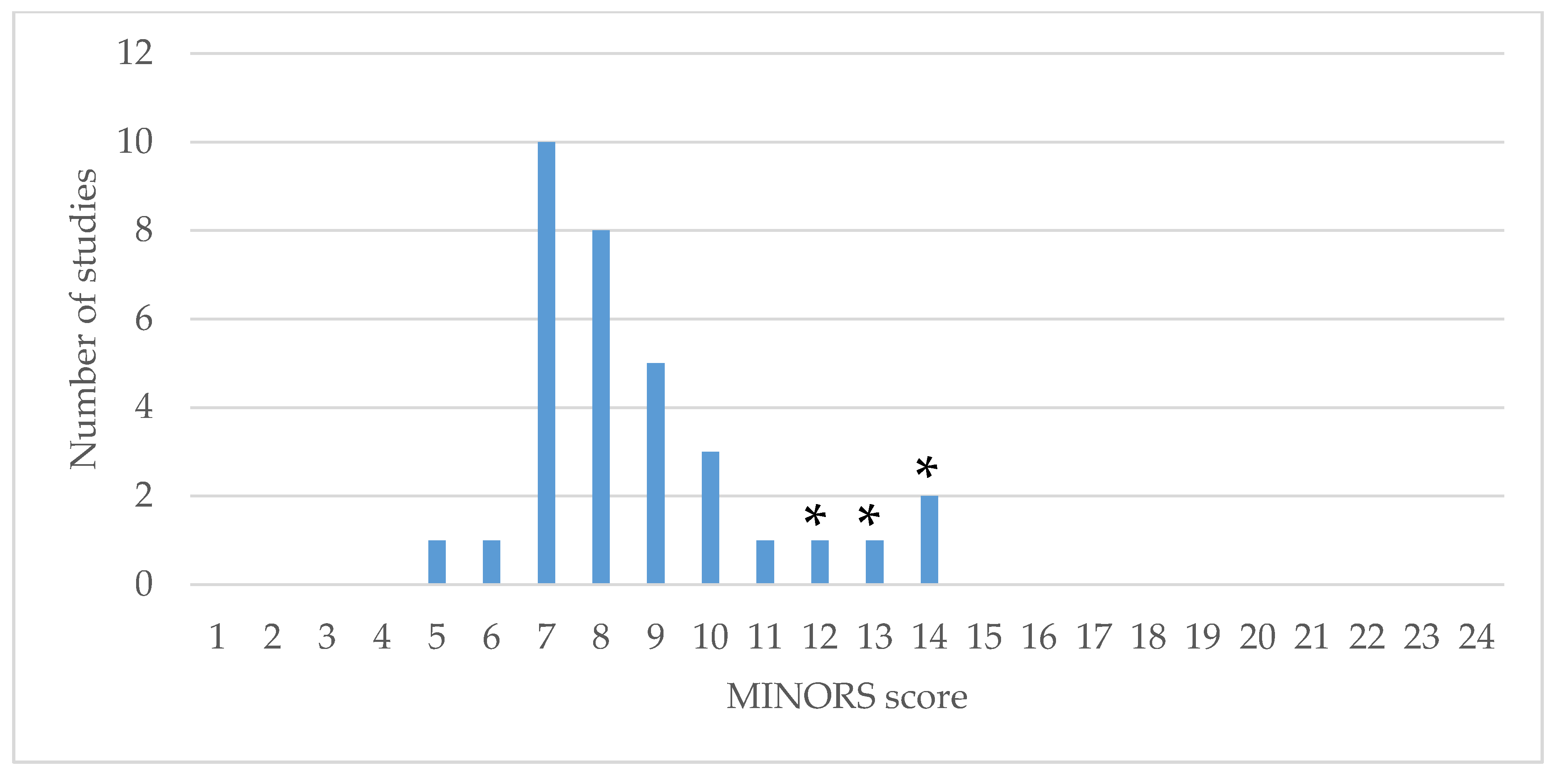
3.4. Study Quality
4. Discussion
5. Conclusions
Supplementary Materials
Author Contributions
Funding
Institutional Review Board Statement
Informed Consent Statement
Data Availability Statement
Conflicts of Interest
References
- Shore, B.J.; Thomason, P.; Reid, S.M.; Shrader, M.W.; Graham, H.K. Cerebral palsy. In Lovell and Winter’s Pediatric Orthopaedics, 8th ed.; Flynn, J.M., Weinstein, S., Eds.; Wolters Kluwer Health: Philadelphia, PA, USA, 2020; pp. 509–590. [Google Scholar]
- Rethlefsen, S.A.; Blumstein, G.; Kay, R.M.; Dorey, F.; Wren, T.A. Prevalence of specific gait abnormalities in children with cerebral palsy revisited: Influence of age, prior surgery, and Gross Motor Function Classification System level. Dev. Med. Child Neurol. 2017, 59, 79–88. [Google Scholar] [CrossRef] [PubMed]
- Boulay, C.; Pomero, V.; Viehweger, E.; Glard, Y.; Castanier, E.; Authier, G.; Halbert, C.; LucJouve, J.; Chabrol, B.; Bollini, G.; et al. Dynamic equinus with hindfoot valgus in children with hemiplegia. Gait Posture 2012, 36, 108–112. [Google Scholar] [CrossRef] [PubMed]
- Davids, J.R.; Rogozinski, B.M.; Hardin, J.W.; Davis, R.B. Ankle dorsiflexor function after plantar flexor surgery in children with cerebral palsy. J. Bone Jt. Surg. Am. 2011, 93, e1381–e1387. [Google Scholar] [CrossRef] [PubMed]
- Gracies, J.-M. Pathophysiology of spastic paresis. II: Emergence of muscle overactivity. Muscle Nerve 2005, 31, 552–571. [Google Scholar] [CrossRef] [PubMed]
- Panteliadis, C.P. Cerebral Palsy: A Multidisciplinary Approach; Springer: New York, NY, USA, 2018. [Google Scholar]
- Rathinam, C.; Bateman, A.; Peirson, J.; Skinner, J. Observational gait assessment tools in paediatrics—A systematic review. Gait Posture 2014, 40, 279–285. [Google Scholar] [CrossRef]
- Baker, R.; McGinley, J.L.; Schwartz, M.H.; Beynon, S.; Rozumalski, A.; Graham, H.K.; Tirosh, O. The gait profile score and movement analysis profile. Gait Posture 2009, 30, 265–269. [Google Scholar] [CrossRef]
- Dreher, T.; Buccoliero, T.; Wolf, S.I.; Heitzmann, D.; Gantz, S.; Braatz, F.; Wenz, W. Long-term results after gastrocnemius-soleus intramuscular aponeurotic recession as a part of multilevel surgery in spastic diplegic cerebral palsy. J. Bone Jt. Surg. Am. 2012, 94, 627–637. [Google Scholar] [CrossRef]
- Galli, M.; Cimolin, V.; Crivellini, M.; Albertini, G. Long-term evaluation of isolated gastrocnemius fascia lengthening in children with cerebral palsy using gait analysis. J. Pediatr. Orthop. Part B 2009, 18, 228–233. [Google Scholar] [CrossRef]
- Svehlik, M.; Kraus, T.; Steinwender, G.; Zwick, E.B.; Saraph, V.; Linhart, W.E. The Baumann procedure to correct equinus gait in children with diplegic cerebral palsy: Long-term results. J. Bone Jt. Surg. Ser. B 2012, 94, 1143–1147. [Google Scholar] [CrossRef]
- Lofterod, B.; Fosdahl, M.A.; Terjesen, T. Can persistent drop foot after calf muscle lengthening be predicted preoperatively? J. Foot Ankle Surg. 2009, 48, 631–636. [Google Scholar] [CrossRef]
- Slim, K.; Nini, E.; Forestier, D.; Kwiatkowski, F.; Panis, Y.; Chipponi, J. Methodological index for non-randomized studies (MINORS): Development and validation of a new instrument. ANZ J. Surg. 2003, 73, 712–716. [Google Scholar] [CrossRef] [PubMed]
- Baddar, A.; Granata, K.; Damiano, D.L.; Carmines, D.V.; Blanco, J.S.; Abel, M.F. Ankle and knee coupling in patients with spastic diplegia: Effects of gastrocnemius-soleus lengthening. J. Bone Jt. Surg. Ser. A 2002, 84, 736–744. [Google Scholar] [CrossRef] [PubMed]
- Borton, D.C.; Walker, K.; Pirpiris, M.; Nattrass, G.R.; Graham, H.K. Isolated calf lengthening in cerebral palsy. Outcome analysis of risk factors. J. Bone Jt. Surg. Br. 2001, 83, 364–370. [Google Scholar] [CrossRef]
- Firth, G.B.; Passmore, E.; Sangeux, M.; Thomason, P.; Rodda, J.; Donath, S.; Selber, P.; Graham, H.K. Multilevel surgery for equinus gait in children with spastic diplegic cerebral palsy medium-term follow-up with gait analysis. J. Bone Jt. Surg. Ser. A 2013, 95, 931–938. [Google Scholar] [CrossRef] [Green Version]
- Galli, M.; Cimolin, V.; Crivellini, M.; Albertini, G. Gait analysis before and after gastrocnemius fascia lengthening in children with cerebral palsy. J. Appl. Biomater. Biomech. 2005, 3, 98–105. [Google Scholar]
- Lofterod, B.; Terjesen, T. Local and distant effects of isolated calf muscle lengthening in children with cerebral palsy and equinus gait. J. Child. Orthop. 2008, 2, 55–61. [Google Scholar] [CrossRef] [Green Version]
- Pilloni, G.; Pau, M.; Costici, F.; Condoluci, C.; Galli, M. Use of 3D gait analysis as predictor of Achilles tendon lengthening surgery outcomes in children with cerebral palsy. Eur. J. Phys. Rehabil. Med. 2019, 55, 250–257. [Google Scholar] [CrossRef]
- Tinney, A.; Thomason, P.; Sangeux, M.; Khot, A.; Graham, H.K. The transverse Vulpius gastrocsoleus recession for equinus gait in children with cerebral palsy. Bone Jt. J. 2015, 97, 564–571. [Google Scholar] [CrossRef] [Green Version]
- Tylkowski, C.M.; Horan, M.; Oeffinger, D.J. Outcomes of gastrocnemius-soleus complex lengthening for isolated equinus contracture in children with cerebral palsy. J. Pediatr. Orthop. 2009, 29, 771–778. [Google Scholar] [CrossRef]
- Wren, T.A.; Do, K.P.; Kay, R.M. Gastrocnemius and soleus lengths in cerebral palsy equinus gait—Differences between children with and without static contracture and effects of gastrocnemius recession. J. Biomech. 2004, 37, 1321–1327. [Google Scholar] [CrossRef]
- Fujita, H.; Fusagawa, H.; Nishibu, H.; Nosaka, T.; Matsuyama, T.; Iba, K.; Yamashita, T. Motion analysis and surgical results of anterior transfer of flexor hallucis longus for equinovarus gait in children with hemiplegia. J. Orthop. Sci. 2020. [Google Scholar] [CrossRef] [PubMed]
- Park, C.I.; Park, E.S.; Kim, H.W.; Rha, D.W. Soft tissue surgery for equinus deformity in spastic hemiplegic cerebral palsy: Effects on kinematic and kinetic parameters. Yonsei Med. J. 2006, 47, 657–666. [Google Scholar] [CrossRef] [PubMed]
- Saraph, V.; Zwick, E.B.; Uitz, C.; Linhart, W.; Steinwender, G. The Baumann procedure for fixed contracture of the gastrosoleus in cerebral palsy. Evaluation of function of the ankle after multilevel surgery. J. Bone Jt. Surg. Br. 2000, 82, 535–540. [Google Scholar] [CrossRef]
- Steinwender, G.; Saraph, V.; Zwick, E.B.; Uitz, C.; Linhart, W. Fixed and dynamic equinus in cerebral palsy: Evaluation of ankle function after multilevel surgery. J. Pediatr. Orthop. 2001, 21, 102–107. [Google Scholar] [CrossRef] [PubMed]
- Engsberg, J.R.; Oeffinger, D.J.; Ross, S.A.; White, H.D.; Tylkowski, C.M.; Schoenecker, P.L. Comparison of three heel cord surgeries in children with cerebral palsy. J. Appl. Biomech. 2005, 21, 322–333. [Google Scholar] [CrossRef] [PubMed] [Green Version]
- Etnyre, B.; Chambers, C.S.; Scarborough, N.H.; Cain, T.E. Preoperative and postoperative assessment of surgical intervention for equinus gait in children with cerebral palsy. J. Pediatr. Orthop. 1993, 13, 24–31. [Google Scholar] [PubMed]
- Kim, D.W.; Kim, H.W.; Yoon, J.Y.; Rhee, I.; Oh, M.K.; Park, K.B. Endoscopic transverse gastrocsoleus recession in children with cerebral palsy. Front. Pediatr. 2020, 8, 112. [Google Scholar] [CrossRef]
- Klausler, M.; Speth, B.M.; Brunner, R.; Tirosh, O.; Camathias, C.; Rutz, E. Long-term follow-up after tibialis anterior tendon shortening in combination with Achilles tendon lengthening in spastic equinus in cerebral palsy. Gait Posture 2017, 58, 457–462. [Google Scholar] [CrossRef]
- Klotz, M.C.; Heitzmann, D.W.; Wolf, S.I.; Niklasch, M.; Maier, M.W.; Dreher, T. The influence of timing of knee recurvatum on surgical outcome in cerebral palsy. Res. Dev. Disabil. 2016, 48, 186–192. [Google Scholar] [CrossRef]
- Rutz, E.; Baker, R.; Tirosh, O.; Romkes, J.; Haase, C.; Brunner, R. Tibialis anterior tendon shortening in combination with Achilles tendon lengthening in spastic equinus in cerebral palsy. Gait Posture 2011, 33, 152–157. [Google Scholar] [CrossRef]
- Skaaret, I.; Steen, H.; Huse, A.B.; Holm, I. Comparison of gait with and without ankle-foot orthoses after lower limb surgery in children with unilateral cerebral palsy. J. Child. Orthop. 2019, 13, 180–189. [Google Scholar] [CrossRef] [PubMed]
- Vuillermin, C.; Rodda, J.; Rutz, E.; Shore, B.J.; Smith, K.; Graham, H.K. Severe crouch gait in spastic diplegia can be prevented: A population-based study. J. Bone Jt. Surg. Ser. B 2011, 93, 1670–1675. [Google Scholar] [CrossRef] [PubMed] [Green Version]
- Kay, R.M.; Rethlefsen, S.A.; Ryan, J.A.; Wren, T.A. Outcome of gastrocnemius recession and tendo-achilles lengthening in ambulatory children with cerebral palsy. J. Pediatr. Orthop. B 2004, 13, 92–98. [Google Scholar] [PubMed]
- Rose, S.A.; DeLuca, P.A.; Davis, I.R.B.; Ounpuu, S.; Gage, J.R. Kinematic and kinetic evaluation of the ankle after lengthening of the gastrocnemius fascia in children with cerebral palsy. J. Pediatr. Orthop. 1993, 13, 727–732. [Google Scholar] [CrossRef]
- Cimolin, V.; Galli, M.; Vimercati, S.L.; Albertini, G. Use of the Gait deviation index for the assessment of gastrocnemius fascia lengthening in children with cerebral palsy. Res. Dev. Disabil. 2011, 32, 377–381. [Google Scholar] [CrossRef] [PubMed]
- Schwartz, M.H.; Rozumalski, A. The Gait deviation index: A new comprehensive index of gait pathology. Gait Posture 2008, 28, 351–357. [Google Scholar] [CrossRef]
- Patikas, D.; Wolf, S.I.; Schuster, W.; Armbrust, P.; Dreher, T.; Doderlein, L. Electromyographic patterns in children with cerebral palsy: Do they change after surgery? Gait Posture 2007, 26, 362–371. [Google Scholar] [CrossRef]
- Wren, T.A.; Do, K.P.; Hara, R.; Dorey, F.J.; Kay, R.M.; Otsuka, N.Y. Gillette Gait Index as a gait analysis summary measure: Comparison with qualitative visual assessments of overall gait. J. Pediatr. Orthop. 2007, 27, 765–768. [Google Scholar] [CrossRef] [Green Version]
- Baker, R.; McGinley, J.L.; Schwartz, M.; Thomason, P.; Rodda, J.; Graham, H.K. The minimal clinically important difference for the Gait profile score. Gait Posture 2012, 35, 612–615. [Google Scholar] [CrossRef]
- Jahn, J.; Vasavada, A.N.; McMulkin, M.L. Calf muscle-tendon lengths before and after Tendo-Achilles lengthenings and gastrocnemius lengthenings for equinus in cerebral palsy and idiopathic toe walking. Gait Posture 2009, 29, 612–617. [Google Scholar] [CrossRef]
- Klotz, M.C.; Wolf, S.I.; Heitzmann, D.; Krautwurst, B.; Braatz, F.; Dreher, T. Reduction in primary genu recurvatum gait after aponeurotic calf muscle lengthening during multilevel surgery. Res. Dev. Disabil. 2013, 34, 3773–3780. [Google Scholar] [CrossRef] [PubMed]
- Rajagopal, A.; Kidzinski, L.; McGlaughlin, A.S.; Hicks, J.L.; Delp, S.L.; Schwartz, M.H. Pre-operative gastrocnemius lengths in gait predict outcomes following gastrocnemius lengthening surgery in children with cerebral palsy. PLoS ONE 2020, 15, e0233706. [Google Scholar] [CrossRef] [PubMed]
- Rutz, E.; McCarthy, J.; Shore, B.J.; Shrader, M.W.; Veerkamp, M.; Chambers, H.; Davids, J.R.; Kay, R.M.; Narayanan, U.; Novacheck, T.F.; et al. Indications for gastrocsoleus lengthening in ambulatory children with cerebral palsy: A Delphi consensus study. J. Child. Orthop. 2020, 14, 1–10. [Google Scholar] [CrossRef] [PubMed]
- Firth, G.B.; McMullan, M.; Chin, T.; Ma, F.; Selber, P.; Eizenberg, N.; Wolfe, R.; Graham, K. Lengthening of the gastrocnemius-soleus complex: An anatomical and biomechanical study in human cadavers. J. Bone Jt. Surg. Am. 2013, 95, 1489–1496. [Google Scholar] [CrossRef] [PubMed]
- Maurer, J.D.; Ward, V.; Mayson, T.A.; Davies, K.R.; Alvarez, C.M.; Beauchamp, R.D.; Black, A.H. Classification of midfoot break using multi-segment foot kinematics and pedobarography. Gait Posture 2014, 39, 1–6. [Google Scholar] [CrossRef] [PubMed]
- Boyd, R.N.; Graham, H.K. Objective measurement of clinical findings in the use of botulinum toxin type A for the management of children with cerebral palsy. Eur. J. Neurol. 1999, 6, s23–s35. [Google Scholar] [CrossRef]
- Shore, B.J.; White, N.; Kerr Graham, H. Surgical correction of equinus deformity in children with cerebral palsy: A systematic review. J. Child. Orthop. 2010, 4, 277–290. [Google Scholar] [CrossRef] [Green Version]
- Rodda, J.; Graham, H.K. Classification of gait patterns in spastic hemiplegia and spastic diplegia: A basis for a management algorithm. Eur. J. Neurol. 2001, 8, 98–108. [Google Scholar] [CrossRef]


Publisher’s Note: MDPI stays neutral with regard to jurisdictional claims in published maps and institutional affiliations. |
© 2021 by the authors. Licensee MDPI, Basel, Switzerland. This article is an open access article distributed under the terms and conditions of the Creative Commons Attribution (CC BY) license (http://creativecommons.org/licenses/by/4.0/).
Share and Cite
Ma, N.; Sclavos, N.; Passmore, E.; Thomason, P.; Graham, K.; Rutz, E. Three-Dimensional Gait Analysis in Children Undergoing Gastrocsoleus Lengthening for Equinus Secondary to Cerebral Palsy. Medicina 2021, 57, 98. https://doi.org/10.3390/medicina57020098
Ma N, Sclavos N, Passmore E, Thomason P, Graham K, Rutz E. Three-Dimensional Gait Analysis in Children Undergoing Gastrocsoleus Lengthening for Equinus Secondary to Cerebral Palsy. Medicina. 2021; 57(2):98. https://doi.org/10.3390/medicina57020098
Chicago/Turabian StyleMa, Norine, Nicholas Sclavos, Elyse Passmore, Pam Thomason, Kerr Graham, and Erich Rutz. 2021. "Three-Dimensional Gait Analysis in Children Undergoing Gastrocsoleus Lengthening for Equinus Secondary to Cerebral Palsy" Medicina 57, no. 2: 98. https://doi.org/10.3390/medicina57020098