

The Effect of Operational Factors on Phase Formation Patterns in the Light-Water Reactor Pressure Vessel Steels

1
National Research Center “Kurchatov Institute”, Moscow 123098, Russia
2
Division of Nuclear Physics and Technologies, National Research Nuclear University “MEPhI”, Moscow 115409, Russia
*
Author to whom correspondence should be addressed.
Metals 2023, 13(9), 1586; https://doi.org/10.3390/met13091586
Submission received: 1 August 2023
/
Revised: 31 August 2023
/
Accepted: 8 September 2023
/
Published: 12 September 2023
(This article belongs to the Special Issue Study on Phase Transformation and Deformation of Metallic Materials)
Abstract
:This paper presents the results of atom probe tomography studies on radiation-induced phase formation in light-water reactor pressure vessel steels after neutron irradiation under various conditions in comparison with the literature data. The given irradiation conditions are fluence (10–100) × 1022 m−2, flux (5–2700) × 1014 m−2s−1 and irradiation temperature (50–400) °C. The composition of the studied steels varies in a wide range for the elements significantly affecting radiation and thermal resistance of steels: Ni in the range of 0.2–6.0 wt.%, Mn–0.03–1.1 wt.%, Cu–0.01–0.16 wt.%, and P–0.01–0.03 wt.%. The number density, volume fraction, size, composition, and nucleation sites of precipitates are determined. The regularities of the effect of various operational factors on the phase formation in these steels have been analyzed and revealed. The study shows that in materials with high copper content, Cu-rich precipitates are formed by a radiation-enhanced mechanism. In materials with low copper content, their formation upon irradiation at 300 °C occurs by a radiation-induced mechanism since the main nucleation sites are point defect clusters formed in cascades. At the same time, the density, volume fraction, and composition of the precipitates depend on the steel composition (Ni and Mn content). In the steel with increased Ni content up to 5 wt.% but with ultra-low Mn content ≤ 0.03 wt.%, it is possible to suppress the formation of Ni-Si-Mn precipitates under irradiation.
1. Introduction
One of the key components of pressurized water reactors, the integrity of which must be maintained throughout the life of a nuclear power plant, both during normal operation and in the event of a maximum design basis accident, is the reactor pressure vessel (RPV). It is a thick-walled cylindrical pressure vessel made of solid one-piece shells—base metal forgings joined together by welds. Inside the RPV are all the fuel and structural components that ensure the reactor operation. Pressurized water, which is heated while passing through the reactor core (from 290 °C to 320 °C for VVER-1000), is pumped through the pressure vessel from bottom to top. The RPV is also a barrier to the release of radioactive fission products. During a basic design accident, cold water is pumped into the hot vessel, and resulting thermal stresses must not lead to vessel destruction. Replacement of the RPVs is not economically viable, and it is the RPV life that determines the service life of the entire reactor. The RPV is made of quenched and tempered low-alloy heat-resistant steel with a tempered bainite structure (Fe-Cr-Mn-Mo-Ni-Si-C system, depending on the reactor type). Nickel is added to improve the steel fracture toughness and hardenability, silicon to deoxidize, and manganese to bind the sulfur impurity in MnS. The presence of copper admixture in the first RPV generations is due to the use of copper-coated welding wire. The steels of the existing RPVs usually contain Cu (0.02–0.4), Ni (0.2–1.7), Mn (0.6–2.0), Si (0.2–0.6), and P (0.003–0.04), in wt.%.
During long-time operation (30 or more years), RPV steels are exposed to high-energy neutrons resulting from fuel fission, up to total accumulated fluences of 2.4 × 1024 m−2 (up to 0.1–0.5 dpa), and elevated temperature (~300 °C). This leads to degradation of the mechanical properties of the RPV materials during the service life and can lead to premature unacceptable failure of the RPV. The degradation of properties is mostly manifested in the loss of ductility and a noticeable shift of the interval of the ductile-to-brittle fracture transition, typical for steels with bcc lattice, to a higher temperature area, known as radiation embrittlement [1,2]. It is the shift of the critical embrittlement temperature that is the key industry parameter used to determine the degree of the RPV radiation embrittlement and service life. The most critical elements of the RPV are the welds opposite the core, which are subject to the greatest radiation embrittlement.
Overall, the degree of RPV steel degradation is determined by structural changes during operation. Therefore, it is important to understand how the RPV steel microstructure changes under irradiation. It depends on many parameters related both to the material itself (material type and composition, previous heat treatment, grain size, impurity content, initial phases, dislocation density, etc.) and to the operating conditions (neutron energy, temperature and duration of irradiation, accumulated irradiation dose (fluence), rate of damage dose accumulation (flux)).
Neutron irradiation leads to the formation of atomic collision cascades, which creates a combination of point defects (vacancies and interstitials) and clusters of these defects in the crystal lattice. These are the initial conditions for the diffusive evolution of alloys under irradiation. Subsequently, defects can recombine, migrate, agglomerate in the form of large clusters, or annihilate at various sinks (free surfaces, grain boundaries, or dislocations), resulting in significant changes in the microstructure and properties of steel. Although individual point defects alone do not cause significant hardening, they are fundamental to a wide range of irradiation effects. Thus, for RPV steels under typical irradiation, clustering of point defects leads to the formation of dislocation loops of mostly interstitial type. Moreover, point defects play a vital role in the long-term evolution of nanostructured features through the transport of solute atoms. Radiation-enhanced diffusion is the result of overconcentration of these defects. Irradiation can be considered on two different time scales. The instantaneous parameter is the neutron flux (flux, φ) expressed in units of m−2s−1, while the integral parameter is the fluence (φt, in units of m−2). Since the concentrations of point defects depend on the neutron flux, the same fluence obtained at different flux levels over different time intervals can lead to very different irradiation effects (flux effect). Typical dose accumulation rates (fluxes) for samples irradiated as part of existing RPVs do not allow obtaining in a short time the values of fast neutron fluence required for proactive prediction. Therefore, accelerated irradiations in research reactors at higher fluxes are used. This raises the question of the effect of flux on the results of microstructural studies and mechanical tests.
Numerous studies have shown that RPV steel radiation embrittlement is caused both by the formation of precipitates and radiation defects—dislocation loops, which act as barriers to the movement of dislocations and lead to radiation hardening (hardening mechanism) and grain-boundary embrittlement due to the weakening of grain-boundary cohesion caused by the formation of grain-boundary segregation of impurity and alloying elements (non-hardening mechanism) [2,3,4,5,6].
The major embrittling elements are Cu, P, Ni, and Mn, and to a lesser extent also, Si [7,8]. In steels with a high-Cu content, copper-enriched high-density precipitates are formed, while in steels with a low Cu content but alloyed with Ni and Mn, precipitates based on Ni, Mn, and Si are formed [2]. In addition, elements P, as well as Ni and Mn, contribute to the grain-boundary embrittlement [4,9]. The difference in the content of impurity and alloying elements in RPV steels of various origins determines differences in the degree and kinetics of their radiation embrittlement. It is worth noting that the chemical composition of the base metal (BM) and weld metal (WM) varies between RPV manufacturers, different RPVs of the same manufacturer, and even in a particular RPV, especially for WM, whose composition may vary in different areas.
Once the negative effect of copper and phosphorus impurities on radiation embrittlement for first-generation RPVs was discovered, their concentration in RPV steels was controlled and limited. However, for second-generation RPV steels alloyed with nickel and manganese, further studies showed an increase in embrittlement in steels containing these elements at the upper limit of grade composition. Therefore, for the next generations of steels with extended service life, the developers follow either the path of reducing the nickel content or, if it is necessary to further increase the strength properties and fracture toughness (e.g., when increasing the size of the vessel), on the contrary, increasing the nickel content with simultaneous limitation of the concentration of other embrittling elements (Mn, Si).
With the development of materials research methods, namely, the invention of high-resolution analytical research methods—high-resolution transmission electron microscopy and atom probe tomography (APT), there was a qualitative step forward in the level of research and improvement of quantitative analysis. This allowed obtaining additional data on the radiation embrittlement mechanisms, verifying or refuting various hypotheses put forward earlier, and, along with the development of modeling methods, this improved models of phase formation and segregation kinetics in RPV steels.
The review of systematic studies of a wide range of RPV materials under various irradiation conditions allows for predicting the behavior of the material under neutron irradiation conditions based on the mechanisms of redistribution of impurity and alloying elements. Moreover, this allows for developing recommendations both on the potential of extending the service life of materials of operating reactors (including the use of compensatory measures—recovery annealing of the most critical elements) and on the development of new materials for new-generation reactors.
The impact of the non-hardening mechanism is found mainly in steels with high nickel content [10]. Since the segregation kinetics for RPV steels is of a damped nature, the contribution of the non-hardening mechanism should reach saturation during the long operation of reactor vessels (more than 30 years in case of RPV service life extension) [10]. Therefore, the main contributor to the change in properties should be the hardening mechanism [1,2]. At the same time, the density of radiation defects does not depend on the steel composition and is significantly lower than that of radiation-induced precipitates [2,11], which makes the contribution of the latter crucial for long service life. In this regard, this paper investigates the peculiarities of phase formation in various RPV steels depending on operational factors. For this purpose, experimental results of APT studies obtained over the last decade for RPV steels of operating Russian reactors, as well as Russian-made steels being developed, in comparison with foreign ones, have been accumulated and analyzed to evaluate the effect of operational factors on phase formation in RPV steels.
2. Materials and Methods
2.1. Materials
The phase formation kinetics was studied for RPV materials of decommissioned VVER-440 reactors, operating VVER-1000 reactors, as well as for advanced steels being developed for next-generation reactors. In order to reveal the effect of dose accumulation rate on phase formation in RPV steels, VVER-1000 RPV samples irradiated at different dose rates were studied—as part of the vessels of operating reactors under the surveillance specimen program and in the research reactor.
Russian-made RPV steels mainly differ from Western RPV steels in high Cr content (usually containing about 2 wt.% Cr), higher P content, lower (in most cases) Cu content, and, as a rule, lower (VVER-440) or higher (VVER-1000) Ni content.
Samples of advanced RPV steels with different Ni and Mn content for next-generation reactors after accelerated irradiation in a material test reactor (MTR) have also been studied. In order to reveal the effect of irradiation temperature, steel samples after accelerated irradiation at different temperatures have been investigated.
Table 1 shows the intervals of the studied material composition, and Table 2 shows the states and irradiation conditions of the studied samples. The studied materials are divided into groups according to the content of the major precipitate-forming elements. The table shows the content of the key chemical elements in the studied steel.
2.2. Atom Probe Tomography Studies
The APT method [12,13] used in this study has the unique ability to identify and quantify individual chemical elements in three dimensions with the highest available spatial resolution, which allows for the detection of inhomogeneities in the distribution of elements and to obtain quantitative data on the distribution and composition of very small precipitates (less than 3–5 nm), clusters and segregation at grain boundaries, interphase boundaries and dislocation loops in three-dimensional space.
When the APT method is used, as well as other methods, there are challenges and inaccuracies that are related both to the method directly and to the material under study. The main artifacts are the effects of preferential evaporation and local magnification due to the difference in the evaporation fields of different atoms (e.g., the Cu evaporation field is lower than the Fe one), matrix atoms aggregating into clusters during reconstruction due to trajectory aberration, inability to distinguish some isotopes due to peak overlap, and changes in the position of atoms during reconstruction due to the migration of some elements along the needle surface (e.g., P, Si, and C). Due to the high locality of the method (a typical studied volume of the sample is ~(50 × 50 × 100–200) nm3), the results obtained may be unrepresentative of the volume of the overall material, so a sufficient volume of the studied material should be accumulated.
The above-mentioned drawbacks were addressed by optimizing the acquisition conditions (temperature, detection rate, rules fraction), as well as by careful and thorough analysis and interpretation of the obtained data based on the results of round-robin experiments performed by the world’s experts in the field of APT research of reactor materials [14].
The studies were performed by atom probe tomography using an atom probe with a local electrode CAMECA® Instruments Inc. LEAP 4000 HR. Needle-shaped specimens were made by the standard electropolishing method using a 10% solution of perchloric acid in acetic acid on a Simplex ElectroPointer from blank parts machined from previously tested Charpy specimens [12]. Data accumulation was carried out in pulsed voltage mode at the following parameters: temperature 50 K, pulse repetition rate 200 kHz, detection rate 0.1–0.2, and pulse fraction of 15–20%. Reconstruction and data processing were carried out using the program CAMECA® IVAS® 3.6.12. All overlapping isotopes were separated by standard methods when decoded on the mass spectrum using peak deconvolution [12,13].
Cluster identification was performed using the maximum separation method (MSM [12,15]) with the following processing parameters determined from the integrated analyses according to [15]:
- Core atoms: Cu, P, Ni, Si, Mn for high-Cu steels or Ni, Si, Mn for low Cu steels;
- Order (of nearest neighbors) 8 (based on the IVAS nearest neighbor distribution);
- dmax (maximum distance between atoms of the selected type in the cluster) = 0.7–0.8 nm;
- Nmin (minimum number of atoms of the selected type in the cluster) = 11–40;
- der = L = dmax.
Clusters located at the boundary were considered in the total density with a factor of ½ and were not considered in the analysis of size and composition.
The number density of precipitates was calculated as:
where Nclusters detected is the number of clusters detected in the dataset, Nedge clusters is the number of clustersdetected at the edge of the analyzed volume, and Vdataset is the volume of the reconstructed dataset.
The volume fraction was calculated according to the formula:
where Ncluster atoms is the number of atoms in all of the clusters and Ntotal atoms is the total number of atoms in the dataset.
The average size (diameter) D was estimated from the equivalent radius [12]:
where Nions is the number of atoms in the cluster, Vat = 0.0118 nm3—atomic volume, and Q—detection efficiency (36% for LEAP 4000HR).
When determining the matrix composition, the measurement error is determined by the variation of values from sample to sample (variation of chemical composition).
The composition of precipitates was estimated for the nucleus by the averaged IVAS cluster concentration profile. The study [16] shows that the Fe content in precipitates does not exceed 6 at%, so the removal of the entire iron content from the precipitate composition is a reasonable assumption, although not quite physically correct. Therefore, only the precipitate-forming atoms (Ni, Mn, Si, Cu, and P) were considered when analyzing the findings.
Noteworthy is that APT studies of all the samples were performed on the same equipment, under identical imaging conditions, using a unified method of processing and presenting the results. At the same time, the heterogeneity of the material chemical composition necessitated the study of a large number of samples and precipitates (the studied volume for each state made up at least 1 × 10⁶ nm3 for at least 4–6 datasets). This allowed for improving the reliability of the revealed regularities of changes in the nanostructure of the studied materials when analyzing the results.
3. Results and Discussion
3.1. High-Cu Materials
In the first generations of reactor vessels, the copper content exceeded the limit of its solubility in solid solution, which led to an unacceptably high level of radiation hardening and embrittlement of RPV steels due to the formation of a high density of radiation-induced copper-based precipitates. All RPVs were either decommissioned or compensatory measures were taken to ensure further operation, i.e., recovery annealing of the weld opposite the core with the most severe radiation embrittlement. The phase formation kinetics in Russian high-Cu steels in ‘irradiation–annealing–irradiation’ cycles is reviewed in [17].
Studies of high-Cu weld metal, irradiated at 250 °C for 20 years in the RPV and demonstrating a high level of radiation embrittlement, revealed the formation of a high density of copper-enriched precipitates. Table 3 presents the results of APT studies of high-Cu RPV steels after long-term operation.
Figure 1 and Figure 2 summarize the results of the authors’ APT studies of high-Cu RPV steels irradiated in the operating RPVs, as well as published results of APT studies of high-Cu steels irradiated in the RPV in terms of the number density and volume fraction of Cu-rich precipitates [18,19,20,21,22].
Figure 3 shows a 3D map of the distribution of elements comprising copper-enriched precipitates in high-Cu steels.
Table 3 shows that the number density of precipitates in high-Cu, low-Ni BM with Cu content of 0.11 wt.% is significantly lower than in high-Cu WM with Cu content of 0.16 wt.%, which is due to a higher Cu content in the latter. At the same time, the sizes of precipitates in BM with lower Cu content are higher. This is in agreement with the literature data [23,24].
For high-Cu steels, copper-enriched precipitates were also detected in the state after long-term thermal conditioning at the irradiation temperature (after 45 years of operation). Comparison of the material after thermal conditioning with the same material exposed to irradiation at operating temperature for the same time shows the difference not only in the number, density, and size of precipitates (significantly lower values for thermal conditioning without irradiation) but also in the composition of the precipitates.
The paper [17] showed that copper depletion in the matrix of RPV steel, irradiated for 45 years, amounted to more than 70% and phosphorus about 50%, while for steel, which was at the same time exposed to the temperature without irradiation, this value was 3% for copper, and there was no depletion of other elements.
Figure 4 shows the distribution of the major precipitate elements in the studied high-Cu materials and 3D APT maps of grain-boundary in high-Cu BM.
The diagram shows that the precipitates formed under prolonged thermal exposure (290 °C) comprise only copper and are characterized by low number density and size, whereas in the case of irradiation (270 °C), the precipitate composition also includes Ni, Mn, Si, and P. The higher volume fraction of precipitates of different compositions in the irradiated state compared to thermal exposure indicates the radiation-enhanced nature (due to an increase of the point defects number) of thermodynamically driven copper-enriched precipitates. In addition, the concentration of phosphorus in precipitates in irradiated WM (with high Mn content) is higher than in BM, in which phosphorus is spent on the formation of grain-boundary segregations (see Figure 4b). This is due to the peculiarities of grain-boundary segregation of phosphorus in materials with different structures and initial phase compositions [17].
Thus, the formation of copper-enriched precipitates occurs in high-Cu RPV steels, which is due to both thermal diffusion (after prolonged thermal exposure) and radiation-stimulated diffusion of impurity elements (during irradiation). Although the exact value of the solubility limit of copper at 300 °C is unknown, it is most likely ~0.007 at.% [25], so the matrix is supersaturated with copper. At the same time, copper-enriched phases are formed under irradiation at RPV operating temperatures (about 300 °C) at Cu contents above (0.05–0.09) wt.%, depending on alloying [17,18,26]. Precipitates in these steels are formed by a radiation-enhanced mechanism (thermodynamically conditioned and enhanced due to radiation-enhanced diffusion, as well as due to new nucleation sites in the form of point defect clusters formed in cascades) and have damped kinetics due to the depletion of matrix copper content [1,2]. Increasing the copper content from 0.1 to 0.3 wt.% leads to higher precipitate density and a greater hardening effect; however, the damping trend remains unchanged.
Experimental and modeling results show that the formation of Cu clusters, vacancy clusters, and Cu-V complexes in bcc-Fe is thermodynamically driven [27,28]. To compensate for lattice distortions, a large Cu cluster (composed of oversized atoms) is preferentially bonded to a vacancy to form a Cu-V complex instead of forming a pure Cu cluster. At the same time, Si atoms are attracted only by vacancies, which should affect the obviously higher Si concentration in precipitates formed in low-Cu RPV steels than in high-Cu RPV steels (if Cu concentration in precipitates is not taken into account). Moreover, since irradiation creates many more vacancies than thermal aging, copper-enriched phases formed under irradiation should have higher Si concentrations due to the higher vacancy concentration in them. These differences in Si concentration can be seen in Figure 4 and were confirmed in [26] for high-Cu RPV steels. Stable copper-enriched phases are formed in supersaturated solid solution through two intermediate phases: BCC structure coherent with the matrix → 9R structure with high twin density → FCC structure without twins. The maximum strength is exhibited by BCC precipitates [29].
Copper-enriched precipitates in irradiated steels also contain Ni and Mn, with copper in the core, whereas Mn and Ni are concentrated in the shell (core-shell structure) [26,30,31]. This indicates the existence of a Ni- and Mn-enriched interface. The existence of such a Cu–Ni/Mn–Fe buffer transition is energetically favored due to the sharp Cu/Fe interface, since due to the vanishingly small solubility of Cu in Fe, the interface energy for copper-enriched clusters in the iron matrix is relatively high, whereas Ni and Mn are much better soluble in Fe and at the same time have energetically favorable interactions with Cu, further reducing the interface energy [31,32]. It is worth noting that although copper-enriched and Ni-Mn-Si precipitates are usually considered as two different types, experimental results obtained by the APT method show that, if the Cu concentration in copper-enriched precipitates is not taken into account, the relative content of Mn and Ni in them is approximately the same [33,34].
3.2. Low-Cu RPV Materials in Operating Reactors (Low Cu, High/Medium Ni, Medium Si)
In order to provide the required strength and fracture toughness at the given dimensions of the vessel to ensure hardenability throughout the entire thickness of the RPV wall, a significant amount of Ni was introduced into the composition of RPV steels. Its content for BM (medium Ni medium Mn medium Si) usually does not exceed 1.35 wt.%, while for WM (high Ni high Mn), it is in the range of 1.4–2.0 wt.%. Manganese varies between 0.4 and 0.5 wt.% in BM and 0.7 and 1.1 wt.% in WM.
Studies have shown that weld metal (high Ni high Mn) embrittlement kinetics is higher than expected. The shifts were progressively greater with higher nickel content in the material. Contributing to this was increasing embrittlement not only by the hardening mechanism but also by the non-hardening mechanism. The features of segregation processes in VVER-1000 RPV steels are investigated in detail in [4,10] and are not reviewed in detail in this study.
The study has shown that Ni, Mn, and Si-based precipitates are formed for low-Cu steels, which are known as late blooming phases [1] and are referred to as MNS, NMS, and NSM in various papers, depending on the precipitate composition [11,35]. To avoid confusion, we will refer to them as NSM precipitates. In the case of NSM precipitates, unlike copper-enriched precipitates, we address here the formation of clusters rich in nickel, manganese, and silicon in materials where none of the elements exceeds the solubility limit in Fe-based binary alloys (Si > 8 at.%, Mn~3 at.%, Ni~5 at.% at 300 °C). The mechanism of solute cluster formation in this case is still debatable. Is it an irradiation-enhanced process dictated by thermodynamics or an irradiation-induced process that would not occur without irradiation? Current models of radiation-induced precipitate formation are described and are based on two major conflicting theories of NSM precipitate formation and growth [1,36,37,38,39].
The first theory is that NSM precipitates are radiation-enhanced (due to radiation-enhanced diffusion), i.e., thermodynamically stable intermetallic compounds such as G or Γ2 phases, which are predicted by equilibrium thermodynamic models for the RPV low operating temperature [36]. The thermodynamically predicted volume fractions of equilibrium precipitates and their compositions are in agreement with atom probe tomography data for steels irradiated to very high fluences [33]. In addition, X-ray diffraction and scattering experiments [40] showed intermetallic crystal structures of Γ2 or G phase in irradiated RPV steels, which is consistent with the thermodynamic calculations of CALPHAD [36]. In favor of this theory is the fact that the thermal stability of NSM precipitates in RPV steels at locally low Fe content and the presence of irradiation-induced radiation defects [41] was predicted by thermodynamic models and simulations [36,42] and confirmed experimentally [40,43,44]. The stability of NSM precipitates has been confirmed both indirectly by annealing irradiated samples at a higher temperature (425 °C), as a result of which some precipitates are preserved and enlarged [43], and directly after long isothermal exposures at temperatures in the range of 330–405 °C, when their formation is observed [45]. Despite the thermodynamic stability of precipitates (G and Γ2 phases), in the absence of a sufficient amount of Cu at RPV operating temperatures, the driving force for the formation of NSM precipitates is small, and their homogeneous nucleation of precipitates in a defect-free matrix is hampered. Therefore, the formation of NMS precipitates requires a large number of irradiation-induced point defects.
The second theory suggests that NSM precipitates are radiation-induced [46] and are formed by radiation-induced segregation (RIS) of Ni, Mn, and Si [47]. In particular, these models suggest that either Mn-Ni clusters are not thermally stable in Fe [48,49] or they are only stable at a combination of low temperatures and high solute concentrations, and thus radiation-induced segregation is required for growth and persistence [37]. Precipitates can be formed both homogeneously (in solid solutions with a lack of defects without driving forces for precipitate release) when RIS leads to local solute concentrations above the limit of their solubility in the matrix and heterogeneously—on sinks. In this case, the sinks can be microstructural features such as dislocation networks, small copper clusters, diffusion-associated Mn dumbbells [48], small dislocation loops, and point defect clusters (mainly interstitial) formed in displacement cascades [37,50], which act as heterogeneous nucleation sites [3,37,46] due to the increased solute concentration on them. The binding energy of defect solute complexes is almost always numerically lower than that of pure solute clusters, so interstitial atoms and vacancies are more likely to act as nucleation sites than pure solute clusters, which facilitates the nucleation of NSM clusters under irradiation. The driving force for the formation of nanoscale solute clusters is the interaction of the solute atoms with their own interstitial defects formed under irradiation [37]. As a result of such an interaction, defect clusters immobilized due to strong binding interactions with solute atoms can turn into nucleation sites, where other solutes gradually accumulate due to transport by point defects [37,50,51], as confirmed experimentally [46,52]. A pinning effect on clusters of self-interstitial atoms has been inferred from experimental data for Mn, P, and Ni [53,54,55] and by modeling for Cr, P, and Ni [56,57,58]. Ab initio computations of solute–loop interactions suggest a similar pinning tendency for Mn, Cu, and Si [51].
It is worth noting that these cascade clusters of interstitial atoms can actually represent proto-prismatic a/2<111> dislocation loops, which are unresolvable in transmission electron microscopy at such sizes [2]. In low-alloyed steels at higher fluence, larger clusters of interstitial atoms are observed in the form of dislocation loops with an unresolvable zero contrast line (of the ‘black dots’ type), which presumably grew from much smaller cascade clusters and thus the loops may be much larger than previously thought [2].
Overall, the formation of phase nuclei requires appropriate temperatures and irradiation doses at which the nucleation rate is greater than the annihilation rate of radiation defects (at temperatures below 400 °C). In addition, the RPV operating times should allow the initial nuclei to grow due to the diffusion of precipitate-forming atoms toward the cluster. Otherwise, radiation-induced precipitates do not emerge [24].
The APT method allowed the detection of not only clusters in the volume but also segregation of elements on all features of the structure (dislocations, carbides/carbonitrides, and grain boundaries [33,36]), indicating a continuous process of solute precipitation at a high density of nucleation sites, triggered and maintained by continuous fluxes of vacancies and self-interstitials and their clustering due to irradiation. This mechanism differs from classical release by nucleation and growth.
In [39], a model of NiMnSiPCu-enriched cluster formation under irradiation was developed using the Objected Kinetic Monte Carlo OKMC method, which allows predicting the values of number density, size, and volume fraction for solute clusters with regard to the chemical composition (Ni, Mn, Si, P, and Cu content in at.%), irradiation conditions (temperature and flux) and cumulative dose (irradiation time equivalent with due regard to the flux). Cr atoms, present in significant amounts in VVER steels, are taken into account through the impact on the diffusion coefficient of self-interstitial defects. This model is intended for materials with XNi < 0.55 (XNi = CNi/(CNi + CMn + CSi + CCu + CP)) and emerges from the results for which, in general, XNi < 0.48. For Russian low-Cu steels in operating reactors, this value is 0.46–0.57 (for most of them, more than 0.5) and lies at the upper limit of the relevant interval. Therefore, the studies of phase formation in steels of different compositions performed in this paper can help to improve the models being developed.
3.2.1. Number Density, Size, and Volume Fraction of Precipitates
Figure 5, Figure 6, Figure 7 and Figure 8 show the APT studies results for RPV samples of operating VVER-1000 reactors (BM—high Ni, high Mn, medium Si, and WM—low-Ni, medium Mn, medium Si): number densities, sizes, and volume fractions.
Figure 6, Figure 7 and Figure 8 show that irradiation leads to the formation of precipitates, the density of which depends not only on the irradiation dose (accumulated fluence) but also on the steel composition. Thus, for BM with significantly lower values of both Ni and Mn concentration in steel, the number density of precipitates is much lower. At the same time, a dose correlation is observed for both BM and WM, but for BM, it is weaker due to the lower absolute values.
The size of precipitates does not seem to strongly depend on the material composition in the studied intervals of high/low Ni, high/medium Mn, medium Si (within the range of scattering caused by local material inhomogeneities), and increasing with dose and showing a tendency to saturation. However, there is a tendency to slightly higher values for BM (see Figure 7), which seems to be related to the lower number density of precipitates in BM. The same was found earlier for high-Cu BM and WM (VVER-440): precipitate sizes in BM samples are significantly higher with their lower density as compared to WM (see Table 3).
The dependences of the number, density, and volume fraction of precipitates presented in Figure 6 and Figure 8 show a correlation with the accumulated fluence due to the large differences in nickel and manganese composition. Therefore, the values of number density and volume fraction of precipitates were normalized by the concentration of precipitate-forming elements in steel (by the value (Ni×Mn/Si)). The choice of the normalizing factor was based on the following dependencies.
Figure 9 shows the dependence of the precipitate volume fraction on the content of Ni, Mn, and Si in the studied steels. Despite the difference in the accumulated fluence, which significantly affects the value of the precipitate volume fraction, the study shows that an increase in the Ni and Mn concentrations in the steel leads to an increase in the precipitate volume fraction.
The correlation between Ni and Mn concentration and the number, density, and volume fraction of precipitates, and consequently radiation hardening, has been shown in numerous papers (e.g., [1,2]).
In [3], for highly irradiated materials with wide alloying intervals for Ni (0.22–3.29 wt.%) and Mn (0.08–1.52 wt.%), a strong synergistic effect of Ni and Mn influence on the formation of NMS precipitates was demonstrated: the volume fraction of NSM precipitates is proportional to Ni1.6Mn0.8, where Ni and Mn are the contents of these elements in steel according to APT data. In steels with high Ni content and low Mn content, the density of NSM precipitates is significantly lower compared to steels with high Mn and Ni content [3]. At ultra-low Mn content ≤ 0.03 wt.% in steels with high Ni content up to 5 wt.%, the formation of NSM precipitates may be suppressed [59]. This may indicate the participation of Mn in nucleation. For example, theoretical results using density functional theory [60] have shown that irradiation early produces a large number of interstitials, which contribute to the nucleation of precipitates as soon as they combine with Mn atoms.
The effect of Si concentration is not so apparent. Figure 9 shows a trend for the volume fraction of precipitates for WM to decrease with Si concentration. Statistical analysis in [61] also showed that within the range of silicon concentrations characteristic for RPV steels (0.2–0.6 wt.%), an increase in its content contributes to a decrease in radiation embrittlement. In addition, in the analytical relationships [62] for determining radiation embrittlement, the silicon content is also included with a minus sign:
where AF = α1exp(α2Ceq). and Ceq = CNi + CMn − αCSi.
3.2.2. Precipitate Nucleation Sites
The APT studies have shown that irradiation leads to the formation of NSM precipitates, which are located both homogeneously in the matrix and on the visible sinks of point defects—dislocations and grain boundaries (see Figure 5 and Figure 12). According to the nucleation sites, they are divided into heterogeneous, nucleated on visible sinks, and homogeneously distributed, formed on the point defect clusters in cascades, which cannot be identified by the APT method. Noteworthy is the absence of precipitates in the zone near grain boundaries (Figure 12), which is caused by the depletion of this zone of precipitate-forming elements (Ni, Mn, Si) due to their grain-boundary segregation.
Figure 13 shows the fraction of precipitates nucleated heterogeneously (mainly on dislocations) in the total fraction of detected precipitates in the studied irradiated medium/high Ni, medium/high Mn, and medium Si steels. The upper estimate of dislocation density for these steels (obtained from the results of transmission electron microscopy studies) is approximately 1 × 10¹⁴ m−2.
Figure 13 shows that for WM, the nucleation sites are mostly point defect clusters (up to 90%). At the same time, the fraction of heterogeneously nucleated precipitates was significantly higher for BM than for WM. Diffusion rates of Mn, Ni, and Si in the matrix seem to be much lower than near heterogeneous nucleation sites. Therefore, for BM, due to lower Ni and Mn concentrations in the steel, a level of RIS on point defect clusters located homogeneously in the matrix, which are nucleation sites for precipitates, is lower than for WM. Therefore, in BM at the initial irradiation stages, precipitate formation occurs predominantly on heterogeneous nucleation sites (dislocations, grain boundaries, and second-phase boundaries). At the same time, there is a trend for the decrease in the fraction of heterogeneously nucleated precipitates in BM with increasing accumulated fluence, which indicates that the nucleation of precipitates on point defect clusters in the matrix is facilitated due to both the increase in nucleation sites and the level of RIS with the irradiation dose. This is in agreement with the literature data. Thus, experimental results [3] and modeling [36] show that for the nucleation of precipitates in steels with low and medium values of Ni concentration, the heterogeneous mechanism is crucial, whereas, for high Ni content (>1.5 at.% Ni), the nucleation in the matrix on point defect clusters prevails.
Paper [36] also showed an upper-limit estimate of the number density of nucleation sites at dislocations and grain boundaries. The upper limit of the site density estimates for heterogeneous precipitate nucleation (1022–1023 m−3 for dislocations and 1023 m−3 for grain boundaries) cannot satisfy the observed high values of the number density of precipitates (up to ~1024 m−3 at very high accumulated fluence [3]). Therefore, heterogeneous nucleation cannot play the decisive role in the usually observed precipitate density (except for cases with low concentration of precipitate-forming elements in the steel and, consequently, low precipitate density), and the main mechanism, in this case, is heterogeneous nucleation on homogeneously located in the matrix point defect clusters formed in cascades.
3.2.3. Composition of Precipitates
Figure 14 shows the results of determining the precipitate composition (the ratio of the main precipitate-forming elements Ni, Si, and Mn, assuming Ni + Mn + Si = 100%) in the studied VVER-1000 RPV samples. It is worth noting that due to the low proportion of heterogeneous precipitates and artifacts in determining their composition (e.g., the appearance of vanadium and carbon in the precipitate nucleation on vanadium carbonitrides), the composition is given for homogeneously located precipitates in the matrix. In addition to certain compositions, known Ni-Si-Mn compounds are also presented.
The study shows that the precipitate compositions occupy narrow ranges of values in the ternary diagram, which differ for BM and WM mainly in the concentration of Mn, which in BM is about twice lower than in WM. The average compositions of precipitates for WM correspond to Ni4Si3Mn2, and for BM—to Ni4Si3Mn. The nickel content in precipitates is about 50 at.%, and the content of manganese and silicon varies in accordance with the material composition. This is in agreement with the findings of [41], which show that in typical low alloy ferritic steels, the clustering of Mn, Ni, and Si is thermodynamically favorable. However, at RPV operating temperatures, thermal effects alone are insufficient for cluster formation and, consequently, for reaching the equilibrium state. Therefore, at the early stages of phase formation, NSM precipitates have an ordered B2 structure, where Ni occupies positions in one sublattice and Mn/Si in another. At the same time, Mn and Si can easily substitute each other according to the concentrations of these elements in the alloy. When Si atoms inside the precipitate are replaced by Mn atoms from the matrix, the Si-Ni bond in the precipitate is replaced by Mn-Ni, and the Mn-Fe bond in the matrix is replaced by Si-Fe, which, according to [61], is energetically favorable. This may indicate that at very high irradiation doses and/or higher temperatures, the observed phases of inaccurate stoichiometric composition can transform to the equilibrium G phase (and also in the case of corresponding alloying—Ni/Mn/Si~16/6/7).
This is consistent with the results obtained in [3,34,55,63]. For materials with a wide range of nickel and manganese concentrations, the study showed that depending on the steel composition, mainly G-(Ni16Si7Mn6) or Γ2 (Ni3SiMn2) phases could be formed (in alloys with (0.7–1.6) wt.% Ni, >1.0 wt.% Mn and (0.4–0.8) wt.% Si) and Ni-silicide type compositions in alloys with low Mn content and high Ni content (>1.6%) [3]. At the same time, the actual compositions of the precipitates vary somewhat depending on the composition of the corresponding alloy, and the precipitates are not claimed to have corresponding crystal structures.
Figure 15 shows the correlation between the concentration of elements in precipitates and the concentration of elements in steel. The chemical composition is calculated in relative units Ni + Mn + Si = 100%. The figure shows that for the studied materials, the precipitate compositions are generally linearly proportional to the material composition.
Figure 16 shows the dependence of copper concentration in precipitates on the Cu content in steels with low Cu and medium/high Ni and Mn content.
Figure 16 shows that for WM with Cu content in steel up to 0.06 wt.%, the Cu content in precipitates does not exceed ~1 at%, while with Cu content in steel 0.07 wt.% and higher, the Cu content in precipitates is much higher—from ~2 to 4.6 at.%. (assuming that the precipitates consist only of Ni, Si, Mn, Cu, and P atoms). Cu-V clusters, in this case, become nucleation sites for precipitates, which can facilitate the nucleation of precipitates.
It is worth noting that some researchers, relying on the same relative concentration of Ni, Si, and Mn (ignoring Cu) [33,34], on the presence of Ni-Si-Mn shell around the copper core in the case of copper-enriched precipitates, as well as on the presence of NSM precipitates near copper-enriched precipitates at high irradiation doses, suggest that the mechanisms of their formation are similar, and the main difference is in the precipitate nucleation. Thus, for copper-enriched precipitates, the nucleation sites are Cu clusters or Cu-vacancy (Cu-V) complexes [27], whereas, for NSM precipitates, potential candidates can be interstitial or vacancy clusters, as well as small solute-defect complexes.
3.3. Effect of Fast Neutron Flux
The radiation resistance of RPV steels is studied using accelerated irradiation, so it is necessary to understand the effect of fast neutron flux on radiation-induced microstructure and mechanical properties. Since it is the irradiation-induced precipitates that mainly contribute to radiation hardening, this section studies the effect of flux on the phase formation of steels with low copper content, medium and high nickel content, medium and high manganese content, and medium silicon content (BM and WM in VVER-1000).
Figure 17, Figure 18 and Figure 19 show the average sizes, number densities, and volume fractions of precipitates in samples irradiated at different flux levels in VVER-1000 reactors in the material test reactor.
Figure 17 shows that the sizes of precipitates after high-flux irradiation are twice lower than after low-flux irradiation, which is associated with a shorter time for diffusion processes of precipitate growth. At the same time, the number density of precipitates is slightly higher after high-flux irradiation (Figure 18). This may be due to both an increase in the number of precipitate nucleation sites and an increase in the RIS level due to a larger number of point defect clusters. The resulting volume fractions of precipitates are somewhat lower in the case of high-flux irradiation (Figure 19).
Figure 20 shows the precipitate compositions in the steels irradiated at high and low flux levels. It can be seen that the precipitate compositions after irradiation at different flux levels do not differ significantly and are determined by the ratio of Ni, Si, and Mn in the steel composition.
Figure 21 shows the evaluation of precipitate nucleation sites in RPV steels irradiated at different flux levels.
Figure 21 shows that in the samples irradiated at high neutron flux, the fraction of precipitates nucleated heterogeneously is still higher in BM but lower than under low-flux irradiation (see empty and filled blue symbols). This is apparently due to the increase in the number of precipitate nucleation sites in the matrix (point defect clusters) under high-flux irradiation [64].
Figure 22 shows the P concentration in precipitates in RPV steels irradiated at different flux levels. Note that ‘high P base’ in Figure 22 is medium Ni, medium Mn, and medium Si steel with untypically high P content among the studied materials of this type (0.009 wt.% compared to 0.006 wt.% in others), which is the reason for high P concentration in precipitates in this case. A higher concentration of phosphorus in precipitates in the samples irradiated at a high neutron flux compared to the samples irradiated at a low flux can be seen. This indicates a higher diffusion of phosphorus at high-flux irradiation (despite the shorter irradiation time) due to a higher concentration of vacancies and interstitials and confirms the results of calculations of [50]. It shows that at typical irradiation temperatures for RPV steel, Cu, Mn, Ni, P, and Si elements will diffuse due to a vacancy drag, and for P and Mn, the dumbbell diffusion (which occurs faster) also plays a big part. Phosphorus of all elements has the maximum diffusional mobility both by vacancy drag and dumbbell mechanism since it forms very stable complexes both with its own interstitials and with vacancies. Moreover, the mixed P dumbbells are dragged by other P atoms in the matrix and can be stable nuclei of dislocation loops and act as point defect sinks, which are further enriched due to the solute dragging mechanism (P, Ni, Si, and Cr).
The studies performed in this work have shown that high-flux irradiation of RPV steels in material test reactors leads to the formation of small precipitates of high density, whereas low-flux irradiation in operating reactors leads to the formation of larger precipitates of lower number density and higher volume fraction. This agrees with the results of earlier experimental studies [65,66,67] using the APT methods, small-angle neutron scattering, and positron annihilation spectroscopy and with the modeling outputs. Thus, the modeling outputs in [68] for FeMnNi alloy irradiated at 300 °C show that the density of defect clusters (SIA clusters and vacancy clusters) increases with a higher flux while the average defect sizes decrease. This is due to the fact that under high-flux irradiation, high doses are achieved with a small thermal component (short time), resulting in smaller cluster sizes compared to low flux. Real irradiation in operating reactors requires a longer time to produce the same dose, which implies a greater influence of the thermal component—and thus clusters of lower number density and larger size are formed.
At the same time, the observed differences in phase formation at different flux levels (increase in number density and decrease in size) do not significantly affect the resulting difference in hardening at different flux levels [64,65] since the effects almost compensate for each other.
If we consider the model of dispersed barrier hardening of irradiated materials, it means that the square root of the product of precipitate number density and size in samples irradiated in a high and low flux reaches similar values at the same fluence, i.e., the resulting resistance to dislocation glide is similar if small clusters with high density (high flux and small thermal component) or larger clusters with lower number density (low flux and large thermal component) are created.
Figure 23 shows the square root of the product of precipitate number density and size for the materials irradiated at different flux levels studied in this work. It can be seen that there is no significant difference between these values for RPV materials irradiated at different flux levels, which should lead to the same level of radiation hardening within the dispersed barrier hardening model.
Thus, the difference in the degree of radiation embrittlement induced by irradiation at different flux levels is observed for materials prone to grain-boundary embrittlement (e.g., WM of VVER-1000 RPVs with increased content of segregating elements—Ni and Mn). It is associated with a lower level of grain-boundary segregation at high flux due to a small thermal component caused by short irradiation time compared to low-flux irradiation, where the thermal component is large [10].
3.4. Effect of Irradiation Temperature on Phase Formation
The irradiation temperature has a significant effect on the occurrence of the processes in steel, both in terms of hardening and non-hardening embrittlement mechanisms and can change the ratio between them.
Depending on the irradiation temperature, the diffusion mobility of different classes of point defects (interstitials and vacancies) changes. Thus, under low-temperature irradiation, interstitials are mobile, and vacancies do not possess diffusional mobility. Under medium-temperature irradiation, both interstitials and vacancies have mobility, but the mobility of the latter is not so high as to provide a sufficient level of recombination, and therefore, the concentration of point defects exceeds the thermal equilibrium one. Under high-temperature irradiation, the mobility of point defects is so high that their enhanced spontaneous recombination leads to the concentration of point defects at a level almost equal to the thermal equilibrium value [24].
Reducing the irradiation temperature of RPV steels leads to a decrease in the migration rate of point defects and atoms, which, on the one hand, reduces the intensity of radiation-induced phase transformations and the degree of RIS and, on the other hand, reduces the probability of annihilation of vacancy-interstitial pairs, increasing the efficiency of radiation damage. Since the formation of precipitates requires both the presence of their nucleation sites, which are radiation defects, and the effective diffusion of alloying atoms that are part of precipitates, it means that there are more and more sites for precipitate nucleation, but radiation-enhanced diffusion is not sufficient for their nucleation and growth.
Increasing the irradiation temperature of RPV steels leads to an increase in the migration rate of point defects and atoms, including the acceleration of the diffusion of impurity elements. On the other hand, the probability of partial recombination of point defects with each other increases, which reduces the efficiency of radiation damage. That is, the sites for precipitate nucleation become less and less, but their growth rate increases. In addition, this may lead to the re-dissolution of the initial hardening phases (carbide phases), which remain stable at lower irradiation temperatures [59].
Thus, the study of medium Ni, medium Mn (1.18–1.34 wt% Ni) RPV steel after irradiation in a material test reactor in the temperature range of 50–400 °C [24] shows that at low temperatures (50–140) °C there is a significantly higher density of dislocation loops with slightly smaller sizes in comparison with the irradiation temperature of 300 °C characteristic for most PWRs; and at higher irradiation temperature (400 °C) dislocation loops are hardly ever formed.
In APT studies, radiation-induced NSM precipitates in [24] were found only in the state after irradiation at 300 °C. The absence of radiation-induced precipitates after irradiation at temperatures of 50–140 °C can be attributed to both the small value of the accumulated fluence (up to 8.5 × 10²² m−2) and the low diffusion mobility of precipitate-forming atoms. Despite the increased diffusional mobility at 400 °C compared to the RPV operating temperature (~300 °C), the annealing of radiation defects does not allow the formation of radiation-induced precipitates at this temperature (400 °C), and the given fast neutron fluence (44 × 1022 m−2).
In paper [59], for steels with ultra-high Ni and ultra-low Mn content, radiation-induced precipitates and carbonitride transformations were not detected under irradiation at 400 °C (and at 330 °C). At the same time, for high Ni, medium Mn steels and ultra-high Ni, high Mn steels, after irradiation at 400 °C, NSM precipitates of very low density and new nanoscale carbonitride phases were detected, differing in size (several times smaller) and density (an order of magnitude more) from the initial carbide and carbonitride phases.
Figure 24 and Figure 25 show the precipitate number densities and sizes in the studied samples of RPV steels with high and ultra-high Ni content and various Mn content after irradiation in the research reactor at 400 °C.
Figure 26 shows a ternary diagram in Ni-Si-Mn coordinates with the compositions of precipitates found in the steels irradiated at 400 °C and the compositions of NSM precipitates in the steels irradiated at 290–300 °C.
The diagram shows that the NSM precipitates detected in steels irradiated at 400 °C are characterized by larger size (1.5–2 times larger) and smaller (10–20 times) density at comparable neutron fluences, as well as different chemical composition (higher Ni and lower Si content), as compared to the previously detected precipitates in VVER-1000 RPV steels irradiated at lower temperatures (290–300 °C) [11]. This study identified interphase boundaries (boundaries of carbide and carbonitride phases) and initial dislocations as the major sites for precipitate nucleation (see Figure 27).
The differences in the characteristics of NSM precipitates in steels irradiated at 400 °C and at 290–300 °C seem to indicate differences in the mechanisms of their formation. The larger size of precipitates is due to the higher diffusional mobility of Ni, Mn, and Si atoms due to higher temperature and greater accumulated fluence at 400 °C. The lower density of precipitates is associated with their nucleation on heterogeneous sinks, since nucleation sites in the form of point defect clusters at the irradiation temperature of 400 °C are apparently absent [24]. The composition of NSM precipitates at 400 °C irradiation temperature correlates with the composition of steel and is close to the equilibrium G and Г2 phases. It appears that under irradiation at 400 °C, precipitate formation is mainly caused by a thermodynamic stimulus, and neutron irradiation accelerates thermodiffusion due to a larger number of point defects. At the same time, radiation-induced precipitates are not formed in steel with ultra-low Mn content.
Thus, despite the relatively high irradiation temperature of 400 °C, the precipitates can emerge in the structure of steels with typical for VVER-1000 RPV steels content of Mn > 0.3 wt.% and with relatively high content of Ni > 1.5 wt.% at neutron fluences comparable to the VVER RPV design fluences. The higher content of both Mn and Ni in the steel leads to an increase in the density of precipitates, which increases with a higher irradiation dose.
In addition to NSM precipitates in the structure of the studied steels irradiated at 400 °C there is a partial dissolution of the initial carbide and carbonitride phases and subsequent formation of new precipitates (see Figure 26) with an increase in the total density of carbonitrides and a decrease in their size, which was not found at lower irradiation temperatures [24,59].
At the same time, for steel with ultra-low Mn content and increased structural stability, due to optimized chemical composition and heat treatment mode, there were no signs of new precipitate formation resulting from irradiation at 400 °C, despite the similar content of carbide-forming elements [59].
4. Conclusions
APT studies of steels with different contents of the major precipitate-forming elements (Cu, Ni, Mn, and Si) irradiated under different conditions (temperature, fluence, and flux) were analyzed to reveal the features and mechanisms of phase formation in RPV steels under the influence of various operational factors.
- In RPV steels with high-Cu content (above the solubility limits in Fe), the formation of Cu-enriched phases occurs by a radiation-enhanced mechanism. The kinetics of phase formation, in this case, have a damped character due to a significant decrease in the Cu supersaturation degree of the matrix;
- In the studied RPV steels with low Cu content, high and medium Ni and Mn content (~1.0–2.0 wt.% Ni and 0.4–1.1 wt.% Mn) and medium Si content (~0.3 wt.%) irradiation leads to the formation of NSM precipitates by a radiation-induced mechanism, mainly on point defect clusters formed in cascades. The phase formation processes are determined by the processes of nucleation and growth of precipitates; however, under irradiation, they differ from the traditional mechanism of nucleation and growth of precipitates. Both the presence of a sufficient number of nucleation sites and a sufficient driving force for phase formation are required. Since the thermodynamic stimulus at RPV operating temperatures is not sufficient for the formation of equilibrium phases, the driving force in the case of irradiation is the interaction of clusters and point defects (mainly interstitials) formed in cascades with solute atoms. These cascade clusters act as sinks for radiation-induced segregation of Ni, Mn, and Si atoms and, at sufficient RIS level, form a precipitate nucleus. Further growth of precipitates is determined by the diffusional mobility of atoms. All this is determined by both the steel composition and the operating parameters (fluence, flux, and irradiation temperature);
- Comparison of the studied materials with different contents of Ni and Mn (~0.7–2 wt.% Ni and 0.4–1.1 wt.% Mn), irradiated under the same conditions, shows higher density of precipitates in materials with higher concentrations of Ni and Mn, caused by a higher level of RIS in them, required for precipitate nucleation on cascade clusters of point defects. At the same time, the study shows that for materials with lower concentrations of Ni and Mn (1.0–1.3 wt.% Ni and 0.4–0.5 wt.% Mn), precipitate nucleation at the initial stage of irradiation occurs heterogeneously mainly on sinks (dislocations) with their fraction decreasing as the fluence increases, since it leads to an increase in the level of RIS;
- The study of materials irradiated in a wide range of fluences (6–101) × 1022m−2 at close flux levels shows that an increase in the fast neutron fluence (and irradiation time) leads both to an increase in nucleation sites number and the level of RIS, which leads to an increase in the density and size of precipitates. The composition of precipitates correlates with the alloy composition, and the studied steels with Ni and Mn content (1.0–2.0 wt.% Ni, 0.4–1.1 wt.% Mn) correspond to the ordered B2 phase. In the B2 phase, Ni atoms occupy positions in one sublattice and Mn and Si in another and can easily substitute each other in accordance with the concentrations of these elements in the alloy;
- Comparison of materials with the same composition irradiated to the same fluence but at different flux levels shows that increasing the fast neutron flux (i.e., decreasing the irradiation time at the same fluence) leads to an increase in nucleation sites number (increasing the number of point defect clusters, at a close RIS level). This determines a higher density of precipitates of smaller sizes due to less time for diffusion processes to occur;
- The study of materials irradiated at different temperatures showed that at low irradiation temperatures (50–130 °C), precipitates are not formed, which is due to the low level of RIS and low diffusional mobility of atoms, while the number of sites for precipitate nucleation increases. At high irradiation temperatures (400 °C), cascade clusters of point defects are hardly formed. Therefore, precipitates can emerge on heterogeneous sinks—dislocations and grain boundaries by thermodynamic driving force along with radiation-enhanced diffusion. The composition of precipitates, in this case, corresponds to equilibrium phases.
Author Contributions
E.K.: conceptualization, validation, supervision, writing—review and editing; S.F.: methodology, investigation, data curation, formal analysis, visualization, writing—original draft preparation. All authors have read and agreed to the published version of the manuscript.
Funding
This research received no external funding.
Data Availability Statement
The data presented in this study are available upon request from the corresponding author.
Acknowledgments
The authors express their gratitude to the team of the Reactor Materials and Technology Department under the guidance of B.A. Gurovich for invaluable assistance in obtaining experimental results and helpful advice.
Conflicts of Interest
The authors declare no conflict of interest.
References
- Odette, G.R.; Yamamoto, T.; Williams, T.J.; Nanstad, R.K.; English, C.A. On the History and Status of Reactor Pressure Vessel Steel Ductile to Brittle Transition Temperature Shift Prediction Models. J. Nucl. Mater. 2019, 526, 151863. [Google Scholar] [CrossRef]
- Williams, T.; Nanstad, R. Low-Alloy Steels. In Structural Alloys for Nuclear Energy Applications; Elsevier: Amsterdam, The Netherlands, 2019; pp. 411–483. [Google Scholar]
- Almirall, N.; Wells, P.B.; Yamamoto, T.; Wilford, K.; Williams, T.; Riddle, N.; Odette, G.R. Precipitation and Hardening in Irradiated Low Alloy Steels with a Wide Range of Ni and Mn Compositions. Acta Mater. 2019, 179, 119–128. [Google Scholar] [CrossRef]
- Fedotova, S.V.; Kuleshova, E.A.; Maltsev, D.A.; Saltykov, M.A. Complex Study of Grain Boundary Segregation in Long-Term Irradiated Reactor Pressure Vessel Steels. J. Nucl. Mater. 2020, 528, 151865. [Google Scholar] [CrossRef]
- Hata, K.; Takamizawa, H.; Hojo, T.; Ebihara, K.; Nishiyama, Y.; Nagai, Y. Grain-Boundary Phosphorus Segregation in Highly Neutron-Irradiated Reactor Pressure Vessel Steels and Its Effect on Irradiation Embrittlement. J. Nucl. Mater. 2021, 543, 152564. [Google Scholar] [CrossRef]
- Roudén, J.; Blomström, J.; Efsing, P.; Berglund, M. Thermal Aging of LAS Weld Metal from Decommissioned Nuclear Components in Swedish PWRs. In Radiation Embrittlement Trend Curves and Equations and Their Use for RPV Integrity Evaluations; ASTM International: West Conshohocken, PA, USA, 2023; pp. 204–216. [Google Scholar]
- Kamboj, A.; Bachhav, M.N.; Dubey, M.; Almirall, N.; Yamamoto, T.; Marquis, E.A.; Odette, G.R. The Effect of Phosphorus on Precipitation in Irradiated Reactor Pressure Vessel (RPV) Steels. J. Nucl. Mater. 2023, 585, 154614. [Google Scholar] [CrossRef]
- Courilleau, C.; Radiguet, B.; Chaouadi, R.; Stergar, E.; Duplessi, A.; Pareige, P. Contributions of Ni-Content and Irradiation Temperature to the Kinetic of Solute Cluster Formation and Consequences on the Hardening of VVER Materials. J. Nucl. Mater. 2023, 585, 154616. [Google Scholar] [CrossRef]
- Styman, P.; Chivers, K.; Long, E.; Ortner, S. Long-Term Aging of NPP Ferritic Steels and Components. In Radiation Embrittlement Trend Curves and Equations and Their Use for RPV Integrity Evaluations; ASTM International: West Conshohocken, PA, USA, 2023; pp. 105–120. [Google Scholar]
- Kuleshova, E.A.; Mal’tsev, D.A.; Fedotova, S.V. Grain Boundary Embrittlement of Steels of VVER-1000 Reactor Vessels Under Long-Term Operation. Met. Sci. Heat Treat. 2019, 61, 463–471. [Google Scholar] [CrossRef]
- Kuleshova, E.A.; Zhuchkov, G.M.; Fedotova, S.V.; Maltsev, D.A.; Frolov, A.S.; Fedotov, I.V. Precipitation Kinetics of Radiation-Induced Ni-Mn-Si Phases in VVER-1000 Reactor Pressure Vessel Steels under Low and High Flux Irradiation. J. Nucl. Mater. 2021, 553, 153091. [Google Scholar] [CrossRef]
- Miller, M.K.; Forbes, R.G. Atom-Probe Tomography; Springer: Boston, MA, USA, 2014; Volume c, ISBN 978-1-4899-7429-7. [Google Scholar] [CrossRef]
- Gault, B.; Chiaramonti, A.; Cojocaru-Mirédin, O.; Stender, P.; Dubosq, R.; Freysoldt, C.; Makineni, S.K.; Li, T.; Moody, M.; Cairney, J.M. Atom Probe Tomography. Nat. Rev. Methods Prim. 2021, 1, 51. [Google Scholar] [CrossRef]
- Dong, Y.; Etienne, A.; Frolov, A.; Fedotova, S.; Fujii, K.; Fukuya, K.; Hatzoglou, C.; Kuleshova, E.; Lindgren, K.; London, A.; et al. Atom Probe Tomography Interlaboratory Study on Clustering Analysis in Experimental Data Using the Maximum Separation Distance Approach. Microsc. Microanal. 2019, 25, 356–366. [Google Scholar] [CrossRef]
- Marquis, E.A.; Hyde, J.M. Applications of Atom-Probe Tomography to the Characterisation of Solute Behaviours. Mater. Sci. Eng. R Rep. 2010, 69, 37–62. [Google Scholar] [CrossRef]
- Edmondson, P.D.; Parish, C.M.; Nanstad, R.K. Using Complimentary Microscopy Methods to Examine Ni-Mn-Si-Precipitates in Highly-Irradiated Reactor Pressure Vessel Steels. Acta Mater. 2017, 134, 31–39. [Google Scholar] [CrossRef]
- Kuleshova, E.A.; Fedotova, S.V.; Zhuchkov, G.M.; Erak, A.D.; Saltykov, M.A.; Dementyeva, M.M.; Alekseeva, E.V. Degradation of RPV Steel Structure after 45 Years of Operation in the VVER-440 Reactor. J. Nucl. Mater. 2020, 540, 152362. [Google Scholar] [CrossRef]
- Edmondson, P.D.; Miller, M.K.; Powers, K.A.; Nanstad, R.K. Atom Probe Tomography Characterization of Neutron Irradiated Surveillance Samples from the R. E. Ginna Reactor Pressure Vessel. J. Nucl. Mater. 2016, 470, 147–154. [Google Scholar] [CrossRef]
- Pareige, P.; Radiguet, B.; Suvorov, A.; Kozodaev, M.; Krasikov, E.; Zabusov, O.; Massoud, J.P. Three-Dimensional Atom Probe Study of Irradiated, Annealed and Re-Irradiated VVER 440 Weld Metals. Surf. Interface Anal. 2004, 36, 581–584. [Google Scholar] [CrossRef]
- Edmondson, P.D.; Massey, C.P.; Sokolov, M.A.; Rosseel, T.M. An Atom Probe Tomography Study of the Through Wall Attenuation Effect on Cu-Rich Precipitate Formation in a Reactor Pressure Vessel Steel. J. Nucl. Mater. 2021, 545, 152740. [Google Scholar] [CrossRef]
- Toyama, T.; Kuramoto, A.; Nagai, Y.; Inoue, K.; Nozawa, Y.; Shimizu, Y.; Matsukawa, Y.; Hasegawa, M.; Valo, M. Effects of Post-Irradiation Annealing and Re-Irradiation on Microstructure in Surveillance Test Specimens of the Loviisa-1 Reactor Studied by Atom Probe Tomography and Positron Annihilation. J. Nucl. Mater. 2014, 449, 207–212. [Google Scholar] [CrossRef]
- Miller, M.K.; Nanstad, R.K.; Sokolov, M.A.; Russell, K.F. The Effects of Irradiation, Annealing and Reirradiation on RPV Steels. J. Nucl. Mater. 2006, 351, 216–222. [Google Scholar] [CrossRef]
- Platonov, P.A.; Chernobaeva, A.A. Formation of Radiation Induced Precipitates in VVER RPV Materials. Int. J. Press. Vessel. Pip. 2016, 148, 36–45. [Google Scholar] [CrossRef]
- Kuleshova, E.A.; Gurovich, B.A.; Bukina, Z.V.; Frolov, A.S.; Maltsev, D.A.; Krikun, E.V.; Zhurko, D.A.; Zhuchkov, G.M. Mechanisms of Radiation Embrittlement of VVER-1000 RPV Steel at Irradiation Temperatures of (50–400) °C. J. Nucl. Mater. 2017, 490, 247–259. [Google Scholar] [CrossRef]
- Salje, G.; Feller-Kniepmeier, M. The Diffusion and Solubility of Copper in Iron. J. Appl. Phys. 1977, 48, 1833–1839. [Google Scholar] [CrossRef]
- Hyde, J.M.; Sha, G.; Marquis, E.A.; Morley, A.; Wilford, K.B.; Williams, T.J. A Comparison of the Structure of Solute Clusters Formed during Thermal Ageing and Irradiation. Ultramicroscopy 2011, 111, 664–671. [Google Scholar] [CrossRef] [PubMed]
- Nagai, Y.; Tang, Z.; Hassegawa, M.; Kanai, T.; Saneyasu, M. Irradiation-Induced Cu Aggregations in Fe: An Origin of Embrittlement of Reactor Pressure Vessel Steels. Phys. Rev. B 2001, 63, 134110. [Google Scholar] [CrossRef]
- Chen, D.; Murakami, K.; Chen, L.; Li, Z.; Sekimura, N. An Investigation of Nucleation Sites for the Formation of Solute Clusters in Ferrite Fe. Nucl. Instrum. Methods Phys. Res. Sect. B Beam Interact. Mater. At. 2020, 478, 182–186. [Google Scholar] [CrossRef]
- Deschamps, A.; Militzer, M.; Poole, W.J. Precipitation Kinetics and Strengthening of a Fe-0.8wt%Cu Alloy. ISIJ Int. 2001, 41, 196–205. [Google Scholar] [CrossRef]
- Chen, L.; Nishida, K.; Murakami, K.; Liu, L.; Kobayashi, T.; Li, Z.; Sekimura, N. Effects of Solute Elements on Microstructural Evolution in Fe-Based Alloys during Neutron Irradiation Following Thermal Ageing. J. Nucl. Mater. 2018, 498, 259–268. [Google Scholar] [CrossRef]
- Styman, P.D.; Hyde, J.M.; Wilford, K.; Morley, A.; Smith, G.D.W. Precipitation in Long Term Thermally Aged High Copper, High Nickel Model RPV Steel Welds. Prog. Nucl. Energy 2012, 57, 86–92. [Google Scholar] [CrossRef]
- Wagner, A.; Ulbricht, A.; Bergner, F.; Altstadt, E. Influence of the Copper Impurity Level on the Irradiation Response of Reactor Pressure Vessel Steels Investigated by SANS. Nucl. Instrum. Methods Phys. Res. Sect. B Beam Interact. Mater. At. 2012, 280, 98–102. [Google Scholar] [CrossRef]
- Wells, P.B.; Yamamoto, T.; Miller, B.; Milot, T.; Cole, J.; Wu, Y.; Odette, G.R. Evolution of Manganese–Nickel–Silicon-Dominated Phases in Highly Irradiated Reactor Pressure Vessel Steels. Acta Mater. 2014, 80, 205–219. [Google Scholar] [CrossRef]
- Fujii, K.; Fukuya, K.; Hojo, T. Concomitant Formation of Different Nature Clusters and Hardening in Reactor Pressure Vessel Steels Irradiated by Heavy Ions. J. Nucl. Mater. 2013, 443, 378–385. [Google Scholar] [CrossRef]
- Jenkins, B.M.; Douglas, J.O.; Almirall, N.; Riddle, N.; Bagot, P.A.J.; Hyde, J.M.; Odette, G.R.; Moody, M.P. The Effect of Composition Variations on the Response of Steels Subjected to High Fluence Neutron Irradiation. Materialia 2020, 11, 100717. [Google Scholar] [CrossRef]
- Ke, H.; Wells, P.; Edmondson, P.D.; Almirall, N.; Barnard, L.; Odette, G.R.; Morgan, D. Thermodynamic and Kinetic Modeling of Mn-Ni-Si Precipitates in Low-Cu Reactor Pressure Vessel Steels. Acta Mater. 2017, 138, 10–26. [Google Scholar] [CrossRef]
- Castin, N.; Bonny, G.; Bakaev, A.; Bergner, F.; Domain, C.; Hyde, J.M.; Messina, L.; Radiguet, B.; Malerba, L. The Dominant Mechanisms for the Formation of Solute-Rich Clusters in Low-Cu Steels under Irradiation. Mater. Today Energy 2020, 17, 100472. [Google Scholar] [CrossRef]
- Mamivand, M.; Wells, P.; Ke, H.; Shu, S.; Odette, G.R.; Morgan, D. CuMnNiSi Precipitate Evolution in Irradiated Reactor Pressure Vessel Steels: Integrated Cluster Dynamics and Experiments. Acta Mater. 2019, 180, 199–217. [Google Scholar] [CrossRef]
- Castin, N.; Bonny, G.; Konstantinović, M.J.; Bakaev, A.; Bergner, F.; Courilleau, C.; Domain, C.; Gómez-Ferrer, B.; Hyde, J.M.; Messina, L.; et al. Multiscale Modelling in Nuclear Ferritic Steels: From Nano-Sized Defects to Embrittlement. Mater. Today Phys. 2022, 27, 100802. [Google Scholar] [CrossRef]
- Sprouster, D.J.; Sinsheimer, J.; Dooryhee, E.; Ghose, S.K.; Wells, P.; Stan, T.; Almirall, N.; Odette, G.R.; Ecker, L.E. Structural Characterization of Nanoscale Intermetallic Precipitates in Highly Neutron Irradiated Reactor Pressure Vessel Steels. Scr. Mater. 2016, 113, 18–22. [Google Scholar] [CrossRef]
- King, D.J.M.; Burr, P.A.; Middleburgh, S.C.; Whiting, T.M.; Burke, M.G.; Wenman, M.R. The Formation and Structure of Fe-Mn-Ni-Si Solute Clusters and G-Phase Precipitates in Steels. J. Nucl. Mater. 2018, 505, 1–6. [Google Scholar] [CrossRef]
- Bonny, G.; Domain, C.; Castin, N.; Olsson, P.; Malerba, L. The Impact of Alloying Elements on the Precipitation Stability and Kinetics in Iron Based Alloys: An Atomistic Study. Comput. Mater. Sci. 2019, 161, 309–320. [Google Scholar] [CrossRef]
- Almirall, N.; Wells, P.B.; Ke, H.; Edmondson, P.; Morgan, D.; Yamamoto, T.; Odette, G.R. On the Elevated Temperature Thermal Stability of Nanoscale Mn-Ni-Si Precipitates Formed at Lower Temperature in Highly Irradiated Reactor Pressure Vessel Steels. Sci. Rep. 2019, 9, 9587. [Google Scholar] [CrossRef]
- Almirall, N.; Wells, P.B.; Pal, S.; Edmondson, P.D.; Yamamoto, T.; Murakami, K.; Odette, G.R. The Mechanistic Implications of the High Temperature, Long Time Thermal Stability of Nanoscale Mn-Ni-Si Precipitates in Irradiated Reactor Pressure Vessel Steels. Scr. Mater. 2020, 181, 134–139. [Google Scholar] [CrossRef]
- Jenkins, B.M.; Styman, P.D.; Riddle, N.; Bagot, P.A.J.; Moody, M.P.; Smith, G.D.W.; Hyde, J.M. Observation of Mn-Ni-Si-Rich Features in Thermally-Aged Model Reactor Pressure Vessel Steels. Scr. Mater. 2021, 191, 126–130. [Google Scholar] [CrossRef]
- Meslin, E.; Radiguet, B.; Loyer-Prost, M. Radiation-Induced Precipitation in a Ferritic Model Alloy: An Experimental and Theoretical Study. Acta Mater. 2013, 61, 6246–6254. [Google Scholar] [CrossRef]
- Messina, L.; Malerba, L.; Olsson, P. Stability and Mobility of Small Vacancy-Solute Complexes in Fe-MnNi and Dilute Fe-X Alloys: A Kinetic Monte Carlo Study. Nucl. Instrum. Methods Phys. Res. Sect. B Beam Interact. Mater. At. 2015, 352, 61–66. [Google Scholar] [CrossRef]
- Ngayam-Happy, R.; Becquart, C.S.; Domain, C.; Malerba, L. Formation and Evolution of MnNi Clusters in Neutron Irradiated Dilute Fe Alloys Modelled by a First Principle-Based AKMC Method. J. Nucl. Mater. 2012, 426, 198–207. [Google Scholar] [CrossRef]
- Ngayam-Happy, R.; Becquart, C.S.; Domain, C. First Principle-Based AKMC Modelling of the Formation and Medium-Term Evolution of Point Defect and Solute-Rich Clusters in a Neutron Irradiated Complex Fe-CuMnNiSiP Alloy Representative of Reactor Pressure Vessel Steels. J. Nucl. Mater. 2013, 440, 143–152. [Google Scholar] [CrossRef]
- Messina, L.; Schuler, T.; Nastar, M.; Marinica, M.-C.; Olsson, P. Solute Diffusion by Self-Interstitial Defects and Radiation-Induced Segregation in Ferritic Fe–X (X=Cr, Cu, Mn, Ni, P, Si) Dilute Alloys. Acta Mater. 2020, 191, 166–185. [Google Scholar] [CrossRef]
- Domain, C.; Becquart, C.S. Solute—<111> Interstitial Loop Interaction in α-Fe: A DFT Study. J. Nucl. Mater. 2018, 499, 582–594. [Google Scholar] [CrossRef]
- Marquis, E.A. Atom Probe Tomography Applied to the Analysis of Irradiated Microstructures. J. Mater. Res. 2014, 30, 1222–1230. [Google Scholar] [CrossRef]
- Pareige, C.; Kuksenko, V.; Pareige, P. Behaviour of P, Si, Ni Impurities and Cr in Self Ion Irradiated Fe-Cr Alloys—Comparison to Neutron Irradiation. J. Nucl. Mater. 2015, 456, 471–476. [Google Scholar] [CrossRef]
- Gómez-Ferrer, B.; Heintze, C.; Pareige, C. On the Role of Ni, Si and P on the Nanostructural Evolution of FeCr Alloys under Irradiation. J. Nucl. Mater. 2019, 517, 35–44. [Google Scholar] [CrossRef]
- Belkacemi, L.T.; Meslin, E.; Décamps, B.; Radiguet, B.; Henry, J. Radiation-Induced Bcc-Fcc Phase Transformation in a Fe 3%Ni Alloy. Acta Mater. 2018, 161, 61–72. [Google Scholar] [CrossRef]
- Terentyev, D.; Malerba, L.; Barashev, A.V. On the Correlation between Self-Interstitial Cluster Diffusivity and Irradiation-Induced Swelling in Fe–Cr Alloys. Philos. Mag. Lett. 2005, 85, 587–594. [Google Scholar] [CrossRef]
- Meslin, E.; Fu, C.-C.; Barbu, A.; Gao, F.; Willaime, F. Theoretical Study of Atomic Transport via Interstitials in Dilute Fe−P Alloys. Phys. Rev. B 2007, 75, 094303. [Google Scholar] [CrossRef]
- Osetsky, Y.; Anento, N.; Serra, A.; Terentyev, D. The Role of Nickel in Radiation Damage of Ferritic Alloys. Acta Mater. 2015, 84, 368–374. [Google Scholar] [CrossRef]
- Kuleshova, E.; Fedotov, I.; Maltsev, D.; Fedotova, S.; Zhuchkov, G.; Potekhin, A. Phase Formation Features of Reactor Pressure Vessel Steels with Various Ni and Mn Content under Conditions of Neutron Irradiation at Increased Temperature. Metals 2023, 13, 654. [Google Scholar] [CrossRef]
- Yu, L.; Guan, H.; Tian, J.; Jing, Y.; Huang, S. Formation and Stability of the Early Stage Solute and Defect Clusters in Cu-Free Ferritic Steels during Irradiation. J. Nucl. Mater. 2021, 557, 153297. [Google Scholar] [CrossRef]
- Takamizawa, H.; Nishiyama, Y. Bayesian Analysis of Japanese Pressurized Water Reactor Surveillance Data for Irradiation Embrittlement Prediction. J. Press. Vessel. Technol. 2021, 143, 051502. [Google Scholar] [CrossRef]
- Russian Standard GOST R. 59115.14-2021; Rules for Strength. Assessment of Equipment and Pipelines of Nuclear Power Installations. Strength Assessment of Pressure Water Reactor Vessel. Russian Standardization Institute: Moscow, Russia, 2021; 62p.
- Murakami, K. Influence of Copper Precipitates on Clustering Behavior of Alloying Elements Observed in Japanese Reactor Pressure Vessel Surveillance Materials Using Atom Probe Tomography. J. Nucl. Mater. 2020, 542, 152508. [Google Scholar] [CrossRef]
- Boåsen, M.; Efsing, P.; Ehrnstén, U. On Flux Effects in a Low Alloy Steel from a Swedish Reactor Pressure Vessel. J. Nucl. Mater. 2017, 484, 110–119. [Google Scholar] [CrossRef]
- Ulbricht, A.; Hernández-Mayoral, M.; Oñorbe, E.; Etienne, A.; Radiguet, B.; Hirschmann, E.; Wagner, A.; Hein, H.; Bergner, F. Effect of Neutron Flux on an Irradiation-Induced Microstructure and Hardening of Reactor Pressure Vessel Steels. Metals 2022, 12, 369. [Google Scholar] [CrossRef]
- Toyama, T.; Yamamoto, T.; Ebisawa, N.; Inoue, K.; Nagai, Y.; Odette, G.R. Effects of Neutron Flux on Irradiation-Induced Hardening and Defects in RPV Steels Studied by Positron Annihilation Spectroscopy. J. Nucl. Mater. 2020, 532, 152041. [Google Scholar] [CrossRef]
- Slugeň, V.; Sojak, S.; Egger, W.; Krsjak, V.; Simeg Veternikova, J.; Petriska, M. Radiation Damage of Reactor Pressure Vessel Steels Studied by Positron Annihilation Spectroscopy—A Review. Metals 2020, 10, 1378. [Google Scholar] [CrossRef]
- Li, J.; Zhang, C.; Martin-Bragado, I.; Yang, Y.; Wang, T. Insights from an OKMC Simulation of Dose Rate Effects on the Irradiated Microstructure of RPV Model Alloys. Nucl. Eng. Technol. 2023, 55, 958–967. [Google Scholar] [CrossRef]
Figure 1.
The density of precipitates as a function of fast neutron fluence in the studied high-Cu low-Ni steels (red and blue symbols) in comparison with the literature data [18,19,20,21,22].

Figure 2.
The volume fraction of precipitates as a function of fast neutron fluence in investigated high-Cu steels (red and blue symbols) in comparison with the literature data.
Figure 2.
The volume fraction of precipitates as a function of fast neutron fluence in investigated high-Cu steels (red and blue symbols) in comparison with the literature data.

Figure 3.
A 3D map of the distribution of elements in high-Cu, low-Ni steels: BM (a) and WM (b).

Figure 4.
Distribution of major elements in precipitates in high-Cu steels after irradiation and thermal exposure (a) and 3D APT maps of high-Cu, low-Ni BM sample containing a grain boundary (b).
Figure 4.
Distribution of major elements in precipitates in high-Cu steels after irradiation and thermal exposure (a) and 3D APT maps of high-Cu, low-Ni BM sample containing a grain boundary (b).

Figure 5.
The 3D maps of atom distribution in RPV steel samples after reactor irradiation to fluence ~5 × 10²³ m−3: low Ni, medium Mn, medium Si (a) and high Ni, high Mn, medium Si (b). Precipitates are additionally visualized by isosurfaces Ni + Si + Mn = 9% and dislocations—by the black short-dashed lines.

Figure 6.
Precipitate number densities in RPV steels high/low Ni, high/medium Mn, and medium Si as a function of fluence (F).
Figure 6.
Precipitate number densities in RPV steels high/low Ni, high/medium Mn, and medium Si as a function of fluence (F).

Figure 7.
Precipitate sizes in high/low Ni, high/medium Mn, and medium Si steels as a function of fluence (F).
Figure 7.
Precipitate sizes in high/low Ni, high/medium Mn, and medium Si steels as a function of fluence (F).

Figure 8.
Volume fractions of precipitates in high/low Ni, high/medium Mn, and medium Si steels as a function of fluence (F).
Figure 8.
Volume fractions of precipitates in high/low Ni, high/medium Mn, and medium Si steels as a function of fluence (F).

Figure 9.
Dependence of precipitate volume fraction on the content of major precipitate-forming elements in the studied steels: Ni (a), Mn (b), and Si (c).
Figure 9.
Dependence of precipitate volume fraction on the content of major precipitate-forming elements in the studied steels: Ni (a), Mn (b), and Si (c).


Figure 10.
Normalized precipitate number density in medium/high Ni, medium/high Mn, and medium Si RPV steels (divided by (CNiCMn)/CiSi) as a function of fluence (F).
Figure 10.
Normalized precipitate number density in medium/high Ni, medium/high Mn, and medium Si RPV steels (divided by (CNiCMn)/CiSi) as a function of fluence (F).

Figure 11.
Normalized precipitate volume fraction in medium/high Ni, medium/high Mn, and medium Si RPV steels (divided by (CNiCMn)/CiSi) as a function of fluence (F).
Figure 11.
Normalized precipitate volume fraction in medium/high Ni, medium/high Mn, and medium Si RPV steels (divided by (CNiCMn)/CiSi) as a function of fluence (F).

Figure 12.
APT map of precipitate distribution in irradiated high Ni, high Mn, and medium Si steel. There is a precipitate-denuded zone near the grain boundary. Precipitates are visualized by isosurfaces.
Figure 12.
APT map of precipitate distribution in irradiated high Ni, high Mn, and medium Si steel. There is a precipitate-denuded zone near the grain boundary. Precipitates are visualized by isosurfaces.

Figure 13.
Fraction of precipitates nucleated heterogeneously from the total number of precipitates in the studied irradiated medium/high Ni, medium/high Mn, and medium Si steels as a function of fluence (F).
Figure 13.
Fraction of precipitates nucleated heterogeneously from the total number of precipitates in the studied irradiated medium/high Ni, medium/high Mn, and medium Si steels as a function of fluence (F).

Figure 14.
Relative composition of NSM precipitates in the studied medium/high Ni, medium/high Mn, and medium Si RPV steels.
Figure 14.
Relative composition of NSM precipitates in the studied medium/high Ni, medium/high Mn, and medium Si RPV steels.

Figure 15.
Dependence of relative concentration of elements in precipitates on their relative concentration in steel.
Figure 15.
Dependence of relative concentration of elements in precipitates on their relative concentration in steel.

Figure 16.
Copper content in precipitates as a function of copper content in steel (Cu bulk composition).
Figure 16.
Copper content in precipitates as a function of copper content in steel (Cu bulk composition).

Figure 17.
Average precipitate size in the studied medium/high Ni, medium/high Mn, and medium Si RPV steels irradiated at different flux levels.
Figure 17.
Average precipitate size in the studied medium/high Ni, medium/high Mn, and medium Si RPV steels irradiated at different flux levels.

Figure 18.
Number densities of precipitates in the studied medium/high Ni, medium/high Mn, and medium Si RPV steels irradiated at different flux levels: absolute (a) and normalized (b) values.
Figure 18.
Number densities of precipitates in the studied medium/high Ni, medium/high Mn, and medium Si RPV steels irradiated at different flux levels: absolute (a) and normalized (b) values.

Figure 19.
Volume fractions of precipitates in the studied medium/high Ni, medium/high Mn, and medium Si RPV steels irradiated at different flux levels: absolute (a) and normalized (b) values.
Figure 19.
Volume fractions of precipitates in the studied medium/high Ni, medium/high Mn, and medium Si RPV steels irradiated at different flux levels: absolute (a) and normalized (b) values.

Figure 20.
Relative composition of NSM precipitates in the studied medium/high Ni, medium/high Mn, and medium Si RPV steels irradiated at different flux levels.
Figure 20.
Relative composition of NSM precipitates in the studied medium/high Ni, medium/high Mn, and medium Si RPV steels irradiated at different flux levels.

Figure 21.
The fraction of precipitates nucleated heterogeneously in the total number of precipitates in the studied medium/high Ni, medium/high Mn, and medium Si RPV steels irradiated at different flux levels.
Figure 21.
The fraction of precipitates nucleated heterogeneously in the total number of precipitates in the studied medium/high Ni, medium/high Mn, and medium Si RPV steels irradiated at different flux levels.

Figure 22.
Phosphorus content in precipitates located homogeneously in the matrix in the studied RPV samples of medium/high Ni, medium/high Mn, and medium Si steels irradiated at different flux levels, depending on the accumulated fast neutron fluence.
Figure 22.
Phosphorus content in precipitates located homogeneously in the matrix in the studied RPV samples of medium/high Ni, medium/high Mn, and medium Si steels irradiated at different flux levels, depending on the accumulated fast neutron fluence.

Figure 23.
Square root of the product of precipitate number density and size for the materials irradiated at different flux levels studied in this work.
Figure 23.
Square root of the product of precipitate number density and size for the materials irradiated at different flux levels studied in this work.

Figure 24.
Size of precipitates in the studied RPV steel samples irradiated at 400 °C in comparison with RPV steels irradiated at 300 °C.
Figure 24.
Size of precipitates in the studied RPV steel samples irradiated at 400 °C in comparison with RPV steels irradiated at 300 °C.

Figure 25.
Number densities of precipitates in the studied RPV steel samples irradiated at 400 °C in comparison with RPV steels irradiated at 300 °C.
Figure 25.
Number densities of precipitates in the studied RPV steel samples irradiated at 400 °C in comparison with RPV steels irradiated at 300 °C.

Figure 26.
Relative composition of NSM precipitates in the studied RPV steels.

Figure 27.
A 3D map of atom distribution in the studied extra high Ni, high Mn, and low Si steel.


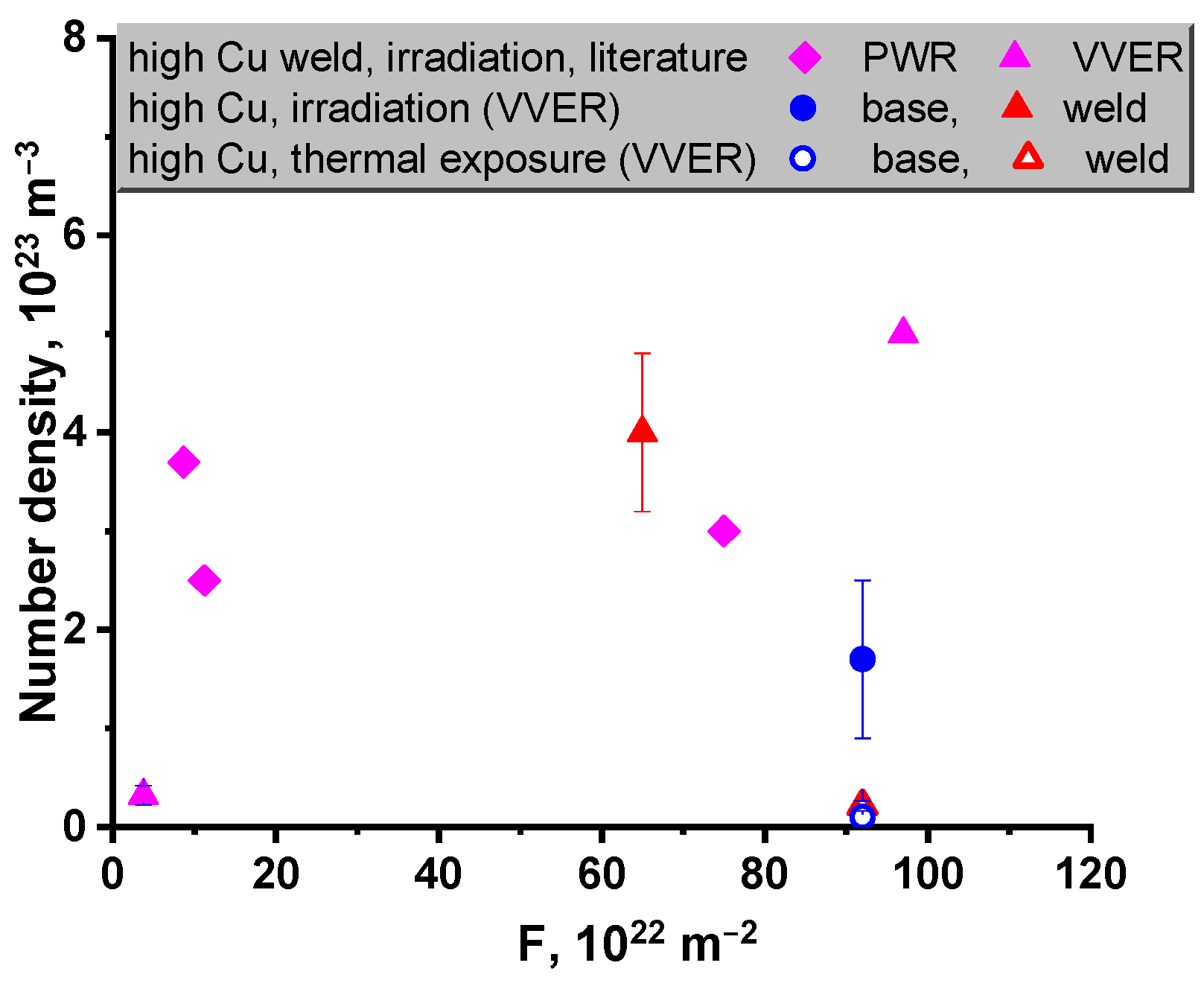
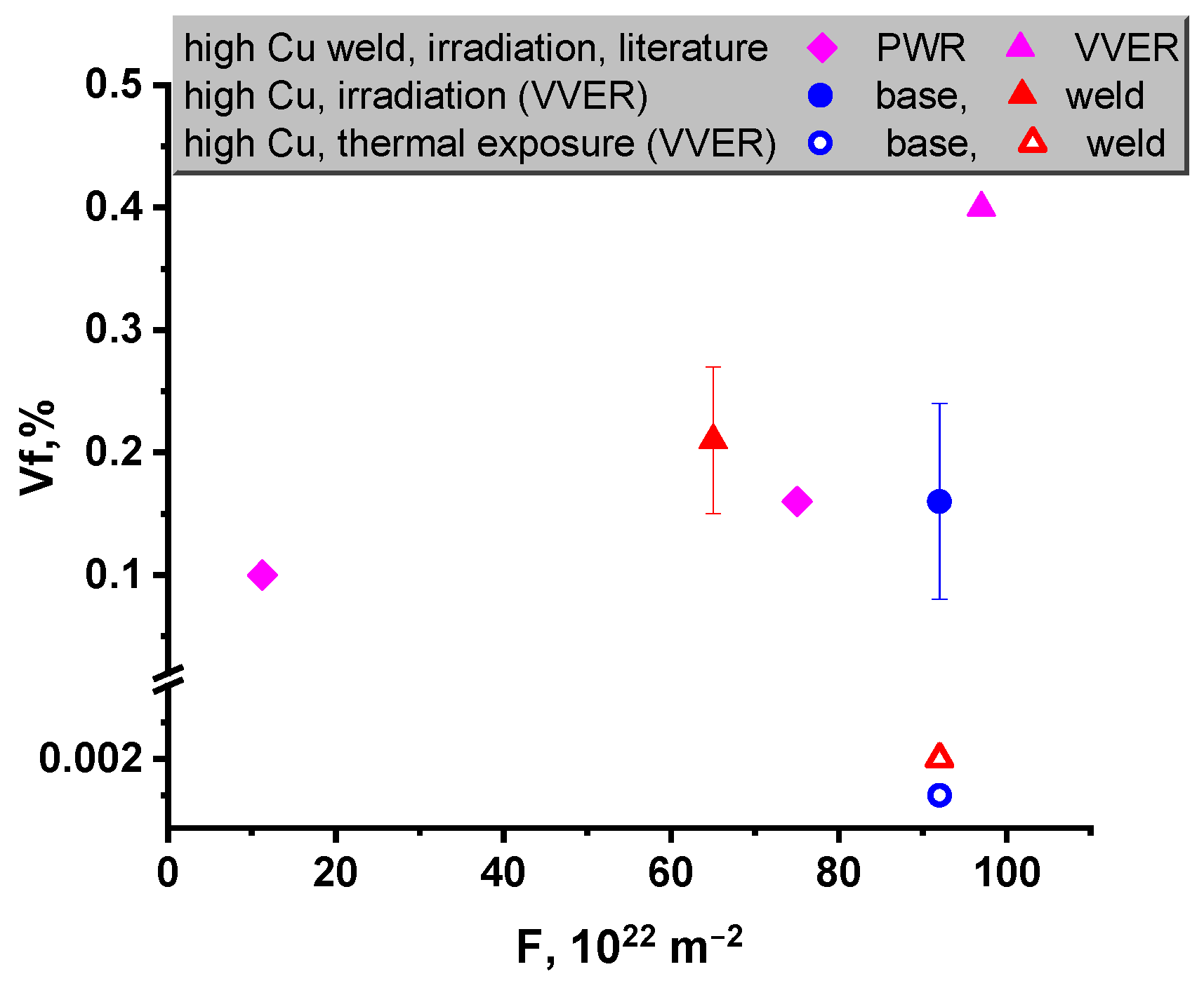
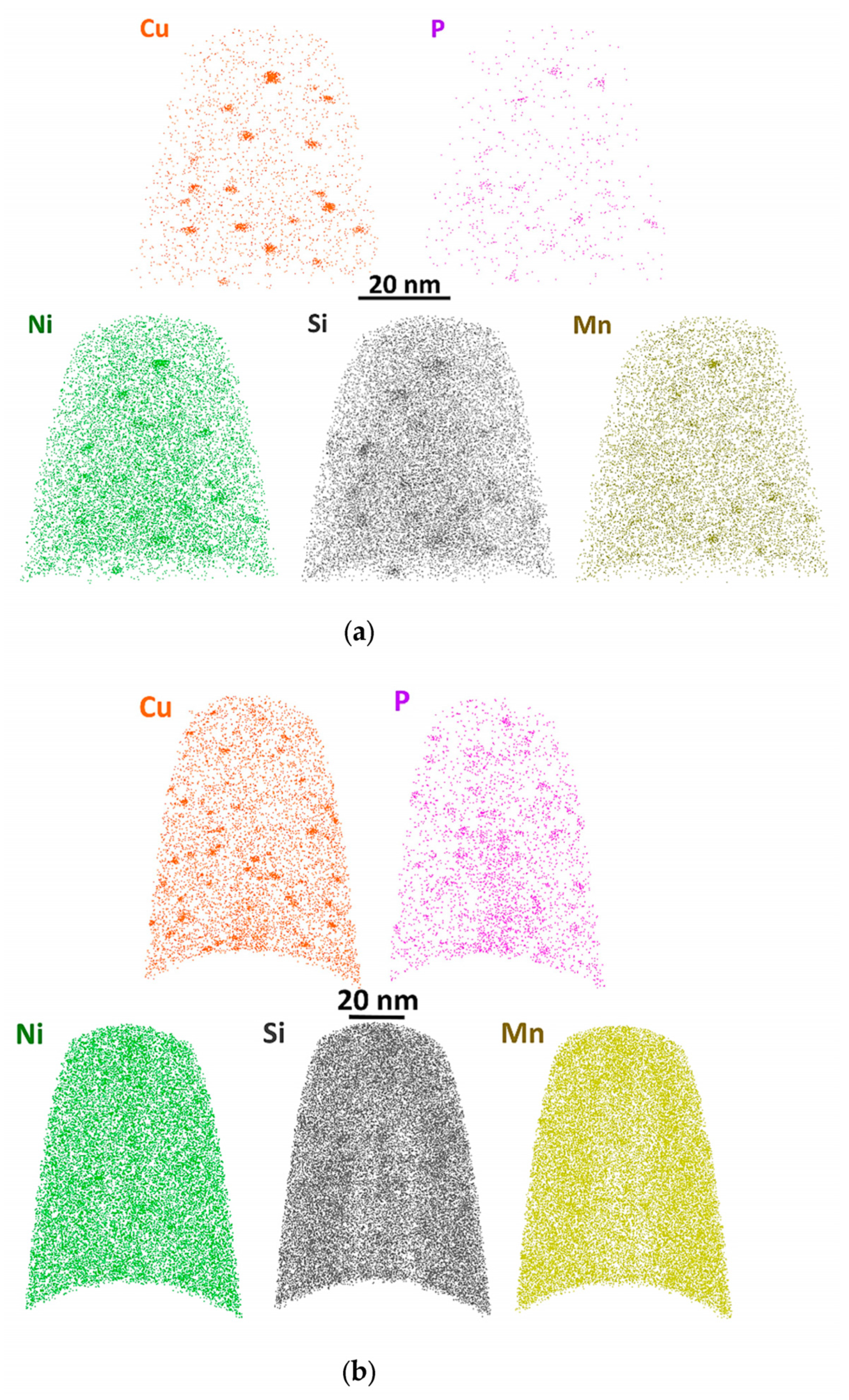
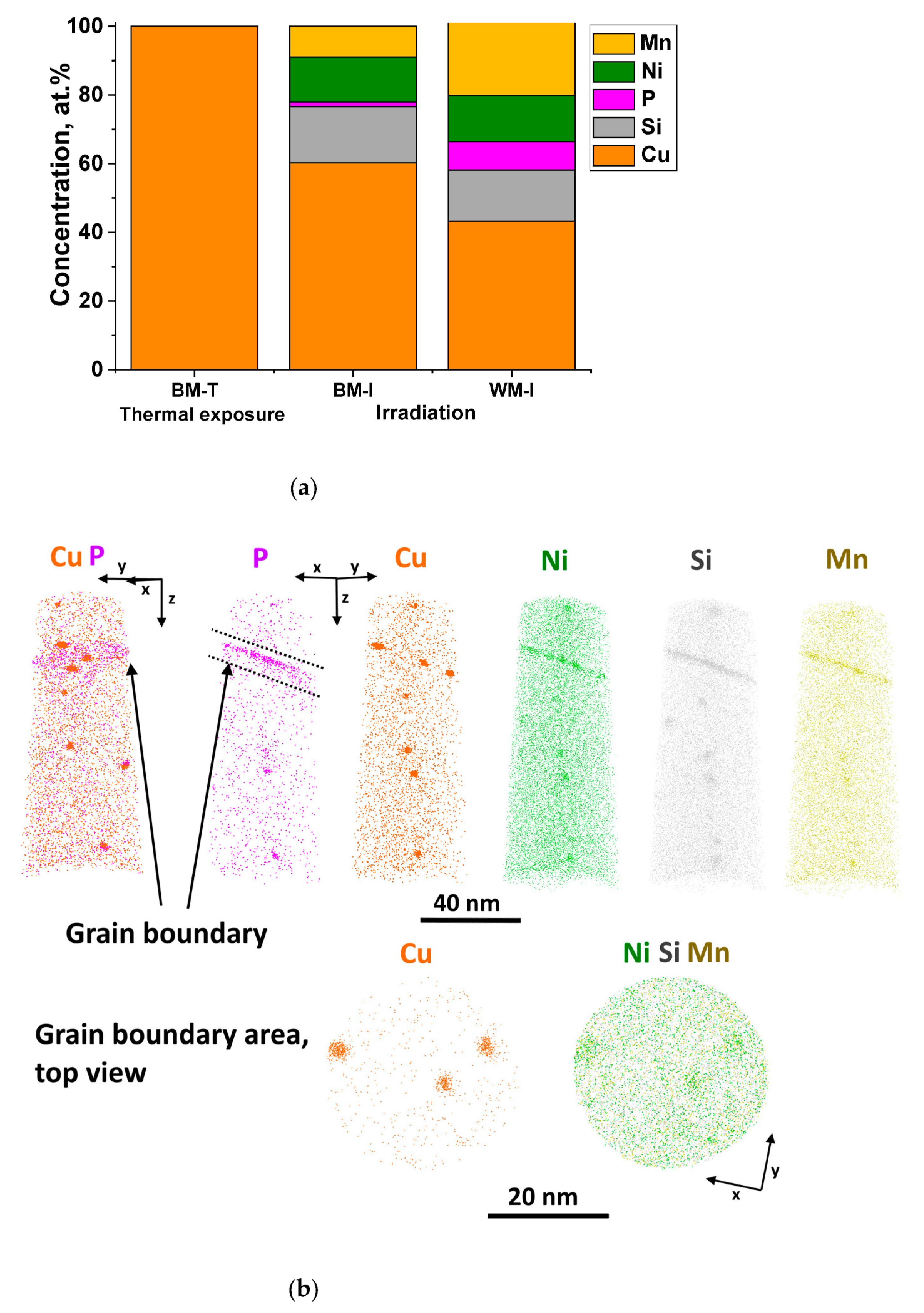
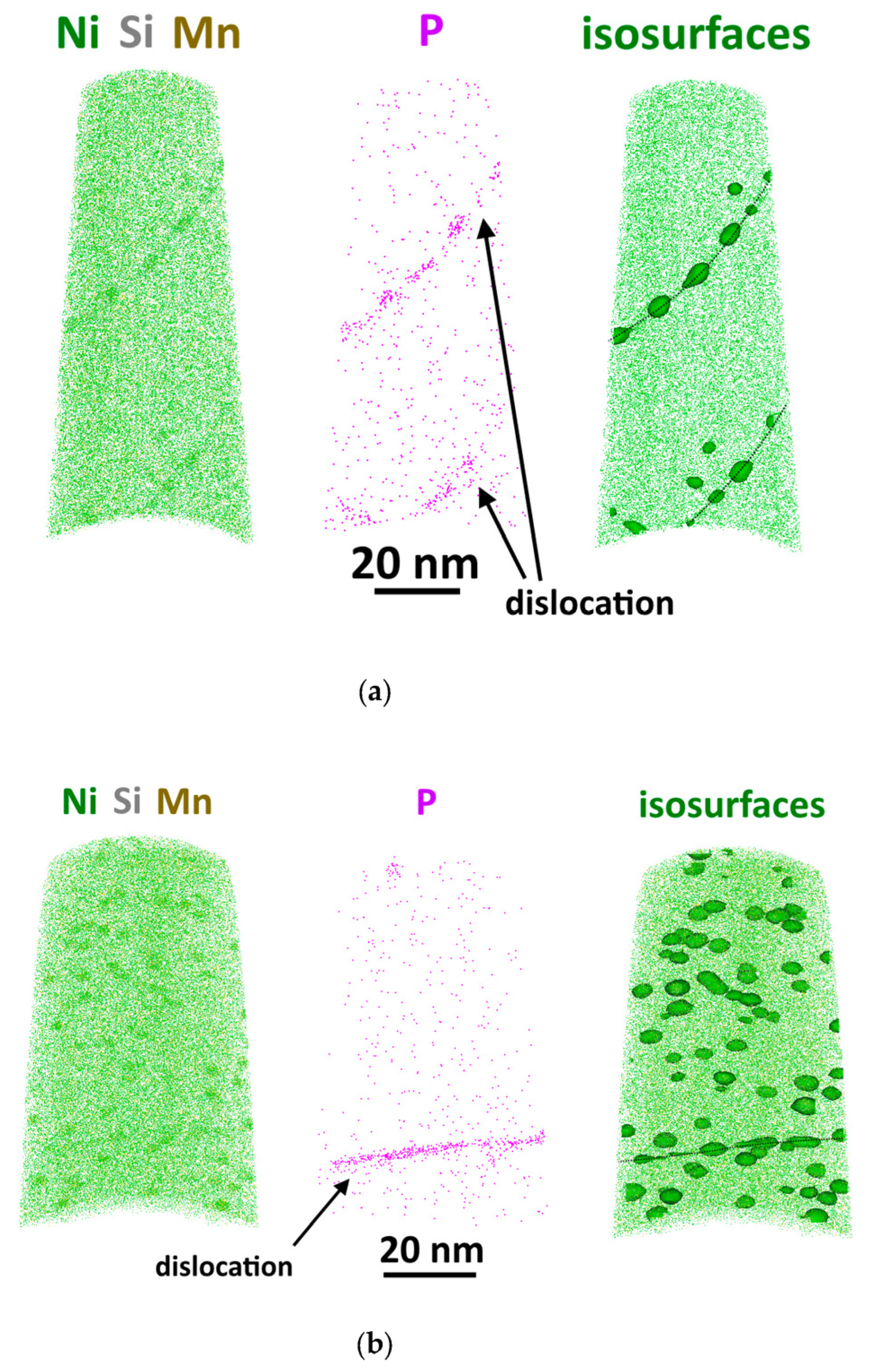
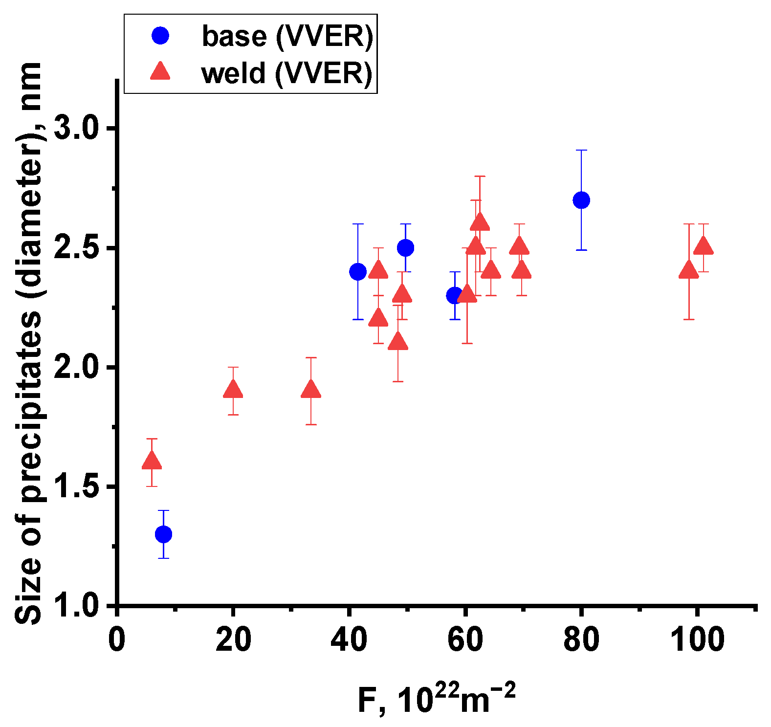
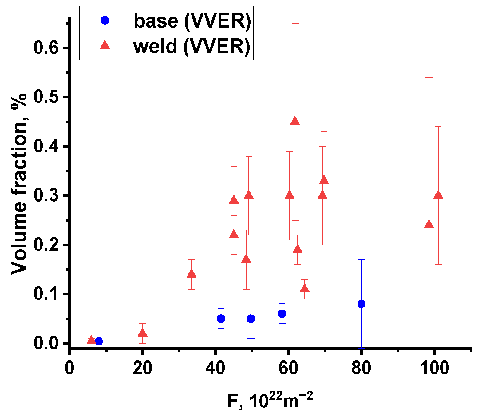
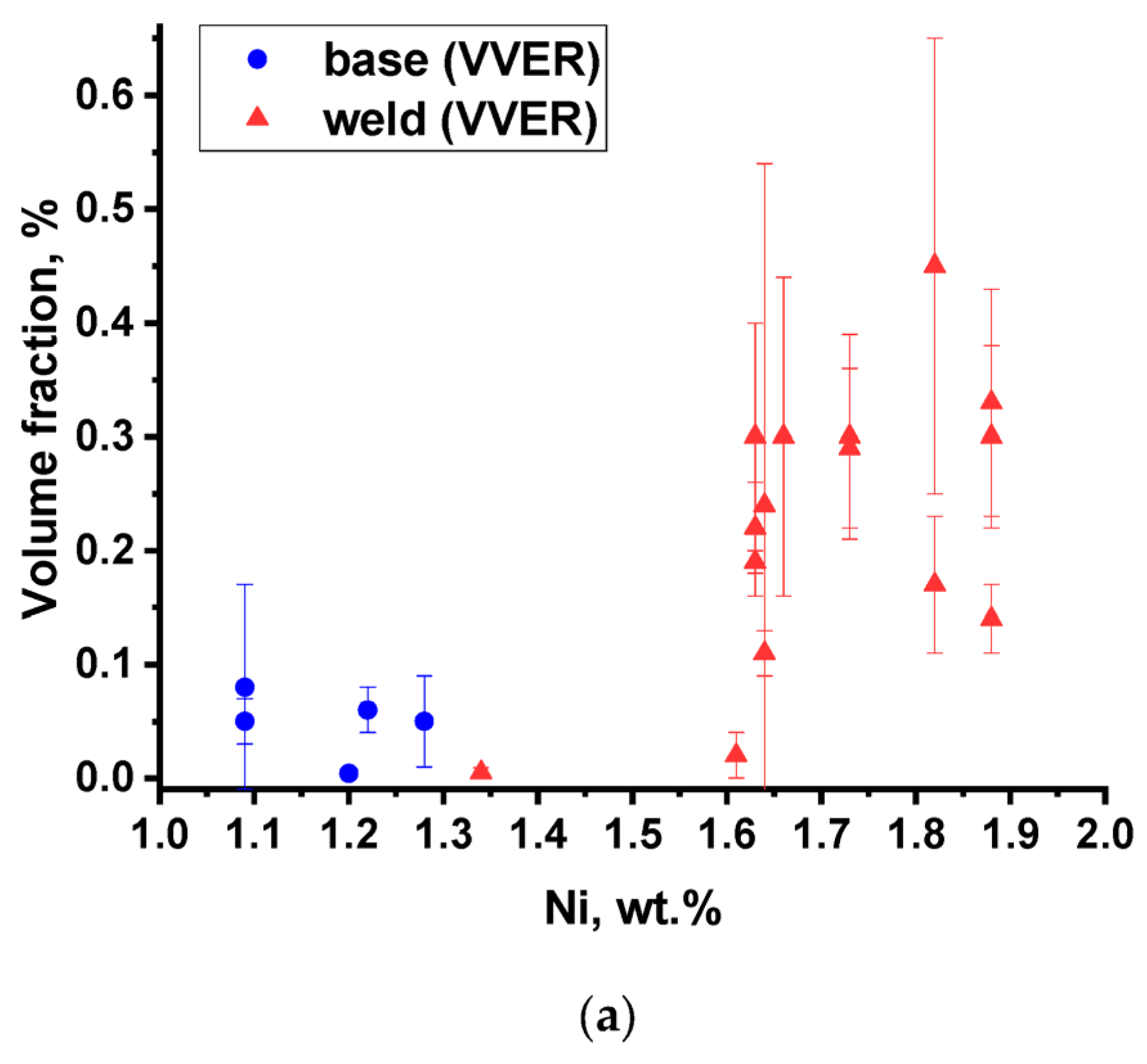
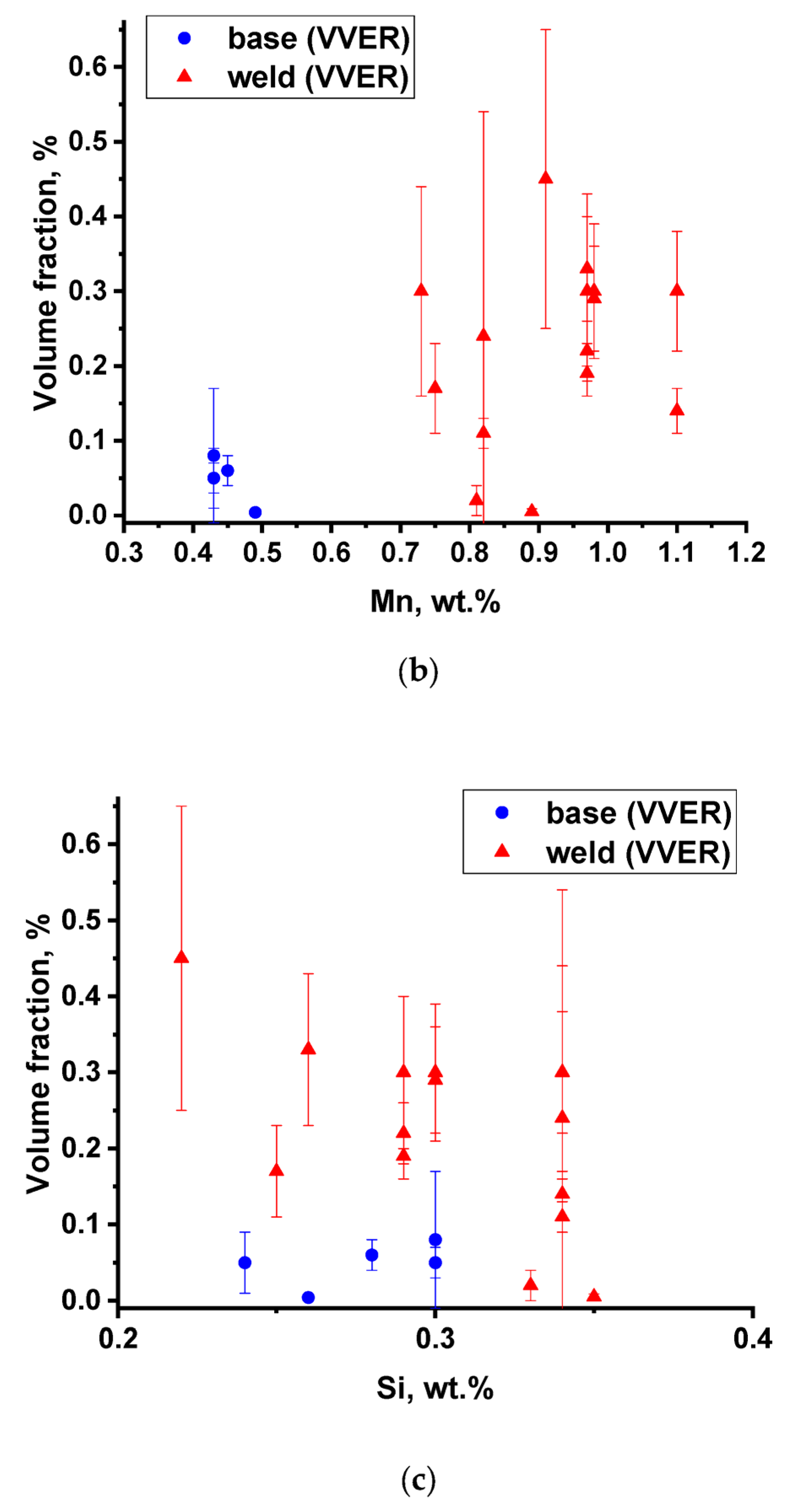
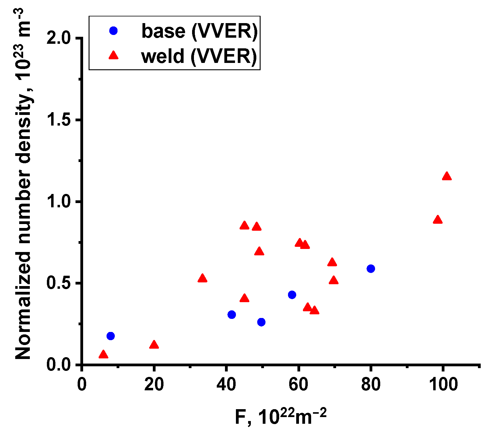
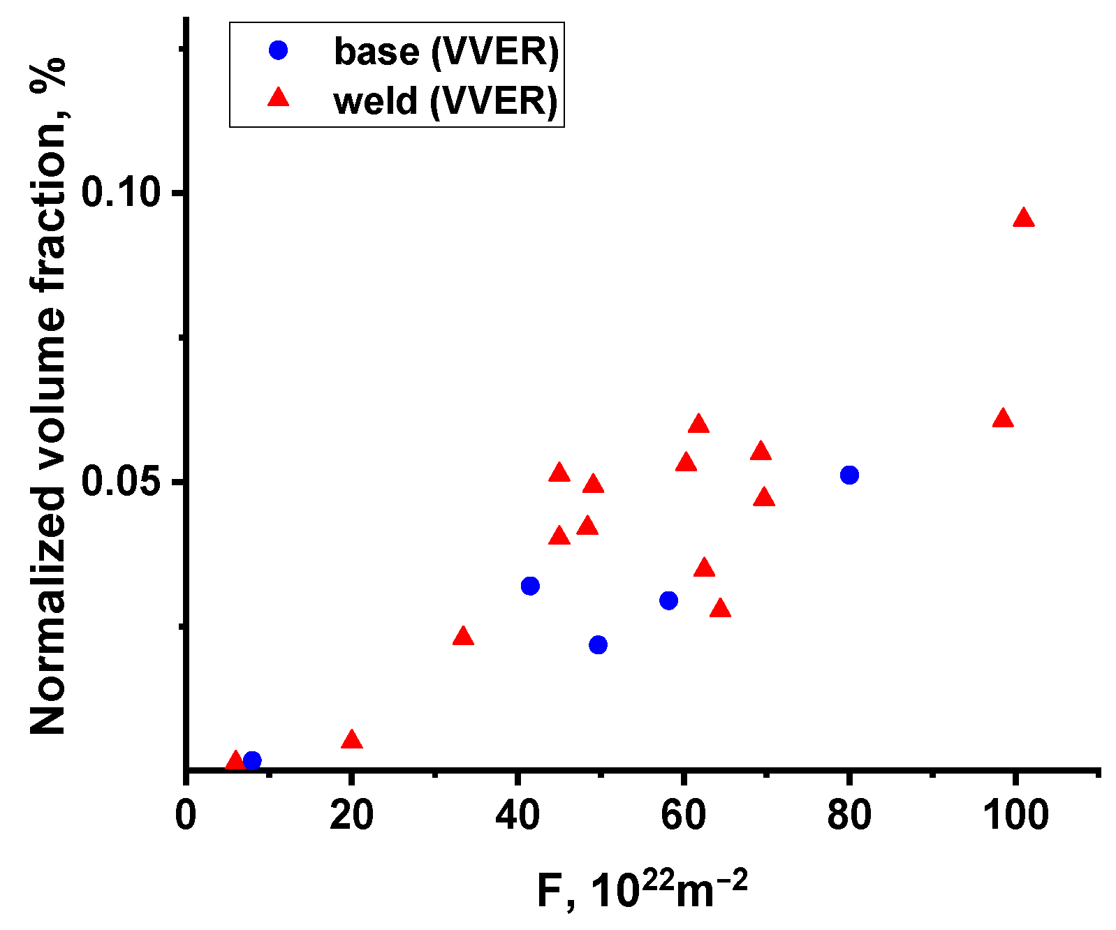
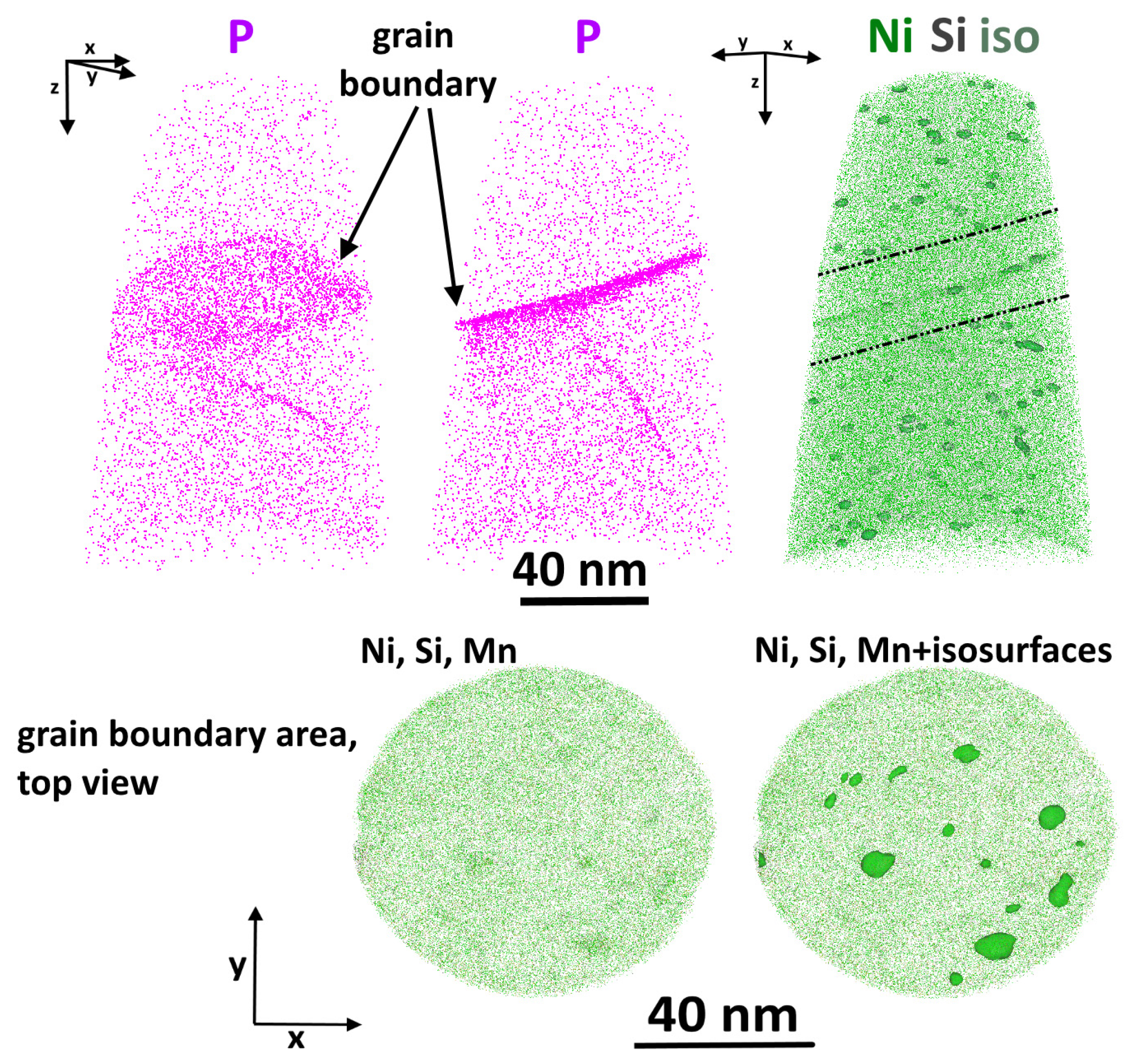
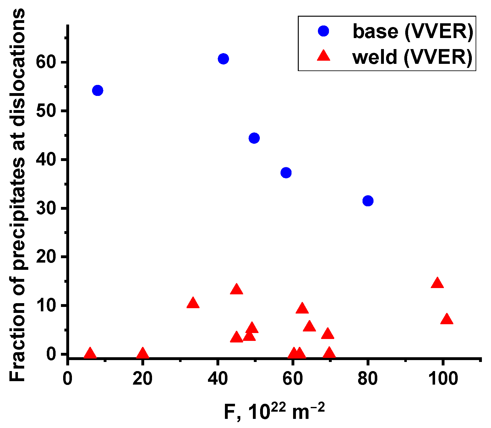
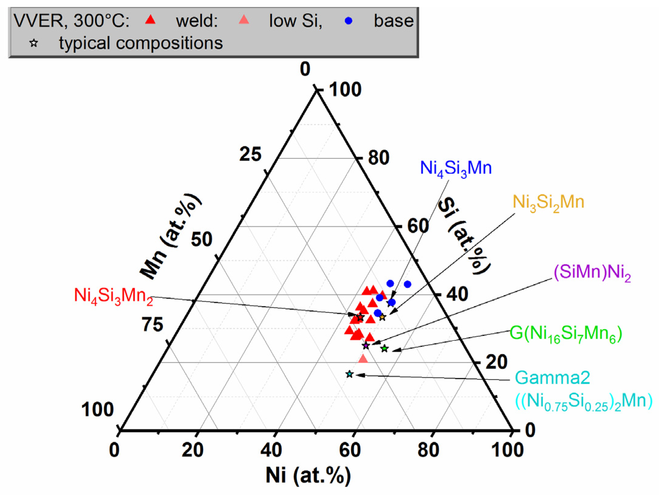
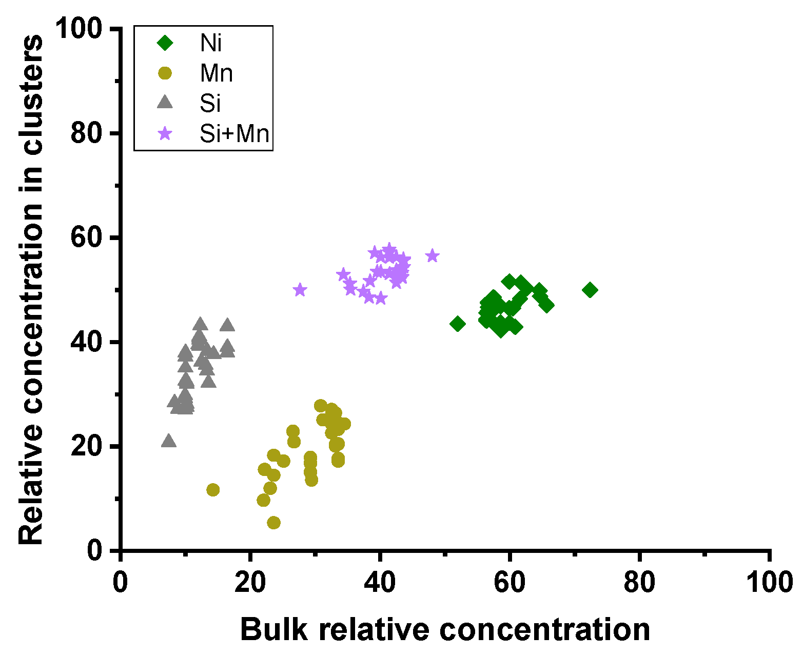
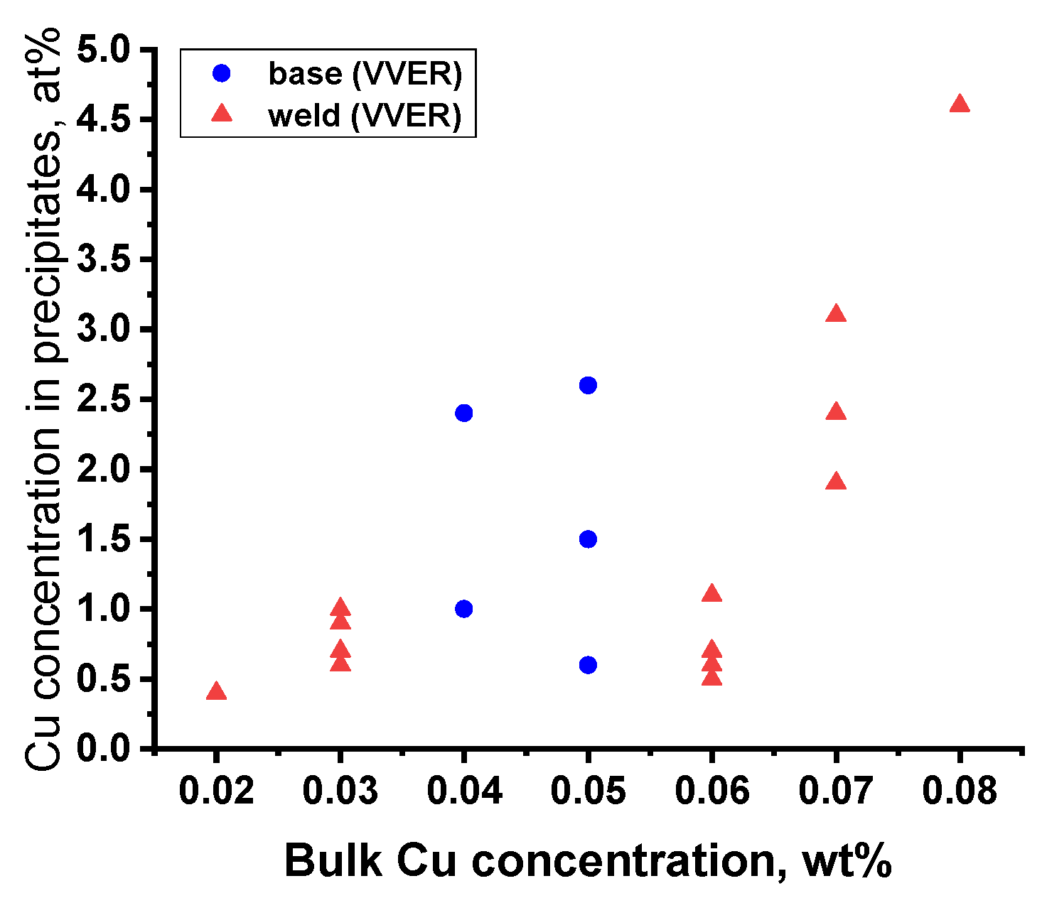
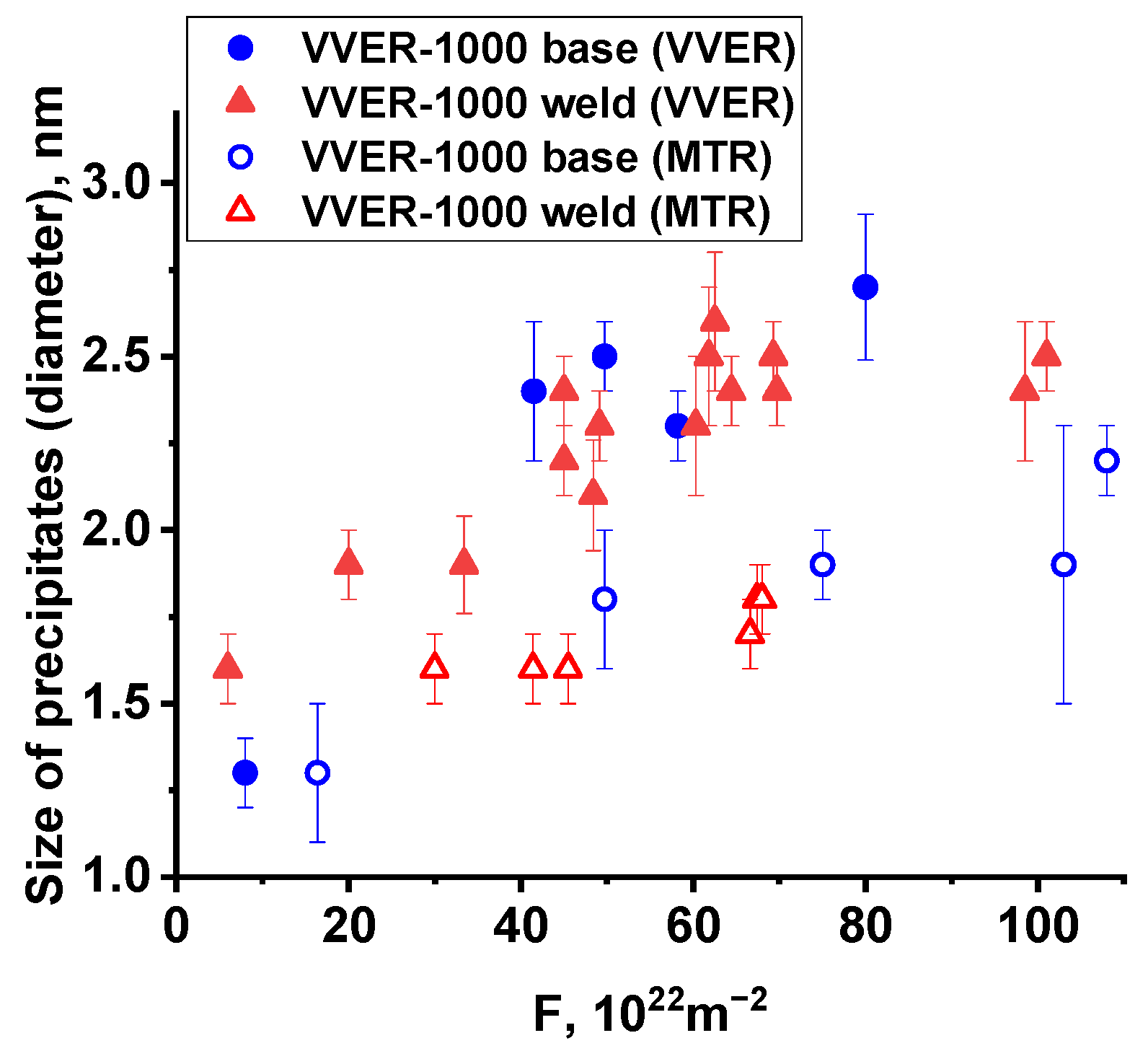
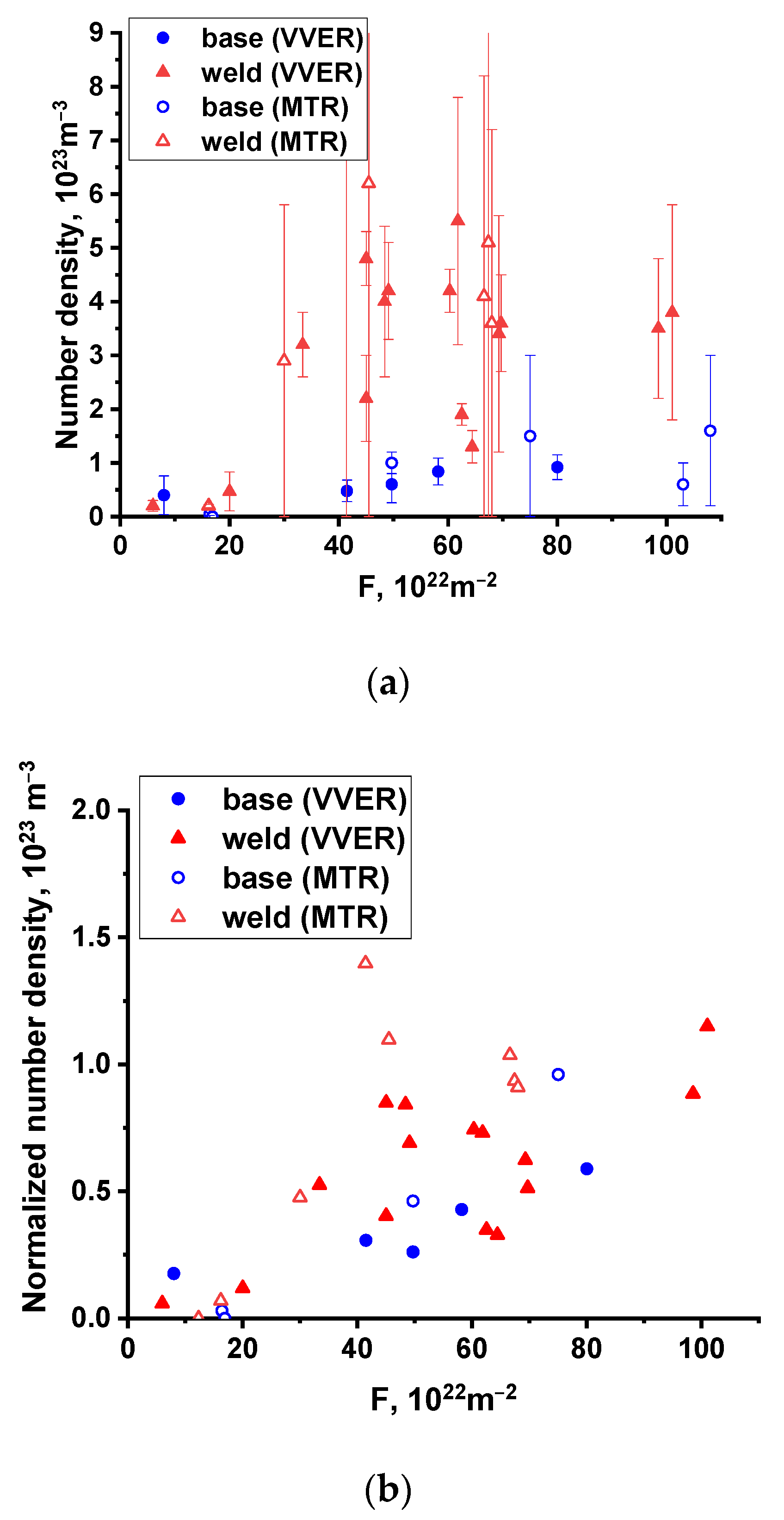
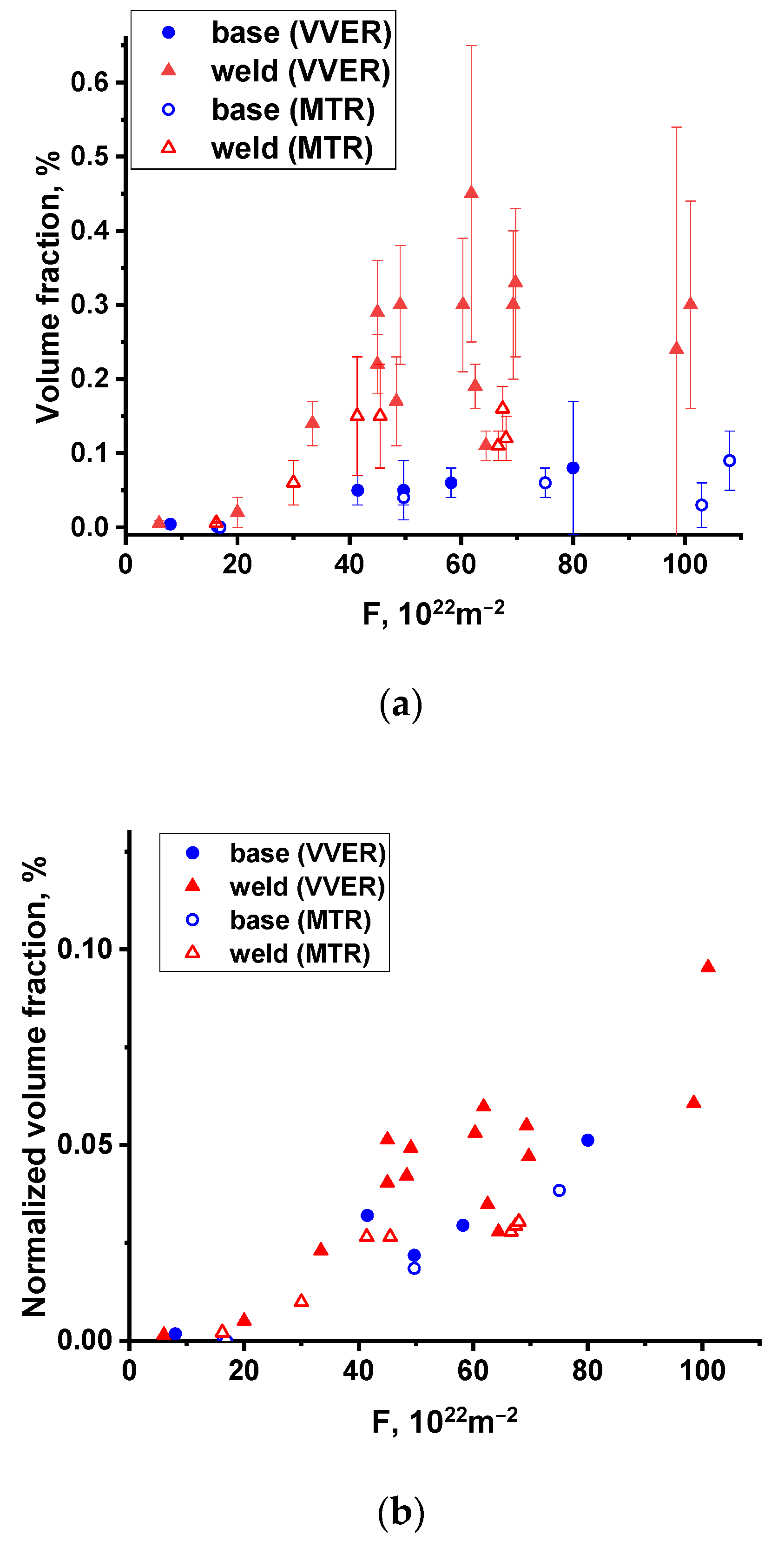
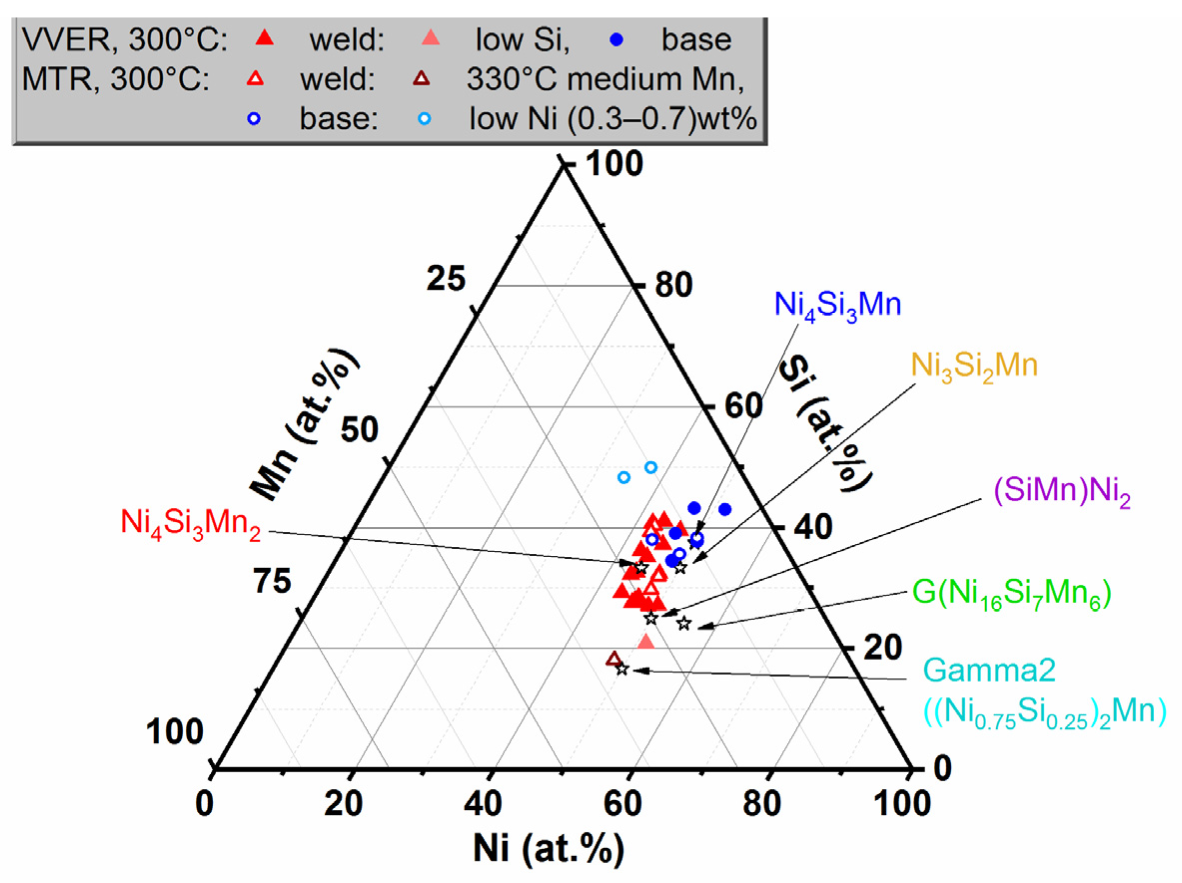
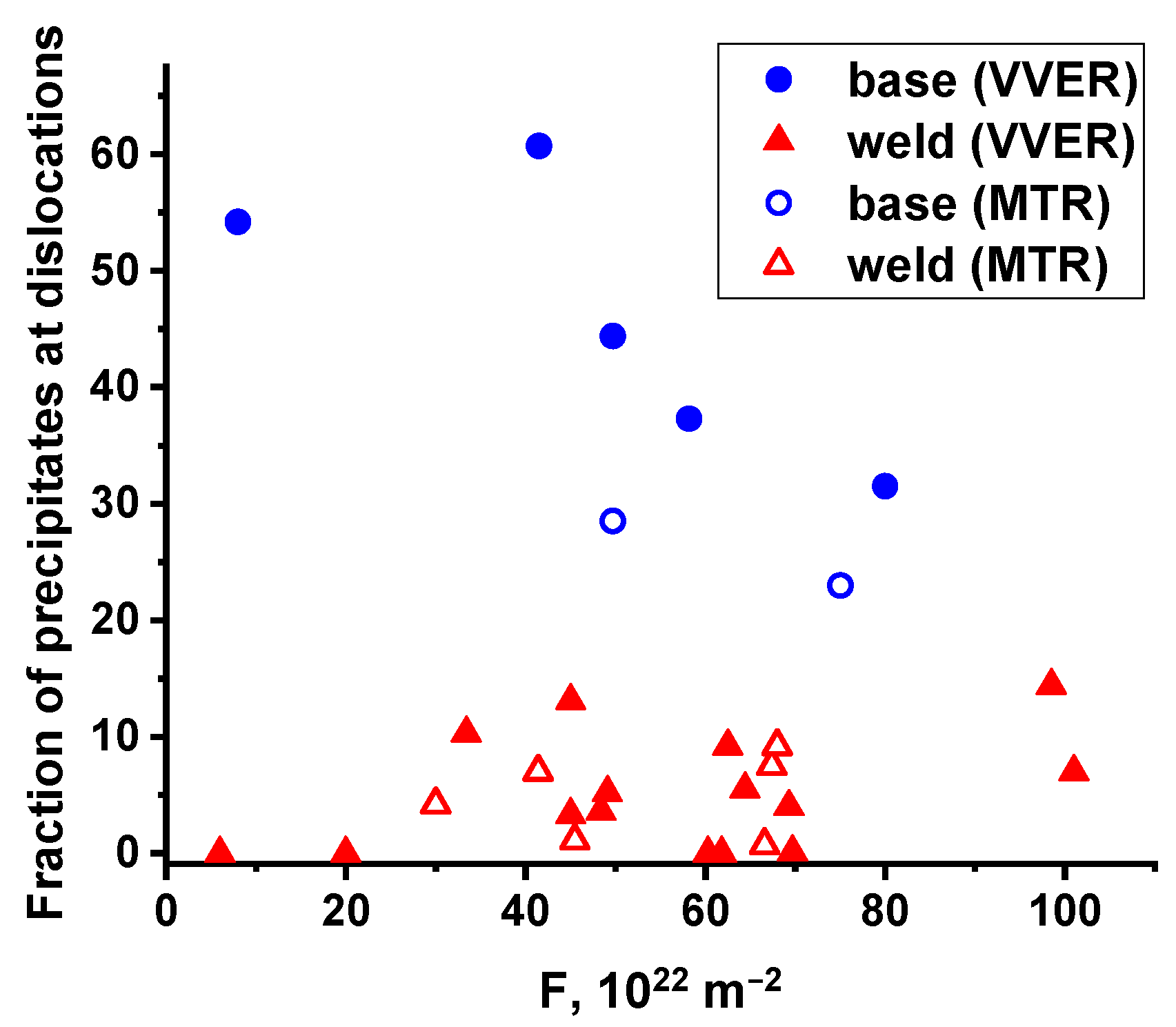
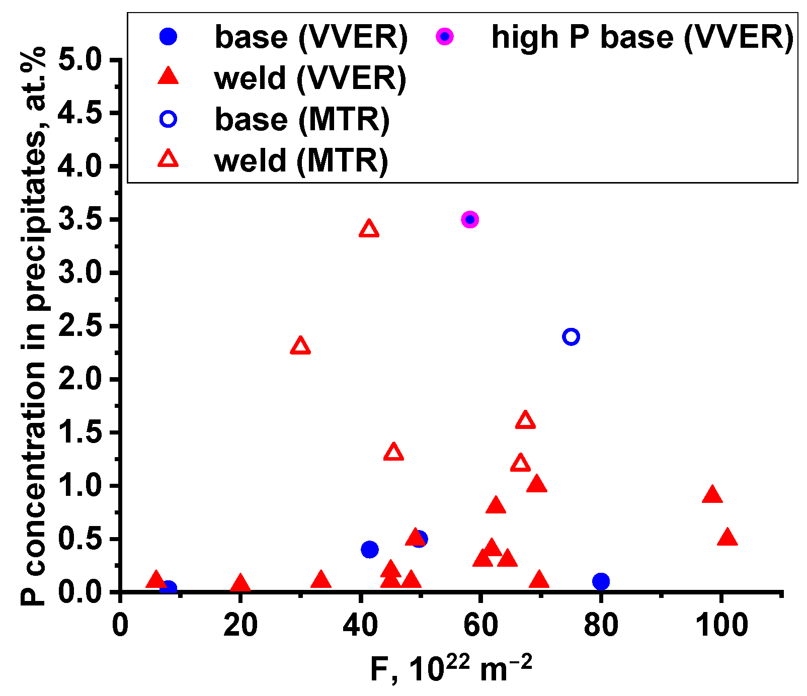
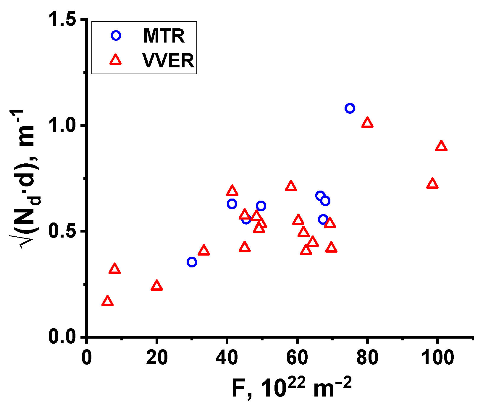
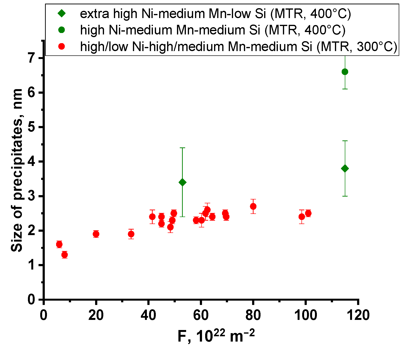
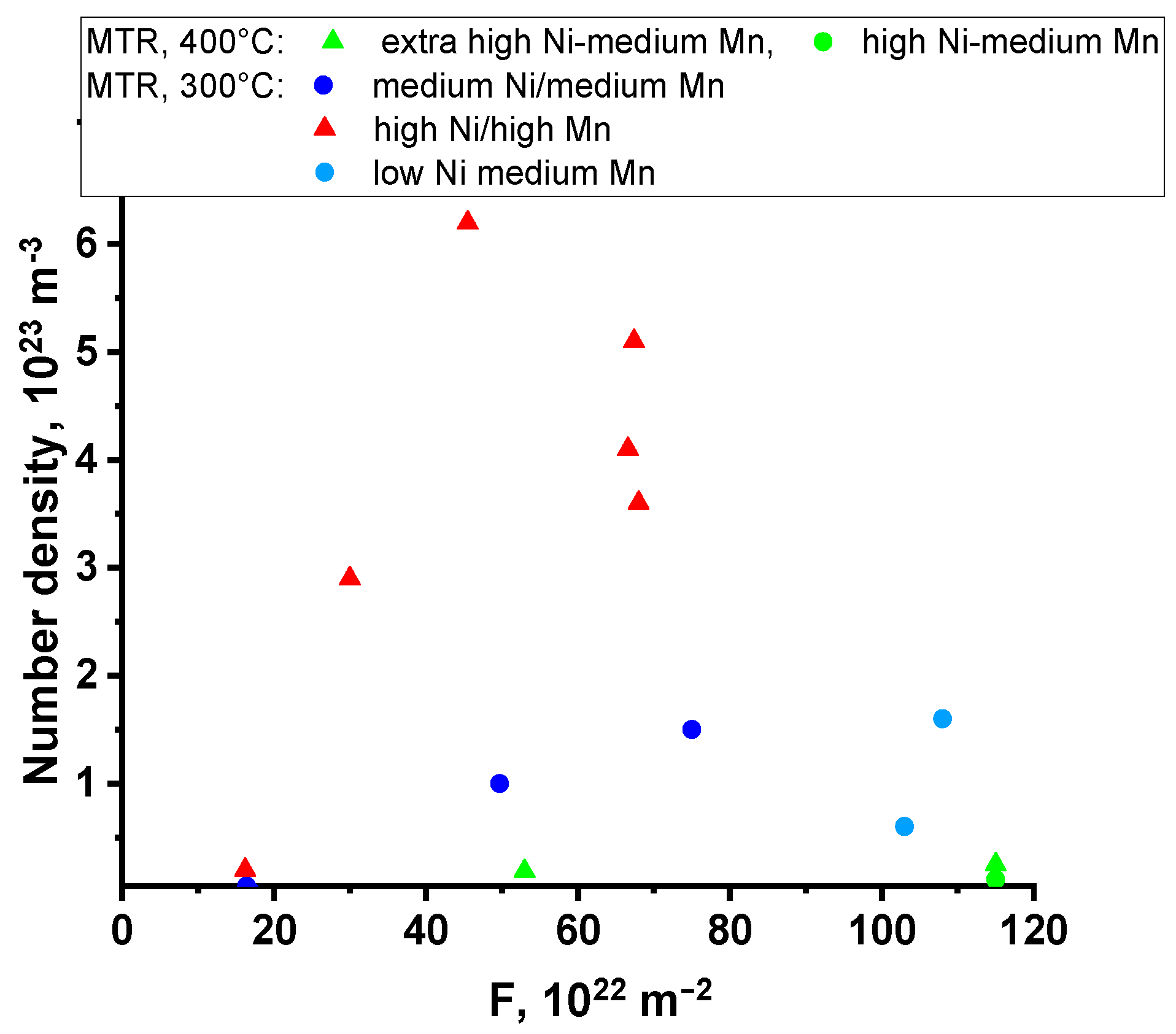
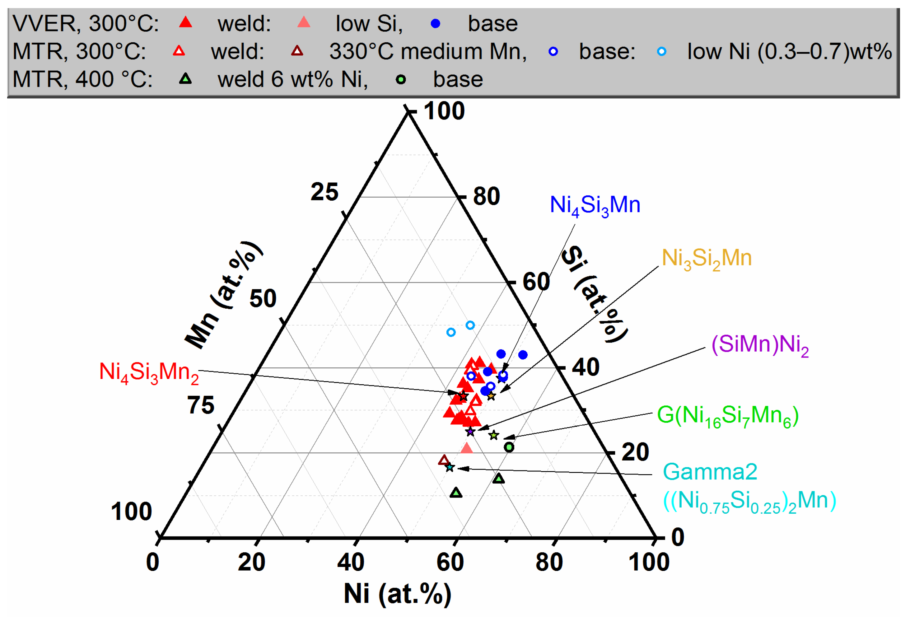
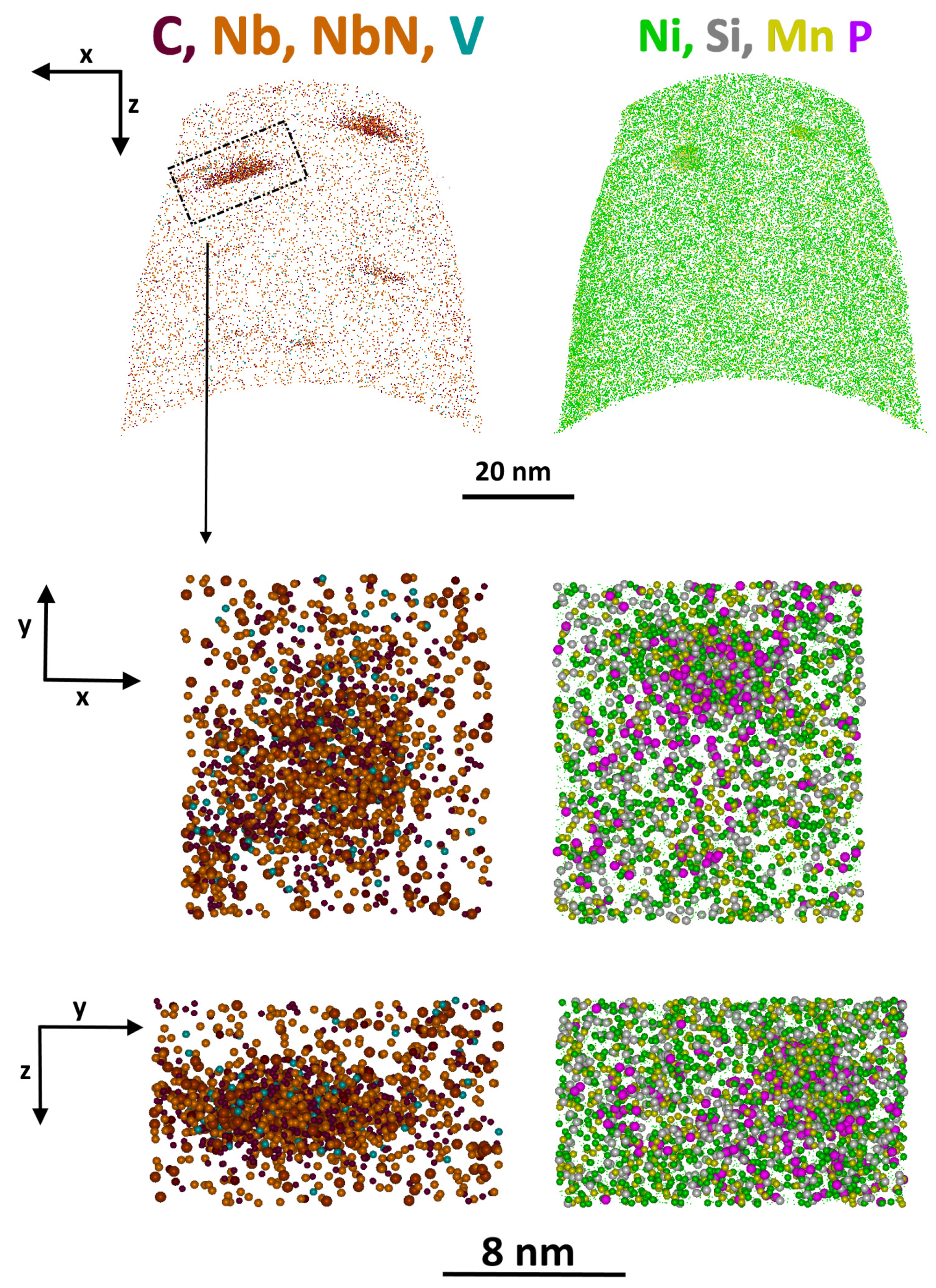
Table 1.
Chemical composition of the studied steels.
| Material Type | Chemical Element Content, wt.%, Fe-Cr-Mo-V-C-Balance | ||||
|---|---|---|---|---|---|
| Cu | Ni | Mn | Si | P | |
| high-Cu, low-Ni BM, WM VVER-440 | 0.11–0.16 | 0.14–0.18 | 0.43–1.05 | 0.3–0.59 | 0.020–0.032 |
| medium Ni, medium Mn, medium Si BM VVER-1000 | 0.04–0.05 | 1.09–1.28 | 0.43–0.49 | 0.24–0.30 | 0.006–0.011 |
| high Ni, high Mn, medium Si WM VVER-1000 | 0.02–0.08 | 1.34–1.88 | 0.73–1.1 | 0.22–0.35 | 0.003–0.011 |
| low-Ni, medium Mn, high Si BM | 0.03 | 0.34–0.71 | 0.26–0.27 | 0.42–0.43 | 0.004–0.005 |
| high Ni, medium Mn, medium Si BM, WM | 0.04 | 1.57–1.61 | 0.31–0.45 | 0.25–0.29 | <0.005 |
| extra high Ni, high Mn, low Si WM | 0.04 | 5.95 | 0.76 | 0.15 | <0.012 |
| extra high Ni, ultra-low Mn, low Si BM | 0.02 | 5.26 | 0.03 | 0.11 | <0.005 |
Table 2.
Studied states and irradiation conditions.
| Material | Reactor | Tirr, °C | Fluence 1, 1022 m−2 | Flux, 1014 m−2s−1 | |
|---|---|---|---|---|---|
| Composition Type | Description | ||||
| high-Cu, low-Ni, medium/high Mn | BM, WM VVER-440 | VVER | 250–270 | 65–92 | 8.0–8.8 |
| medium Ni, medium Mn, medium Si | BM VVER-1000 | VVER | 290 | 8-80 | 5.0–11.5 |
| MTR | 300 | 49.7–75.0 | 550–660 | ||
| 400 | 45 | 600 | |||
| high Ni, high Mn, medium Si | WM VVER-1000 | VVER | 290 | 6–101 | 2.5–25 |
| MTR | 300 | 30–68 | 550–1650 | ||
| low-Ni, medium Mn, high Si | BM | MTR | 300 | 103–108 | 535 |
| high Ni, medium Mn, medium Si | BM, WM | MTR | 330 | 16.4 | 764 |
| 400 | 115 | 2740 | |||
| extra high Ni, high Mn, low Si | WM | MTR | 400 | 53–115 | 821–2740 |
| extra high Ni, ultra-low Mn, low Si | BM | MTR | 330 | 16.9 | 788 |
| 400 | 115 | 2740 | |||
1 Hereinafter, the fluence of fast neutrons (F) is given for neutrons with energy E > 0.5 MeV.
Table 3.
Parameters of precipitates in high-Cu RPV steels based on the results of APT studies.
| Material | Cu Content, wt% | T, °C | F, 1022 m−2 | Exposure Time, Years | Nd, 1022 m−3 | D, nm | Vf, % | Precipitate Composition, at% | ||||
|---|---|---|---|---|---|---|---|---|---|---|---|---|
| Cu | P | Si | Ni | Mn | ||||||||
| High-Cu, low-Ni WM | 0.16 | 250 | 65 | 20 | 4 ± 0.1 | 2.1 ± 0.1 | 0.21 ± 0.06 | 43.3 | 8.2 | 14.9 | 11.5 | 22.1 |
| High-Cu, low-Ni BM | 0.11 | 270 | 92.7 | 45 | 1.7 ± 0.8 | 2.8 ± 0.1 | 0.16 ± 0.06 | 54.6 | 1.2 | 20.7 | 13.5 | 10.0 |
| High-Cu, Low-Ni BM | 0.13 | 290 | 0 | 45 | 0.21 ± 0.05 | 1.0 ± 0.3 | ≤0.002 | 100 | – | – | – | – |
Here T is irradiation temperature, F is fast neutron fluence, Nd, D and Vf are number density, size (diameter) and volume fraction of precipitates.
Disclaimer/Publisher’s Note: The statements, opinions and data contained in all publications are solely those of the individual author(s) and contributor(s) and not of MDPI and/or the editor(s). MDPI and/or the editor(s) disclaim responsibility for any injury to people or property resulting from any ideas, methods, instructions or products referred to in the content. |
© 2023 by the authors. Licensee MDPI, Basel, Switzerland. This article is an open access article distributed under the terms and conditions of the Creative Commons Attribution (CC BY) license (https://creativecommons.org/licenses/by/4.0/).
Share and Cite
MDPI and ACS Style
Fedotova, S.; Kuleshova, E. The Effect of Operational Factors on Phase Formation Patterns in the Light-Water Reactor Pressure Vessel Steels. Metals 2023, 13, 1586. https://doi.org/10.3390/met13091586
AMA Style
Fedotova S, Kuleshova E. The Effect of Operational Factors on Phase Formation Patterns in the Light-Water Reactor Pressure Vessel Steels. Metals. 2023; 13(9):1586. https://doi.org/10.3390/met13091586
Chicago/Turabian StyleFedotova, Svetlana, and Evgenia Kuleshova. 2023. "The Effect of Operational Factors on Phase Formation Patterns in the Light-Water Reactor Pressure Vessel Steels" Metals 13, no. 9: 1586. https://doi.org/10.3390/met13091586
Note that from the first issue of 2016, this journal uses article numbers instead of page numbers. See further details here.