Article

Genomic Features of the Bundle-Forming Heliobacterium Heliophilum fasciatum

by
Kelly S. Bender
1,
Michael T. Madigan
1,
Kyleigh L. Williamson
1,
Marisa H. Mayer
2,
Mary N. Parenteau
2,
Linda L. Jahnke
2,
Paula V. Welander
3,
Sophia A. Sanguedolce
4,
Abigail C. Brown
4 and
W. Matthew Sattley
4,*
1
Microbiology Program, School of Biological Sciences, Southern Illinois University, Carbondale, IL 62901, USA
2
Exobiology Branch, NASA Ames Research Center, Moffett Field, CA 94035, USA
3
Department of Earth System Science, Stanford University, Stanford, CA 94305, USA
4
Division of Natural Sciences, Indiana Wesleyan University, Marion, IN 46953, USA
*
Author to whom correspondence should be addressed.
Microorganisms 2022, 10(5), 869; https://doi.org/10.3390/microorganisms10050869
Submission received: 31 March 2022 / Revised: 18 April 2022 / Accepted: 19 April 2022 / Published: 21 April 2022
(This article belongs to the Special Issue Phototrophic Bacteria)

Abstract

:
Eight species of heliobacteria have had their genomes sequenced. However, only two of these genomes have been analyzed in detail, those from the thermophilic Heliomicrobium (Hmi.) modesticaldum and the alkaliphilic Heliorestis (Hrs.) convoluta. Here we present analyses of the draft genome sequence of a species of heliobacterium that grows optimally at a moderate temperature and neutral pH. The organism, Heliophilum (Hph.) fasciatum, is phylogenetically unique among cultured heliobacteria and was isolated from rice soil, a common habitat for heliobacteria. The Hph. fasciatum genome contains 3.14 Mbp—similar to that of other reported heliobacteria—but has a G+C base ratio that lies between that of Hmi. modesticaldum and Hrs. convoluta. Many of the genomic features of Hmi. modesticaldum and Hrs. convoluta, such as the absence of genes encoding autotrophic pathways, the presence of a superoperonal cluster of photosynthesis-related genes, and genes encoding endospore-specific proteins, are also characteristic of the Hph. fasciatum genome. However, despite the fact that Hph. fasciatum is diazotrophic, classical nif genes encoding the alpha and beta subunits of dinitrogenase (nifDK) present in other heliobacteria could not be identified. Instead, genes encoding several highly divergent NifDK homologs were present, at least one of which likely encodes a functional dinitrogenase and another a methylthio-alkane reductase (MarDK) for sulfur assimilation. A classical NifH (dinitrogenase reductase) homolog was also absent in Hph. fasciatum, but a related protein was identified that likely carries out this function as well as electron delivery to MarDK. The N2-fixing system of Hph. fasciatum is therefore distinct from that of other heliobacteria and may have unusual properties.

1. Introduction

Heliobacteriaceae comprise a family of anoxygenic phototrophic bacteria phylogenetically and physiologically distinct from purple and green bacteria [1,2]. Unlike the latter two groups, heliobacteria do not contain bacteriochlorophyll (Bchl) a, b, c, d, or e, but instead, produce Bchl g as their main pigment, a bacteriochlorophyll structurally most closely related to green plant chlorophyll a [3]. In addition, heliobacteria also differ from purple and green bacteria in that heliobacteria are of gram-positive lineage, are obligate anaerobes, lack all known biochemical pathways for autotrophic growth, and produce heat-resistant endospores [4,5,6].
Several species of heliobacteria have been isolated since the discovery of Heliobacterium (Hbt.) chlorum by Gest and Favinger in the early 1980s [1,7] reviewed in [2,4,8], and described species form two broad groups on the basis of their pH optima for growth: neutrophilic species, with pH optima near 7, and alkaliphilic species, with pH optima near 9. This physiological dichotomy of heliobacteria is mirrored in their phylogeny (Figure 1). Although several species of heliobacteria have had their genomes sequenced, thus far only two species—both extremophiles—have had their genomes analyzed in detail. These include the thermophilic (and neutrophilic) species Heliomicrobium (previously Heliobacterium) [8] modesticaldum [9,10,11] and the alkaliphilic species Heliorestis convoluta [12,13]. These studies have provided the genomic support for the aforementioned unusual physiological and biochemical features of the heliobacteria and confirmed that all characterized species (with the exception of Candidatus “Heliomonas lunata” [14]) are robust diazotrophs [15,16].
In 1996, Ormerod et al. [16] described a new genus of bundle-forming heliobacteria in which a raft of apparently loosely attached rod-shaped cells displayed swimming motility as a unit (Figure 2). Besides exhibiting the usual assortment of heliobacterial physiological properties [5], this organism, named Heliophilum fasciatum (“sun-loving bundle-forming heliobacterium”), differed in several respects from Hmi. modesticaldum and Hrs. convoluta, most notably in its habitat and phylogenetic status (Table 1). By 16S rRNA gene sequencing criteria, Hph. fasciatum lies basal to all known neutrophilic heliobacteria and can be considered a “bridge” species between neutrophilic and alkaliphilic clades (Figure 1). All three of these heliobacteria assimilate acetate and pyruvate during photoheterotrophic growth and only Hrs. convoluta is unable to grow fermentatively in darkness (Table 1). In addition, each of these three species is morphologically distinct, shows distinct pH and temperature optima for growth, and was isolated from geographically well separated and geochemically distinct habitats (Table 1 and Table 2).
Despite the fact that Hph. fasciatum is phylogenetically and phenotypically distinct from Hmi. modesticaldum and Hrs. convoluta (Figure 1 and Table 1), this species has remained in the background of heliobacterial research until its genome was recently sequenced as part of a systematic study of the heliobacteria to support taxonomic revisions in the family [8]. Genomic studies of heliobacteria have lagged behind those of purple and green bacteria, and to date, a total of only eight species of heliobacteria have had their genomes sequenced, six of which only to the draft stage. Sequence data have shown the size of heliobacterial genomes to be quite variable, as the largest heliobacterial genome encodes more than a thousand additional proteins than does the smallest (Table 2). In a comparative study of the genomes of the eight species of heliobacteria listed in Table 2 [8], the authors showed that a phylogenetic tree constructed from the genomes of these species closely mirrored the tree constructed from 16S rRNA gene sequences (Figure 1). In addition, the authors used genomic data along with the comparative sequences of a few key genes and operons to propose that three of the species in the genus Heliobacterium should be grouped into their own genus, Heliomicrobium [8]; these include Hmi. modesticaldum, Hmi. gestii, and Hmi. undosum (Table 2). Analyses of genes encoding particular photocomplex and endospore proteins supported the taxonomic proposals, as did genes encoding carbon monoxide dehydrogenase, an enzyme previously unsuspected in the heliobacteria [8].
Thus far, detailed analyses of the genomes of only Hmi. modesticaldum and Hrs. convoluta have been published [10,11,13]. Here we augment these studies with an analysis of the major gene sets of Hph. fasciatum using data generated from our own sequencing project. Our results focus on genes encoding proteins of major pathways of carbon, nitrogen, and sulfur metabolism, and genes encoding the formation of endospores and flagellar components, comparing them where appropriate with those from other heliobacteria, in particular, the two species with complete genome sequences (Table 1 and Table 2). Our analyses have uncovered several similarities and differences in the genetic capabilities of Hph. fasciatum compared with those of other heliobacteria, including in particular the fact that despite its diazotrophic phenotype, Hph. fasciatum lacks genes encoding the canonical nitrogenase universally present in all other cultured heliobacteria and in most other anoxygenic phototrophic bacteria.

2. Materials and Methods

The Heliophilum fasciatum strain TanzaniaT was obtained from the Leibniz-Institut DSMZ, Braunschweig, Germany, as DSM 11170T. Cells of Hph. fasciatum were grown phototrophically in medium PYE [15], and genomic DNA was isolated using the JetFlex™ Genomic DNA Purification Kit (ThermoFisher Cat No. A30700). The genome was sequenced through the U.S. Department of Energy Joint Genome Institute (JGI) Community Science Program. Whole-genome shotgun sequencing was done with Illumina NovaSeq sequencing (library 300 bp), and the resulting fragments were assembled and annotated by the JGI Integrated Microbial Genomes (IMG) annotation pipeline (IMGAP v.5.0.23 with gene calling program CRT 1.8.2). The genome sequence of Hph. fasciatum TanzaniaT is publically available (listed as Heliophilum fasciatum MTM in the JGI/IMG database) under the genome ID 2929297113. The genome of the exact same strain of Hph. fasciatum was independently sequenced at JGI by another group [8] and is publicly available as genome ID 2795386140; the latter sequence was also accessioned into Genbank as reference sequence NZ_SLXT00000000.1.
Phylogenetic analyses of 16S ribosomal RNA genes and protein sequences were performed using MEGA version X [18,19]. The Hph. fasciatum strain TanzaniaT 16S rRNA gene sequence was aligned with corresponding sequences from other heliobacterial type species and Escherichia coli (J01859), used as the outgroup to root the phylogenetic tree. The tree was drawn using MEGA X according to the parameters described in the legend in Figure 1. For trees of nif-related proteins, Hph. fasciatum NflHDK amino acid sequences were used as queries in blastp [20] searches to identify other heliobacteria homologs. The genetic organization of the Hmi. gestii strain DSM 11169 nif and anf loci were obtained from Genbank genome accession PRJNA599378. Protein phylogenetic analyses were performed on a subset of NifHDK homologs previously designated as belonging to nitrogenase-like protein groups I–IV [21,22]. Following ClustalW [23] analysis, the resulting alignments were assembled into maximum-likelihood trees using the LG + G substitution model [24] with 100 replicates. Note that the files for NifD and NifK homologs were concatenated prior to tree assembly.

3. Results and Discussion

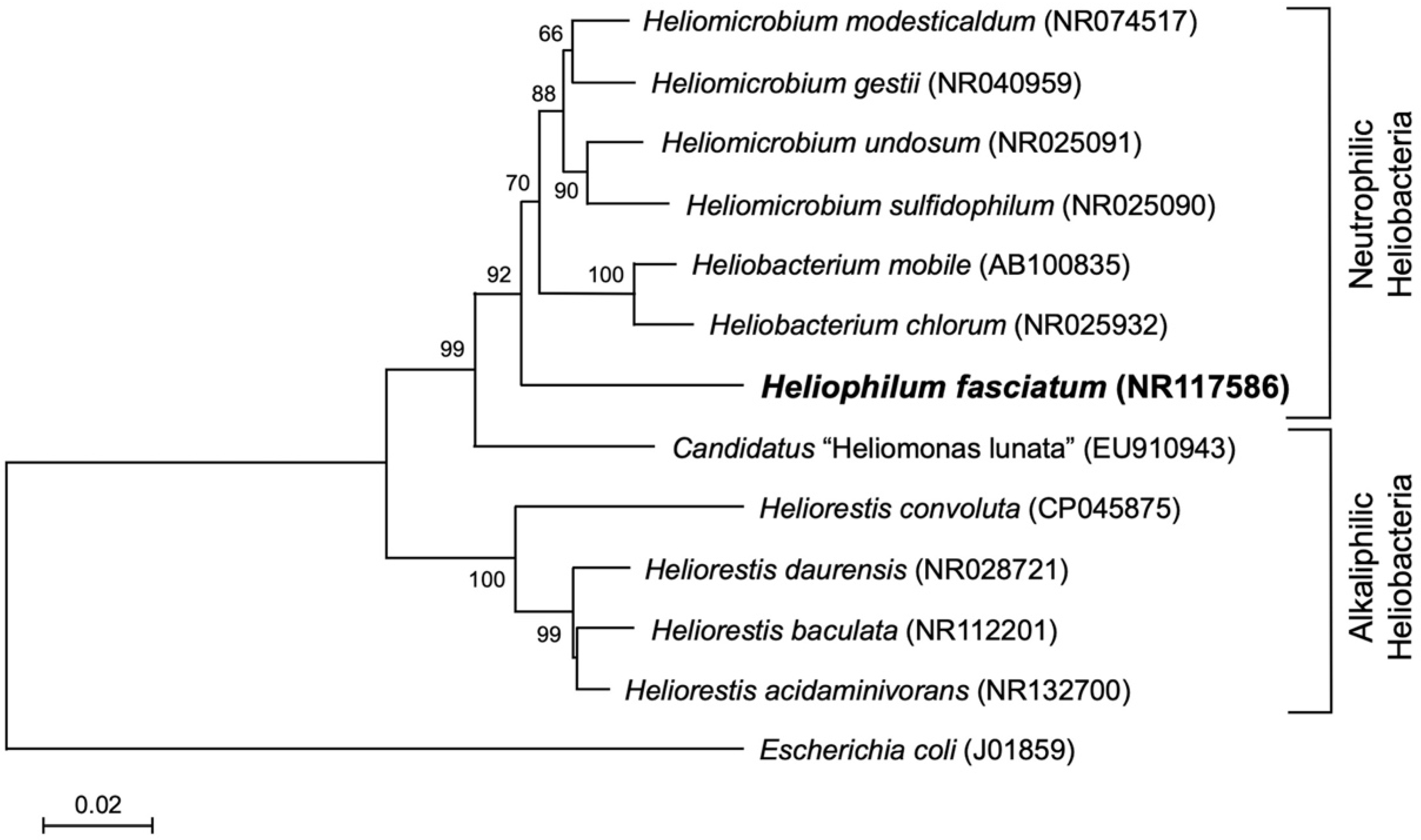
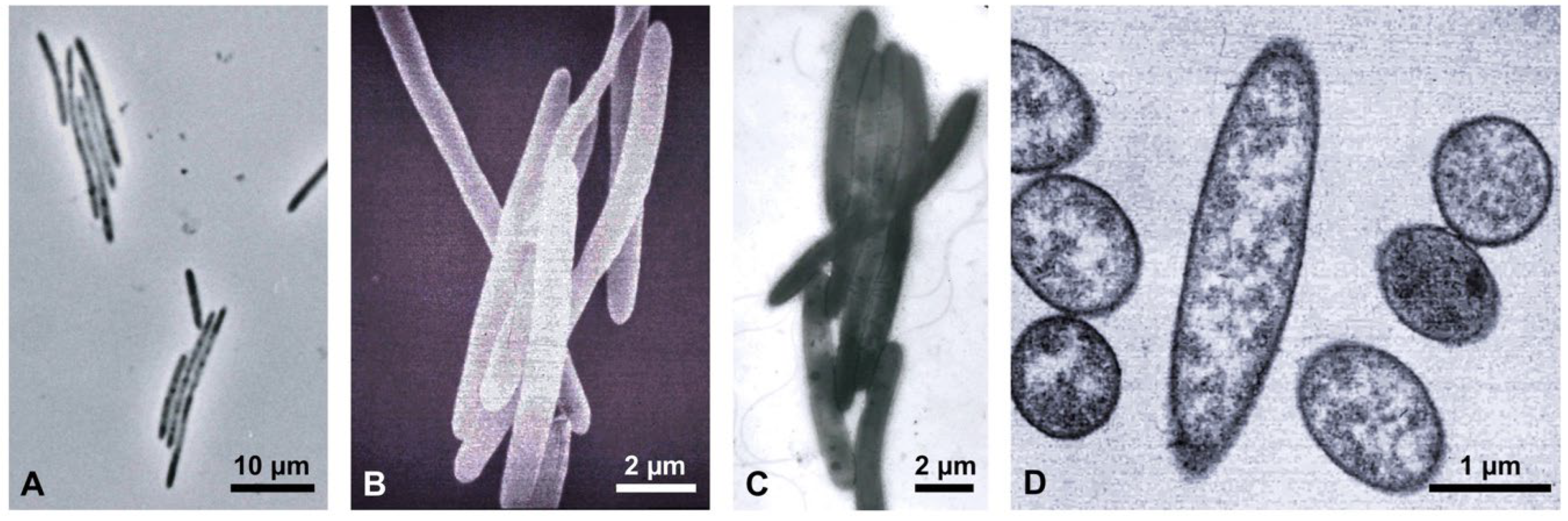
3.1. General Genomic Properties

Table 3 compares genomic statistics of Hph. fasciatum strain TanzaniaT with those of Hmi. modesticaldum and Hrs. convoluta. The genome of Hph. fasciatum consists of a single chromosome and was sequenced to 477X coverage and assembled to yield a total of 75 contigs. The estimated size of the Hph. fasciatum genome is 3,141,306 base pairs, and its G+C base ratio is approximately equidistant from that of Hrs. convoluta, whose genome is near the lowest of genomic G+C ratios in heliobacteria, and Hmi. modesticaldum, whose genome is near the highest (Table 3). The average nucleotide identity between the three species’ genomes clearly reflected their phylogenetic positions (Figure 1), and the Hph. fasciatum genome is clearly the most gene-dense of the three species. Moreover, the percentage of hypothetical proteins encoded by the Hph. fasciatum genome is similar to that of Hmi. modesticaldum, and both of these were significantly lower than that of Hrs. convoluta (Table 3).

3.2. Phototrophy

Genes encoding phototrophy in heliobacteria are organized into an assemblage called the photosynthesis gene cluster (PGC) [10,11]. The arrangement of photosynthesis genes in the PGC of all heliobacteria sequenced thus far is nearly identical [8], and this trend extends to Hph. fasciatum. As in other heliobacteria, the Hph. fasciatum bch genes encoding biosynthetic proteins of the major heliobacterial pigment Bchl g were split into two sub-clusters consisting of bchJGMELNBIDH and bchXZY, with the gene encoding the homodimeric type I (Fe–S type) reaction center protein PshA positioned between bchG and bchM. Other genes, including those encoding proteins for carotenoid and cofactor biosynthesis and major aspects of cell division and electron transport, were arranged identically to their complements in other sequenced heliobacteria, including Hmi. modesticaldum and Hrs. convoluta [10,13].
In heliobacteria, the bchJGMELNBIDH and bchXZY gene clusters are separated by about 20 genes encoding a variety of proteins, most prominently those necessary for cofactor biosynthesis. However, an unusual feature of the PGC of Hph. fasciatum was the insertion between the two bch gene clusters of 11 additional genes encoding proteins unrelated to photosynthesis. This assortment of genes encodes a variety of proteins including the spore-coat protein CotF (discussed later), carbon monoxide dehydrogenase, a nucleotide-binding protein for DNA uptake, a transcription regulator, and a transposase. Based on the heliobacterial genomes currently available and the phylogenetic divergence of Hph. fasciatum from other heliobacteria (Figure 1), this multiple-gene insertion may be unique to the PGC of this particular species.

3.3. Central Carbon Metabolism

Although all heliobacteria presumably exhibit a common mechanism for light-harvesting photochemistry (for a recent summary, see Orf and Redding [5]), carbon source utilization varies somewhat among species of Heliobacteriaceae. Although a few heliobacteria (e.g., Hmi. gestii and Hmi. modesticaldum) are able to use certain carbohydrates as a carbon source [16,17], all heliobacteria catabolize pyruvate as a preferred carbon source, and most others can use yeast extract, lactate, or certain fatty acids for photoheterotrophic growth [4]. In addition to the best photoheterotrophic growth on pyruvate or lactate, good growth of Hph. fasciatum was demonstrated on acetate or butyrate plus CO2, and weaker but measurable growth on ethanol plus CO2 [16]. As is true for all other heliobacteria thus far studied, photoautotrophic growth (light/H2 + CO2) of Hph. fasciatum has not been demonstrated, and therefore, as a group, heliobacteria are phototrophic but not photosynthetic in the usual sense of this word since they lack a complete autotrophic pathway. Besides H2, photoautotrophic growth of Hph. fasciatum using sulfide as an electron donor also was not observed [16]. For Hph. fasciatum, sulfide levels above 0.1 mM were growth inhibitory [16], and thus tests for photoautotrophic growth at concentrations above this were not performed.
The use of lactate as a carbon source by Hph. fasciatum is afforded by the presence of genes encoding both lactate permease and lactate dehydrogenase. In addition, genes encoding alcohol dehydrogenase, aldehyde dehydrogenase, and acetyl-CoA synthetase were all identified in the Hph. fasciatum genome and presumably account for the utilization of ethanol as a growth substrate, an ability shared with Hmi. gestii. Based on the presence of several key genes (e.g., those genes encoding coenzyme A transferase, acyl-(butyryl)-CoA dehydrogenase, enoyl-CoA hydratase, 3-hydroxybutyryl-CoA dehydrogenase, and acetyl-CoA C-acetyltransferase), butyrate metabolism in Hph. fasciatum appears to be identical to that described in Hrs. convoluta [13].
Although photoassimilation of propionate was not tested in the species description of Hph. fasciatum, the ability of this species to use propionate as a carbon source seems likely because genes encoding propionyl-CoA carboxylase, methylmalonyl-CoA epimerase, and methylmalonyl-CoA mutase were all identified in the Hph. fasciatum genome. This genetic profile matches that of Hrs. convoluta and Hrs. acidaminivorans, both of which photoassimilate and grow photoheterotrophically on propionate (notably, this synteny was also observed in the genomes of Hmi. modesticaldum and Hmi. gestii, species that have not been shown to utilize propionate but may also be able to do so). It is likely that this same gene complement also occurs in Hrs. daurensis, a species in which growth on propionate was also observed [25] but for which no genome sequence is available. Although propionate did not support photoheterotrophic growth of either Hrs. baculata [26] or Hmi. sulfidophilum [27], it did serve as a suitable carbon source for most species of Heliorestis, as well as Hmi. undosum [27]. Considering the prevalence of these genes in heliobacteria, propionate assimilation may be more widespread among heliobacteria than previously thought.
According to culture-based studies, all neutrophilic heliobacteria can grow by pyruvate fermentation in darkness [4,5]. By contrast, alkaliphilic heliobacteria, including all species of Heliorestis and Candidatus “Heliomonas lunata” (Figure 1), cannot grow in darkness and thus appear to be obligate photoheterotrophs. Depending on the species of heliobacterium, pyruvate is fermented with or without the production of H2 [28], suggesting that pyruvate is metabolized by either pyruvate:ferredoxin oxidoreductase (H2 produced) [29] or pyruvate:formate lyase (no H2 produced). Genes encoding pyruvate:ferredoxin oxidoreductase are present in the Hph. fasciatum genome, although the biochemistry of pyruvate fermentation in this species has not been studied. However, the absence of key genes encoding a H2-evolving [Fe-Fe] hydrogenase in all genome-sequenced Heliomicrobium species (Table 2) and in Hph. fasciatum suggests that these species do not evolve H2 while fermenting pyruvate. Curiously, however, as in species of Heliomicrobium [5], the genome of Hph. fasciatum contained a homolog of hydA, which encodes the H2-evolving HydA polypeptide of the [Fe-Fe] hydrogenase, but lacked the hydEFG maturase genes required to synthesize a functional enzyme. By contrast, both hydA (two copies) and hydEFG are present in the genomes of Hbt. chlorum and Hbt. mobile and the latter species evolves H2 during pyruvate fermentation [28]. Surprisingly though, Hrs. convoluta and Hrs. acidaminivorans—alkaliphiles that do not ferment pyruvate—also possess two copies of hydA and one copy of hydEFG. Thus, the distribution (or indeed, the very existence) of functional [Fe-Fe] hydrogenases in heliobacteria requires further study. In contrast to an [Fe-Fe] H2-evolving hydrogenase, genes encoding a [Ni-Fe] uptake-type hydrogenase are present in the Hph. fasciatum genome and in all heliobacteria with sequenced genomes (Table 2).
Although the genome of Hph. fasciatum encodes no obvious mechanism for carbohydrate uptake, such as the ribose ABC transporter identified in Hmi. modesticaldum and Hrs. convoluta [10,13], genes encoding complete glycolytic and nonoxidative pentose phosphate pathways were identified. As is true for all other sequenced heliobacteria, genes encoding glucose 6-phosphate dehydrogenase and 6-phosphogluconolactonase were absent in Hph. fasciatum, suggesting that the possession of incomplete Entner–Doudoroff and oxidative pentose phosphate pathways is universal among the Heliobacteriaceae.
Genes encoding enzymes of a complete citric acid cycle (CAC) were identified in Hph. fasciatum. The unusual citrate (re)-synthase identified in the genomes of both Hmi. modesticaldum [10] and Hrs. convoluta [13] was also encoded in the Hph. fasciatum genome. Described in Hmi. modesticaldum by Tang et al. [17], this unorthodox form of citrate synthase may have ancestral lineage within nonphototrophic clostridia, in which the enzyme is also found [5], and is presumably a common feature of heliobacterial central carbon metabolism.
Like other heliobacteria, Hph. fasciatum lacks a gene encoding citrate lyase and is, therefore, incapable of autotrophic growth using the reverse CAC present in green sulfur bacteria. Likewise, key genes of the Calvin–Benson cycle are absent in the Hph. fasciatum genome. Although a gene homologous to the C-terminal domain of the ribulose-bisphosphate carboxylase (RuBisCO) large subunit (cbbL) was identified in the genomes of Hph. fasciatum, Hbt. mobile, Hbt. chlorum, and Hmi. undosum, (but not in the genomes of Hmi. modesticaldum, Hmi. gestii, Hrs. convoluta, and Hrs. acidaminivorans), no gene encoding the corresponding small subunit of RuBisCO (cbbS) was identified. Moreover, as is the case with other heliobacteria, no gene encoding phosphoribulokinase was present in the genome of Hph. fasciatum.
Presumably, anaplerotic CO2 assimilation through the activity of phosphoenolpyruvate (PEP) carboxykinase (encoded by pckA) is an important mechanism of non-autotrophic CO2 fixation for all heliobacteria [5,17]. Interestingly, however, although pckA is present in the genomes of all other heliobacteria for which a sequence is available, no homolog encoding PEP carboxykinase could be identified in the genome of Hph. fasciatum. By contrast, as previously mentioned, the Hph. fasciatum genome does encode pyruvate:ferredoxin oxidoreductase, and in addition to evolving CO2 during pyruvate fermentation, this enzyme can assimilate CO2 by carboxylating acetyl-CoA to pyruvate. To replenish CAC intermediates drawn off for biosynthetic purposes in Hph. fasciatum, oxaloacetate can be synthesized from pyruvate using an encoded ATP-dependent pyruvate carboxylase.
Nearly all heliobacteria require biotin as a growth factor, with the only documented exceptions being Hrs. convoluta and Hrs. acidaminivorans [12,14]. The genomes of both of these species contain the full complement of biotin biosynthesis genes (bioABCDF). By contrast, the genomes of most other sequenced species of heliobacteria contain bioABD but lack genes encoding BioC (malonyl-[acyl-carrier protein] O-methyltransferase) and BioF (8-amino-7-oxononaoate synthase), enzymes required for biotin biosynthesis. Among sequenced heliobacteria, Hph. fasciatum is uniquely deficient in regard to biotin biosynthesis, as its genome contains bioB, which encodes biotin synthase but lacks all other genes of the biotin (bio) operon.

3.4. Nitrogen Metabolism: Utilization of Fixed Nitrogen

Heliophilum fasciatum grows using ammonia, glutamine, yeast extract, or dinitrogen as nitrogen sources; glutamate or aspartate are not used [16]. Two separate and divergent genes encoding ammonium transporter (Amt) proteins are present to import ammonia. Genes encoding the common glutamine synthetase–glutamate synthase pathway are also present, allowing for the incorporation of ammonia into key organic nitrogen compounds. In addition, a gene encoding the substrate-binding domain of an ABC glutamine transporter is present and may account for the organism’s ability to use externally supplied glutamine. While Hph. fasciatum has not been shown to use nitrite as a nitrogen source, and nitrite transporters have not been identified in any heliobacterium [5], a NrfAH-type nitrite reductase is encoded in the Hph. fasciatum genome. By contrast, genes encoding an assimilative nitrate reduction system could not be identified.

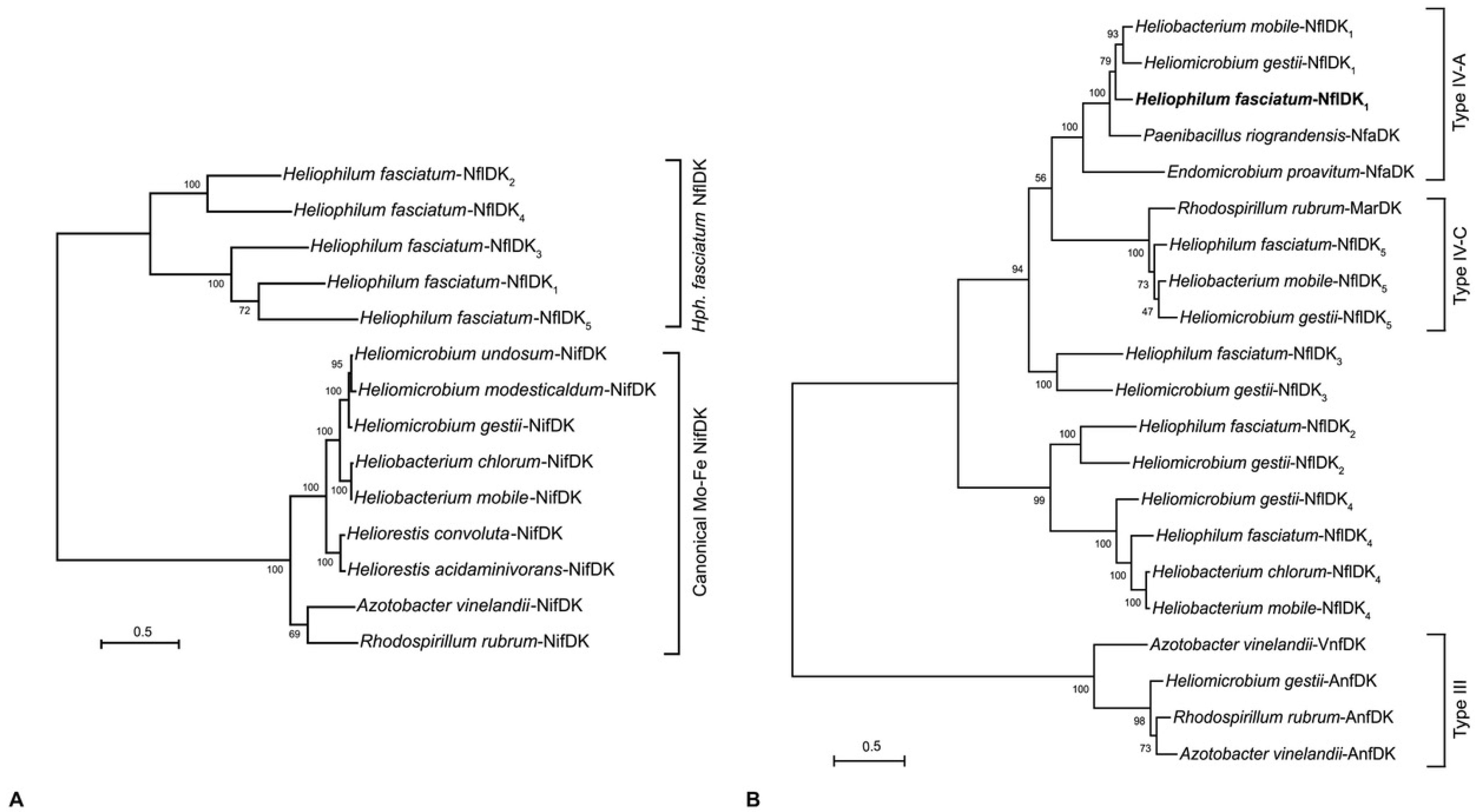
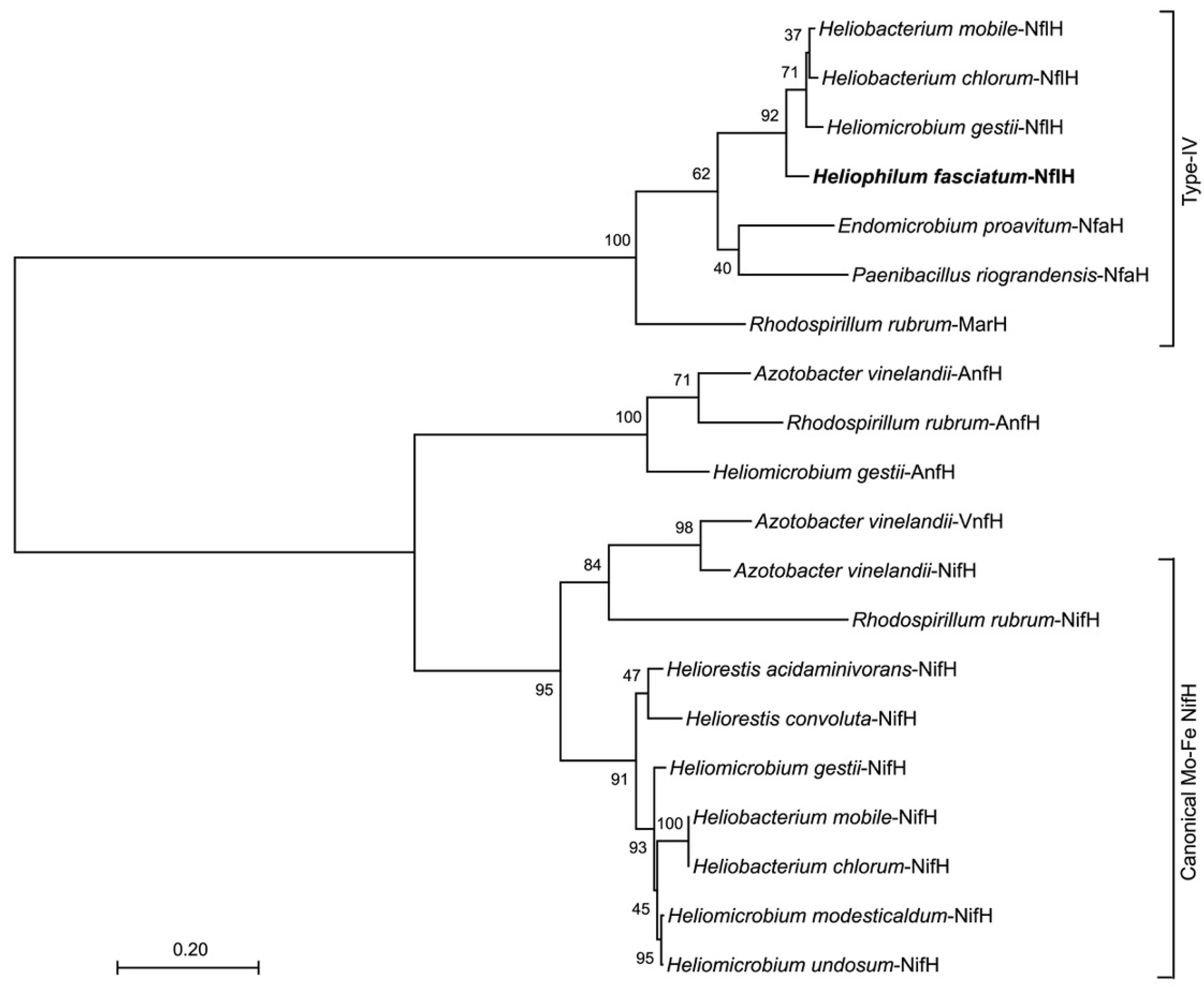
3.5. Nitrogen Metabolism: Nitrogen Fixation

Although the genomics supporting the utilization of fixed nitrogen compounds by Hph. fasciatum is not unusual, such is not true of the genomics supporting its diazotrophic growth. A nitrogenase system has been detected by acetylene reduction in N2-grown cells of Hph. fasciatum, albeit activities are expressed at lower levels than in cells of Heliomicrobium gestii or Heliomicrobium mobile [15,16]. Six-genes encoding NifHDK (dinitrogenase reductase and the α- and β-subunits of dinitrogenase, respectively) and NifENB (nitrogenase assembly and maturase proteins) are present in virtually all diazotrophs that produce a FeMo-cofactor-containing nitrogenase and are considered the minimal gene set required for a diazotrophic phenotype [30]. Curiously, however, experiments to detect genes encoding dinitrogenase reductase (nifH) and the α subunit of dinitrogenase (nifD) in Hph. fasciatum by Southern blot and PCR analyses were unsuccessful, suggesting that a unique enzyme or pathway for nitrogen fixation might exist in this phototroph [31]. The Hph. fasciatum genome supports this hypothesis by revealing genes encoding five distinct NifDK-like proteins that contain the pfam 00148 oxidoreductase domain present in the α- and β-subunits of all known FeMo-cofactor-containing dinitrogenases [30]. All genes encoding these nitrogen fixation-like proteins in Hph. fasciatium (genes and proteins abbreviated nfl and Nfl, respectively, and numbered 1–5) are clustered, with genes encoding NflDK1–4 located at the end of the contig Ga0493917_07 and those encoding NflDK5 positioned near the beginning of contig Ga0493917_01 (Figure 3A). In contrast to this arrangement, a “traditional nif cluster” containing nifI1, nifI2, nifH, nifD, nifK, nifE, nifN, nifX, fdxB, nifB, and nifV present in the genomes of Hmi. modesticaldum [10], Hrs. convoluta [13], and Hmi. gestii (Figure 3B), is absent from the Hph. fasciatum genome. Instead, single copies of nfl genes similar to nifB, nifH, nifX, and fdxB are present in the same chromosomal locus in Hph. fasciatum as genes encoding NflDK1 (Figure 3A). A NifV (homocitrate synthase) homolog is encoded elsewhere in the Hph. fasciatum chromosome; however, homologs of NifE and NifN could not be identified. Additionally, genes encoding an alternative nitrogenase, such as the FeFe-cofactor nitrogenase of Hmi. gestii (Figure 3C), were also absent from Hph. fasciatum.
The apparent absence of genes encoding NifEN in Hph. fasciatum is of particular interest but is not without precedence. FeMo nitrogenases lacking NifE or NifN (or both) are thought to be “ancestral nitrogenases”, enzymes that employ a truncated cofactor assembly pathway and catalyze an activity in addition to N2 reduction [30,32]. For example, the diazotrophic methanogen Methanocaldococcus sp. strain FS406-22 and bacterium Caldicellulosiruptor sp. strain YA01 synthesize nitrogenases that lack NifN but still contain NifE [33,34]. However, the genome of Endomicrobium proavitum strain Rsa215, a diazotrophic free-living termite gut bacterium, is missing genes encoding both NifE and NifN [35] indicating that a functional nitrogenase can be synthesized that lacks these proteins.
Because the NifEN proteins are hypothesized to be encoded by genes originating from a nifDK duplication [36], the Hph. fasciatum NflDK1–5 homologs (Figure 3A) were compared to NifDK and NifEN homologs from other heliobacteria as well as from the model diazotrophs Azotobacter vinelandii and Rhodospirillum rubrum. The results showed that none of the five Hph. fasciatum NflDK proteins formed a clade with NifEN homologs (data not shown) or with NifDK (Figure 4A). Thus, despite possessing genes encoding molybdate transport (ModA) and molybdenum cofactor synthesis (Moa/Moe), Hph. fasciatum is the only known species of heliobacteria to lack both NifEN and an ortholog of the classical NifDK nitrogenase.
A search for NflDK1–5 homologs in other species of heliobacteria identified two such proteins in Hbt. chlorum, four in Hbt. mobile, and five in Hmi. gestii (Figure 4B). In addition to the five Hmi. gestii NflDK homologs, the Hmi. gestii genome encodes a classical NifDK as well as a homolog of the Fe-Fe alternative dinitrogenase (AnfDK) of Azotobacter vinelandii and Rsp. rubrum (Figure 3B,C and Figure 4A,B) (alternative nitrogenases lack Mo but contain Fe-Fe or V-Fe cofactors and are thought to be redundant enzymes that support nitrogen fixation when Mo is limiting [37]). Our discovery of anfDK in Hmi. gestii (Figure 3C) complements a previous finding of an anfH-like gene in this species [32] and physiological evidence for an Fe-Fe nitrogenase system [38]. Moreover, an examination of the chromosomal locus encoding AnfHDK in Hmi. gestii also indicated the presence of a gene encoding the δ-subunit (anfG) of its Fe-Fe nitrogenase (Figure 3C).
Phylogenetic studies of nitrogenases have revealed at least six groups (designated I–VI) of “nif-like” genes distributed throughout Bacteria and Archaea, with members of groups IV–VI predicted to encode alternative functions such as roles in (bacterio)chlorophyll biosynthesis or archaeal methyl coenzyme-M reductase cofactor F430 biosynthesis, as well as various aspects of sulfur metabolism [21,22,31,35,39,40,41,42,43]. Phylogenetic analysis of the Hph. fasciatum NflDK1–5 homologs indicates that they do not form a clade with Nif, Anf, or Vnf nitrogenases (groups I–III). Instead, the NflDK1–5 proteins are more similar to the group IV MarDK, NflDK, and NfaDK (nitrogen fixation IV—subgroup A) proteins of Rhodospirillum rubrum, Paenibacillus riograndensis, and Endomicrobium proavitum, respectively [22,35] (Figure 4B). Whereas the MarDK proteins of Rsp. rubrum do not function as a nitrogenase [22], and the role of the NfaDK proteins in P. riograndensis is unknown (P. riograndensis also possesses canonical Nif and alternative Anf nitrogenases) [44], the E. proavitum NfaDK homolog falls within subgroup IV-A and is predicted to possess all the ligands required for N2 reduction [35]. Moreover, despite lacking homologs to NifEN, cells of E. proavitum incorporate 15N2 into biomass, presumably through the activity of NfaDK, the only nif-like genes present in the genome [35]. Therefore, based on its placement in a clade with E. proavitum NfaDK (Figure 4B) and the absence of genes encoding a classical nitrogenase system (Figure 4A), we predict that diazotrophy in Hph. fasciatum is supported by its NfaDK homolog NflDK1. If true, and if such an enzyme is a less robust nitrogenase than are classical nitrogenases, this could explain the lower nitrogenase activities observed in cells of Hph. fasciatum compared with those of other heliobacterial species [15,16].
While the roles of Hph. fasciatum NflDK2–4 are unknown, it is notable that the Hph. fasciatum, Hmi. gestii, and Hbt. mobile NflDK5 homologs form a clade with the Rhodospirillum rubrum MarDK proteins of the MarBHDK system that synthesizes methionine from volatile organic sulfur compounds (VOSCs) such as dimethyl sulfide and (2-methylthio)ethanol [22] (Figure 4B). MarDK are group IV-C nitrogenase-like proteins encoding the methylthio-alkane reductase that cleaves the C–S bond of VOSCs. Utilization of the MarBHDK pathway to assimilate sulfur from organic sources may compensate for the apparent lack of a sulfite reduction system in Hph. fasciatum (see Section 3.5). Hmi. gestii, Hbt. mobile, and Hbt. chlorum also possess an additional nifH-like (nflH) gene linked to the genes encoding these MarDK homologs. These NifH homologs form a clade with the MarH protein of Rsp. rubrum (Figure 5) and likely encode the electron-delivering subunit of a methylthio-alkane reductase. Hph. fasciatum possesses only one nifH-like gene, and it lies adjacent to the genes encoding NflDK1 in the genome (Figure 3A). Surprisingly, however, this NifH-like protein along with NfaH proteins of P. riograndensis and the E. proavitum nitrogenase form a clade with MarH instead of with NifH from other heliobacteria or AnfH or VnfH of Fe-Fe and V-Fe nitrogenases (Figure 5). All of these proteins possess the same critical residues necessary for reductive activity, which may allow for their cross-reactivity.
How the Hph. fasciatum and E. proavitum genomes have come to share these divergent NifHDK homologs is puzzling. Whereas both microbes are obligate anaerobes and fermentative, one is phototrophic while the other is symbiotic, and thus their ecology is unrelated. Why Hph. fasciatum is the only known heliobacterium to lack a conventional nitrogenase is also an intriguing question. Although we acknowledge that our analysis is not based on a single-contig closed genome, we nevertheless feel, for three reasons, that genes encoding a canonical nitrogenase system in Hph. fasciatum are truly absent. First, sequencing coverage of the Hph. fasciatum genome was high (477X), thus reducing the chance of losing or overlooking multi-gene operons. Second, experiments by others using PCR and Southern blots to detect nifH and nifD in Hph. fasciatum were negative, although both genes were easily identified in other heliobacterial species [31]. Finally, using degenerate primers targeting nifD, anfD, and vnfD [45], no PCR amplicons were obtained from Hph. fasciatum genomic DNA, although a canonical nifD was obtained from two other heliobacterial species used as controls (K.S.B., unpublished results).
Another interesting aspect of the Hph. fasciatum nitrogenase story is the lack of genes encoding obvious nitrogen metabolism regulators. A gene encoding a P-II nitrogen regulatory protein is present upstream of a gene encoding an ammonia transporter (2929297543), but this is not close to the loci encoding the NflDK1–5 proteins. Further studies are thus needed to determine the expression patterns of the Hph. fasciatum NflDK1–5 homologs, as well as mutational and 15N2 incorporation experiments to confirm the nitrogenase activity of Hph. fasciatum NflDK1 and test the N2-reducing ability of mutant derivatives lacking one or more homologs. The significance of encoding multiple NifDK-type proteins is also unknown, but it is not unusual for genomes encoding group IV nitrogenase-like enzymes to contain multiple copies of nifDK-like genes [43]. It is therefore possible that these genes encode important biochemical functions that are yet to be recognized.

3.6. Sulfur Metabolism

Unlike many heliobacteria that require a reduced source of sulfur for biosynthetic purposes [4,5,9], Hph. fasciatum grows in defined media with sulfate as the only sulfur source [16]. In this regard, the Hph. fasciatum genome encodes the widely distributed CysPTWA-mediated sulfate uptake system: CysNDC proteins that make adenylphosphosulfate (APS) and phosphoadenylphosphosulfate (PAPS) from sulfate, and AprAB and CysH that make sulfite from APS and PAPS, respectively. However, the Hph. fasciatum genome lacks homologs of CysJI, the classical alpha-beta sulfite reductase that reduces sulfite to sulfide in a wide variety of bacteria. Two genes were present that encoded a protein weakly similar to CysI (beta subunit of sulfite reductase). These genes (IDs 292929318 and 2929297321), were nearly identical and were annotated as “sulfite reductase beta subunit-like” but were only 20% identical to authentic CysI from Escherichia coli or Bacillus subtilis. Although it is possible that the Hph. fasciatum CysI-like protein participates in sulfite reduction, the absence of a homolog encoding the alpha subunit (CysJ) of sulfite reductase leaves open the question of exactly how sulfite reduction occurs in Hph. fasciatum.
Most heliobacteria will grow in media containing sulfide, and some species are remarkably sulfide tolerant [9,14]. Hph. fasciatum is an exception and is completely growth inhibited by as little as 0.1 mM sulfide [16]. Consistent with this, Hph. fasciatum lacks the common sulfide oxidation system—sulfide:quinone oxidoreductase—of purple and green sulfur bacteria [46]. In addition, Hph. fasciatum lacks sox genes (as do all known heliobacteria), which encode enzymes necessary to oxidize thiosulfate, the other reduced inorganic sulfur compound commonly oxidized as an electron donor by anoxygenic phototrophs. Thus, once sulfide is available in the Hph. fasciatum cell, from either the reduction of sulfate or from organic sources, it is likely to be quickly incorporated into sulfur-containing amino acids. In this regard, Hph. fasciatum is well equipped. Its genome encodes cysteine synthase that makes cysteine from serine and sulfide, as well as all genes necessary to convert aspartate to homocysteine, from which methionine is made. In addition, the genome encodes an ABC transporter for methionine and an ABC system for the uptake of branched-chain amino acids. However, as for methionine, all Hph. fasciatum genes necessary for the biosynthesis of valine, leucine, and isoleucine could be identified.

3.7. Motility

Cells of Hph. fasciatum are large rods (Figure 2) that display an unusual form of motility, unique among heliobacteria. Hph. fasciatum cells form bundles of parallel cells resembling the rafts of Bacillus or Proteus that form in biofilms [47,48]; the Hph. fasciatum bundles are motile as a unit and move with a rolling motion [16]. Bacterial flagella consist of three major components: the basal body, which traverses the cytoplasmic membrane (and if present, the outer membrane), the hook complex, and the filament, the rotating component that imparts cell motility [49]. Although both gram-positive and gram-negative bacterial flagella contain a basal body and hook that connects with the filament, gram-positive species lack the L and R rings that imbed the flagellum into the outer membrane of gram-negative bacteria [50]. In agreement with the Bacillus subtilis genome, only two of the three Flh proteins (FlhAB), highly conserved proteins required for exporting flagellar components across the cytoplasmic membrane [51], were identified in the Hph. fasciatum genome. A gene encoding FlhE, a protein found primarily in gram-negative enteric bacteria that functions to prevent proton leakage during the secretion of flagellar components across the cytoplasmic membrane, is missing, but it has been shown to be unessential for swimming motility [52].
Genes encoding Flg proteins (responsible for forming or regulating the formation of the flagellar rod, P ring, and hook) [53,54] in the Hph. fasciatum genome also followed the Bacillus pattern; genes encoding all eight of the Flg proteins of Bacillus (FlgBCDEFGKLMN) were identified. In addition, an unidentified protein with significant homology to FlgJ, a protein missing in Bacillus, was identified in the Hph. fasciatum genome and shared 50–60% identity with FlgJ from several gram-positive bacteria. FlgJ is a peptidoglycan hydrolase [54] and is found in the genomes of all heliobacteria listed in Table 2. Flg proteins not encoded in the Hph. fasciatum genome include FlgAHI and FlgOPQT, outer membrane proteins or proteins otherwise unessential for motility in gram-positive bacteria [53,54].
Genes encoding most Fli proteins responsible for forming the basal body, hook, filament, and cytoplasmic flagellar components (FliACDEFGHIJKLMNOPQRS) [53,54] were identified in the Hph. fasciatum genome, but the filament chaperone protein FliT, unessential in many motile bacteria, and regulatory proteins FliZY showed less than 50% identity to homologs from Bacillus. Mot proteins form the stator complex in the flagellar basal body [53], and the Hph. fasciatum genome encodes MotA and MotB, as does Bacillus. As expected, MotCD, proteins that form a second stator in Pseudomonas aeruginosa and some other gram-negative bacteria, and MotXY, proteins that form an alternative stator in bacteria that use a Na+-motive force instead of a H+-motive force to power the flagellum, such as Vibrio alginolyticus, were not encoded in the Heliophilum genome.
The Heliophilum genome encoded several chemotaxis proteins, suggesting that the bundled cells can respond to chemical attractants and repellants. Chemotaxis-specific proteins included virtually all “Che” proteins (CheABCDRWXY) plus at least five chemical sensors, the methyl-accepting chemotaxis proteins [55]. The only major Escherichia coli Che gene not identified in Hph. fasciatum was CheZ. This protein has phosphatase activity and functions to dephosphorylate CheY, the protein that controls the direction of flagellar rotation. CheZ is distributed only in certain Proteobacteria, and its function is replaced in gram-positive bacteria by CheC and CheX [56], both of which were encoded in the Hph. fasciatum genome.
Although no obvious answer emerged to explain the bundle-forming phenotype of Hph. fasciatum, this behavior could be a manifestation of biofilm formation. In this connection, cultures of Hph. fasciatum streaked on agar plates form spreading, diffuse colonies [16]. Rafts of cells are common in biofilm-forming bacterial species, and the dynamic nature that exists between free-swimming individual cells and rafts of cells indicates that specific “glue-like” substances are not required to form rafts [47]. Instead, it is likely that rafting is the product of intercellular bundling of flagella to yield cells with their flagella interwoven in phase [47,57]; notably, such flagellar aggregates were observed in the original description of Hph. fasciatum [16]. From an ecological perspective, rafts might help Hph. fasciatum become established on a moist surface, such as a rice plant root, where individual cells could be washed away.

3.8. Sporulation

True to its phylogenetic roots with the endospore-forming Firmicutes, Hph. fasciatum forms endospores—dormant cells highly resistant to physiochemical extremes [58]—complete with the signature molecule of these structures, dipicolinic acid–Ca2+ complexes [16]. Moreover, experiments to hybridize Hph. fasciatum DNA with endospore-specific gene probes from Bacillus subtilis confirmed the genetic capacity for this phototroph (and several other species of heliobacteria) to produce endospores [6].
Over 500 gene products have been linked to endospores in Bacillus subtilis, several of which are unessential for endospore production [59]. Sporulation-specific genes in Hmi. modesticaldum were found to be but a small subset of those found in B. subtilis, although several key sporulation genes were identified. As in Hmi. modesticaldum and Hrs. convoluta, all five sigma factors absolutely essential for the formation of endospores (sigma E, F, G, H, and K) were identified in the Hph. fasciatum genome. An ortholog of the master transcription factor Spo0A, active during the early stages of sporulation [60], is present in all heliobacterial genomes, including that of Heliophilum. However, Spo0M, a key protein that regulates several genes essential for sporulation, is absent from the Hmi. modesticaldum and Hrs. convoluta genomes [10,13]. By contrast, the Hph. fasciatum genome contained a putative spo0M gene (30–40% identity to spo0M from several sporulating bacteria). Hbt. chlorum, Hmi. gestii, and Hmi. undosum but not Hbt. mobile also contained a spo0M homolog, although all of these species encoded proteins less than 40% identical to the Hph. fasciatum protein, suggesting considerable divergence in this protein in those heliobacteria that encode it.
Also present in the Hph. fasciatum genome were genes encoding a variety of small acid-soluble spore proteins (SASPs). These proteins function to protect DNA in endospores from damage by desiccation, ultraviolet radiation, and to some degree, from heat [60,61,62]. Genes encoding the major alpha/beta-type SASPs (sspA and sspB) were present and showed high homology to genes from all other heliobacteria and chemotrophic endospore-forming bacteria. Three different α/β-type proteins were found in the Hph. fasciatum genome compared with just two in the genomes of the two alkaliphiles, Hrs. convoluta and Hrs. acidaminovorans, and four in each of the other five genome-sequenced heliobacterial species that grow optimally at neutral pH (Figure 1). In addition, the Hph. fasciatum genome encoded a minor SASP found in B. subtilis but not encoded in any other heliobacterial genome. This SASP was of the “thioredoxin type” (B. subtilis sspT), a protein encoded only in the genomes of a subset of sporulating bacteria [61]. Although the function of this minor SASP is unknown, it is clearly an endospore-associated protein because its transcription is dependent on the spore-specific sigma factor, σF. The genomes of all heliobacteria other than Hph. fasciatum encoded a different minor SASP, sspH, a gene under the control of σK in the developing forespore and whose product is also of unknown function.
The endospore is covered by a coat consisting of both structural and enzymatic proteins, and in B. subtilis, these proteins are encoded by a variety of genes, including over 20 cot genes [63]. Although Cot proteins are widespread in Bacillus species, in Clostridium species many cot genes are missing [63]. The Hph. fasciatum genome contained a handful of cot homologs. CotJC was encoded in the genomes of all heliobacteria listed in Table 2 and is one of the few Cot proteins universally present in species of Bacillus and Clostridium. The inner spore core protein GerQ is also present in all heliobacterial genomes. Genes encoding the spore-coat proteins CotJA and CotJB were annotated in Hph. fasciatum but the proteins bore no close identity to B. subtilis CotJA and CotJB. Notably, however, the Hph. fasciatum CotJA and CotJB homologs bore significant similarities to CotJA/JB from Clostridium perfringens, suggesting that there may be different forms of these endospore-specific proteins in aerobes compared with anaerobes.
Finally, in contrast to all other heliobacteria, a homolog of CotSA, a protein universally distributed in species of Bacillus and Clostridium [63], was autoannotated in the Hph. fasciatum genome as a cell wall glycosyltransferase with ~30% identity to B. subtilis CotSA. A protein similar to the B. subtilis spore-coat protein CotF was also identified in the Hph. fasciatum genome but is absent from the genomes of other sequenced heliobacteria; CotF is a spore-coat protein widely distributed among endospore-forming bacteria [63]. Genes encoding homologs of the B. subtilis spore-coat proteins CotABCDEGHIKMNOPQRSUVWXYZ or CotIC, NE, NH, NW, and OO could not be identified in the Hph. fasciatum genome. Thus, as regards the genetics of sporulation, Hph. fasciatum has the essential genes necessary to form endospores but, like other heliobacteria with analyzed genomes, is missing many of the unessential genes present in the genome of the well-studied Bacillus subtilis.

4. Conclusions

The genome of Heliophilum fasciatum represents the first detailed analysis of a genome from a species of heliobacteria that grows optimally at moderate temperatures and neutral pH. Although no firm explanation emerged for the mechanism that allows this organism to move about its habitat in loosely attached bundles, the genomics supporting the physiology of this species revealed many things in common with other heliobacteria (e.g., lack of autotrophy, ability to grow in darkness by fermentation, production of endospores, to mention a few), as well as an important feature possibly unique to this species: diazotrophy in the absence of a canonical nitrogenase. Whether nitrogen fixation in the Tanzanian rice soil habitat of Hph. fasciatum [16] is best served by its unusual nitrogenase system remains to be seen. However, the fact that all other heliobacteria with sequenced genomes (some species of which originate from rice soils) contain a canonical nitrogenase (Figure 3, Figure 4 and Figure 5), suggests that the ancestor of Hph. fasciatum may have been incapable of diazotrophy but that eventually, this species acquired an enzyme with nitrogenase-like activity from either duplications or lateral transfers of genes encoding functionally related proteins. Nitrogen fixation by phototrophic purple bacteria in paddy soil environments is well known [64,65]. Hph. fasciatum and other paddy-dwelling heliobacterial species could also be contributing fixed nitrogen to rice plants, perhaps in exchange for organic compounds to fuel their photoheterotrophic metabolism.

Author Contributions

M.T.M., K.S.B., M.H.M., M.N.P., L.L.J., P.V.W. and W.M.S. conceived and designed the experiments. K.S.B. supplied cells of Hph. fasciatum to M.H.M., who isolated genomic DNA and submitted it to JGI for sequencing. All authors analyzed or reviewed the data, with the bulk of the analysis done by K.S.B., M.T.M. and W.M.S. The draft manuscript was prepared by M.T.M., K.S.B. and W.M.S., with other authors providing reviews and edits for the final version. All authors have read and agreed to the published version of the manuscript.

Funding

This work was supported by JGI New Investigator Proposal Award Number CSP 504032 to M.H.M. and P.V.W. and by NASA Exobiology Cooperative Agreement 80NSSC21M0355 to M.T.M.

Institutional Review Board Statement

Not applicable.

Informed Consent Statement

Not applicable.

Data Availability Statement

The Heliophilum fasciatum TanzaniaT genome sequence (listed as Heliophilum fasciatum MTM) is publically available in the JGI/IMG database (https://img.jgi.doe.gov/) under the genome ID 2929297113.

Acknowledgments

The authors appreciate the technical assistance of Erik Velkme and Alexandria Wilcox.

Conflicts of Interest

The authors declare no conflict of interest.

References

  1. Gest, H.; Favinger, J.L. Heliobacterium chlorum, an anoxygenic brownish-green photosynthetic bacterium containing a “new” form of bacteriochlorophyll. Arch. Microbiol. 1983, 136, 11–16. [Google Scholar] [CrossRef]
  2. Madigan, M.T.; Ormerod, J.G. Taxonomy, physiology, and ecology of heliobacteria. In Anoxygenic Photosynthetic Bacteria; Blankenship, R.E., Madigan, M.T., Bauer, C.E., Eds.; Kluwer Academic Publishers: Dordrecht, The Netherlands, 1995; Volume 2, pp. 17–30. ISBN 0-306-47954-0. [Google Scholar]
  3. Brockmann, H.; Lipinski, A. Bacteriochlorophyll g. A new bacteriochlorophyll from Heliobacterium chlorum. Arch. Microbiol. 1983, 136, 17–19. [Google Scholar] [CrossRef]
  4. Sattley, W.M.; Madigan, M.T. The Family Heliobacteriaceae. In The Prokaryotes—Firmicutes and Tenericutes; Rosenberg, E., DeLong, E.F., Lory, S., Stackebrandt, E., Thompson, F., Eds.; Springer: Berlin/Heidelberg, Germany, 2014; pp. 185–196. ISBN 978-3-642-30120-9. [Google Scholar]
  5. Orf, G.S.; Redding, K.E. The Heliobacteria. In Encyclopedia of Biological Chemistry; Joseph, J., Ed.; Elsevier: Oxford, UK, 2021; Volume 2, pp. 352–364. [Google Scholar]
  6. Kimble-Long, L.K.; Madigan, M.T. Molecular evidence that the capacity for endosporulation is universal among phototrophic heliobacteria. FEMS Microbiol. Lett. 2001, 199, 191–195. [Google Scholar] [CrossRef]
  7. Stevenson, A.K.; Kimble, L.K.; Woese, C.R.; Madigan, M.T. Characterization of new phototrophic heliobacteria and their habitats. Photosynth. Res. 1997, 53, 1–12. [Google Scholar] [CrossRef]
  8. Kyndt, J.A.; Salama, D.M.; Meyer, T.E.; Imhoff, J.F. Phylogenetic relationship of phototrophic heliobacteria and systematic reconsideration of species and genus assignments based on genome sequences of eight species. Int. J. Syst. Evol. Microbiol. 2021, 71, 4729. [Google Scholar] [CrossRef]
  9. Kimble, L.K.; Mandelco, L.; Woese, C.R.; Madigan, M.T. Heliobacterium modesticaldum, sp. nov., a thermophilic heliobacterium of hot springs and volcanic soils. Arch. Microbiol. 1995, 163, 259–267. [Google Scholar] [CrossRef]
  10. Sattley, W.M.; Madigan, M.T.; Swingley, W.D.; Cheung, P.C.; Clocksin, K.M.; Conrad, A.L.; Dejesa, L.C.; Honchak, B.M.; Jung, D.; Karbach, L.E.; et al. The Genome of Heliobacterium modesticaldum, a Phototrophic Representative of the Firmicutes Containing the Simplest Photosynthetic Apparatus. J. Bacteriol. 2008, 190, 4687–4696. [Google Scholar] [CrossRef] [Green Version]
  11. Sattley, W.M.; Swingley, W.D. Properties and evolutionary implications of the heliobacterial genome. In Genome Evolution of Photosynthetic Bacteria; Beatty, T.J., Ed.; Academic Press: San Diego, CA, USA, 2013; Volume 66, pp. 67–98. ISBN 9780123979230. [Google Scholar]
  12. Asao, M.; Jung, D.; Achenbach, L.A.; Madigan, M.T. Heliorestis convoluta sp. nov., a coiled, alkaliphilic heliobacterium from the Wadi El Natroun, Egypt. Extremophiles 2006, 10, 403–410. [Google Scholar] [CrossRef]
  13. Dewey, E.D.; Stokes, L.M.; Burchell, B.M.; Shaffer, K.N.; Huntington, A.M.; Baker, J.M.; Nadendla, S.; Giglio, M.G.; Bender, K.S.; Touchman, J.W.; et al. Analysis of the Complete Genome of the Alkaliphilic and Phototrophic Firmicute Heliorestis convoluta Strain HHT. Microorganisms 2020, 8, 313. [Google Scholar] [CrossRef] [Green Version]
  14. Asao, M.; Takaichi, S.; Madigan, M.T. Amino acid-assimilating phototrophic heliobacteria from soda lake environments: Heliorestis acidaminivorans sp. nov. and ‘Candidatus Heliomonas lunata’. Extremophiles 2012, 16, 585–595. [Google Scholar] [CrossRef]
  15. Kimble, L.K.; Madigan, M.T. Nitrogen fixation and nitrogen metabolism in heliobacteria. Arch. Microbiol. 1992, 158, 155–161. [Google Scholar] [CrossRef]
  16. Ormerod, J.G.; Kimble, L.K.; Nesbakken, T.; Torgersen, Y.A.; Woese, C.R.; Madigan, M.T. Heliophilum fasciatum gen. nov. sp. nov. and Heliobacterium gestii sp. nov.: Endospore-forming heliobacteria from rice field soils. Arch. Microbiol. 1996, 165, 226–234. [Google Scholar] [CrossRef] [PubMed]
  17. Tang, K.-H.; Yue, H.; Blankenship, R.E. Energy metabolism of Heliobacterium modesticaldum during phototrophic and chemotrophic growth. BMC Microbiol. 2010, 10, 150. [Google Scholar] [CrossRef] [PubMed] [Green Version]
  18. Kumar, S.; Stecher, G.; Li, M.; Knyaz, C.; Tamura, K. MEGA X: Molecular Evolutionary Genetics Analysis across Computing Platforms. Mol. Biol. Evol. 2018, 35, 1547–1549. [Google Scholar] [CrossRef] [PubMed]
  19. Stecher, G.; Tamura, K.; Kumar, S. Molecular Evolutionary Genetics Analysis (MEGA) for macOS. Mol. Biol. Evol. 2020, 37, 1237–1239. [Google Scholar] [CrossRef]
  20. Altschul, S.F.; Madden, T.L.; Schäffer, A.A.; Zhang, J.; Zhang, Z.; Miller, W.; Lipman, D.J. Gappled BLAST and PSI-BLAST: A new generation of protein database search programs. Nucleic Acids Res. 1997, 25, 3389–3402. [Google Scholar] [CrossRef] [Green Version]
  21. Raymond, J.; Siefert, J.L.; Staples, C.R.; Blankenship, R.E. The Natural History of Nitrogen Fixation. Mol. Biol. Evol. 2004, 21, 541–554. [Google Scholar] [CrossRef] [Green Version]
  22. North, J.A.; Narrowe, A.B.; Xiong, W.; Byerly, K.M.; Zhao, G.; Young, S.J.; Murali, S.; Wildenthal, J.A.; Cannon, W.R.; Wrighton, K.C.; et al. A nitrogenase-like enzyme system catalyzes methionine, ethylene, and methane biogenesis. Science 2020, 369, 1094–1098. [Google Scholar] [CrossRef]
  23. Larkin, M.A.; Blackshields, G.; Brown, N.P.; Chenna, R.; McGettigan, P.A.; McWilliam, H.; Valentin, F.; Wallace, I.M.; Wilm, A.; Lopez, R.; et al. Clustal W and Clustal X version 2.0. Bioinformatics 2007, 23, 2947–2948. [Google Scholar] [CrossRef] [Green Version]
  24. Le, S.Q.; Gascuel, O. An Improved General Amino Acid Replacement Matrix. Mol. Biol. Evol. 2008, 25, 1307–1320. [Google Scholar] [CrossRef] [Green Version]
  25. Bryantseva, I.A.; Gorlenko, V.M.; Kompantseva, E.I.; Achenbach, L.A.; Madigan, M.T. Heliorestis daurensis, gen. nov. sp. nov., an alkaliphilic rod-to-coiled-shaped phototrophic heliobacterium from a Siberian soda lake. Arch. Microbiol. 1999, 172, 167–174. [Google Scholar] [CrossRef] [PubMed]
  26. Bryantseva, I.A.; Gorlenko, V.M.; Kompantseva, E.I.; Tourova, T.P.; Kuznetsov, B.B.; Osipov, G.A. Alkaliphilic heliobacterium Heliorestis baculata sp. nov. and emended description of the genus Heliorestis. Arch. Microbiol. 2000, 174, 283–291. [Google Scholar] [CrossRef] [PubMed]
  27. Briantseva, I.A.; Gorlenko, V.M.; Turova, T.P.; Kuznetsov, B.B.; Lysenko, A.M.; Bykova, S.A.; Gal’Chenko, V.F.; Mitiushina, L.L.; Osipov, G.A. Heliobacterium sulfidophilum sp. nov. and Heliobacterum undosum sp. nov.: Sulfide-oxidizing heliobacteria from thermal sulfidic springs. Mikrobiologia 2000, 69, 396–406. [Google Scholar]
  28. Kimble, L.K.; Stevenson, A.K.; Madigan, M.T. Chemotrophic growth of heliobacteria in darkness. FEMS Microbiol. Lett. 1994, 115, 51–55. [Google Scholar] [CrossRef]
  29. Pickett, M.W.; Williamson, M.P.; Kelly, D.J. An enzyme and13C-NMR study of carbon metabolism in heliobacteria. Photosynth. Res. 1994, 41, 75–88. [Google Scholar] [CrossRef]
  30. Dos Santos, P.C.; Fang, Z.; Mason, S.W.; Setubal, J.C.; Dixon, R. Distribution of nitrogen fixation and nitrogenase-like sequences amongst microbial genomes. BMC Genom. 2012, 13, 162. [Google Scholar] [CrossRef] [Green Version]
  31. Enkh-Amgalan, J.; Kawasaki, H.; Seki, T. NifH and NifD sequences of heliobacteria: A new lineage in the nitrogenase phylogeny. FEMS Microbiol. Lett. 2005, 243, 73–79. [Google Scholar] [CrossRef]
  32. Garcia, A.K.; McShea, H.; Kolaczkowski, B.; Kaçar, B. Reconstructing the evolutionary history of nitrogenases: Evidence for ancestral molybdenum-cofactor utilization. Geobiology 2020, 18, 394–411. [Google Scholar] [CrossRef] [Green Version]
  33. Chen, Y.; Nishihara, A.; Haruta, S. Nitrogen-fixing Ability and Nitrogen Fixation-related Genes of Thermophilic Fermentative Bacteria in the Genus Caldicellulosiruptor. Microbes Environ. 2021, 36, ME21018. [Google Scholar] [CrossRef]
  34. Lie, T.J.; Kuo, Y.P.; Leite, M.; Costa, K.C.; Harwood, C.S.; Leigh, J.A. A genetic study of Nif-associated genes in a hyperthermophilic methanogen. Microbiol. Spectr. 2022, 10, e02093-21. [Google Scholar] [CrossRef]
  35. Zheng, H.; Dietrich, C.; Radek, R.; Brune, A. Endomicrobium proavitum, the first isolate of Endomicrobia class. nov. (phylum Elusimicrobia)—An ultramicrobacterium with an unusual cell cycle that fixes nitrogen with a Group IV nitrogenase. Environ. Microbiol. 2015, 18, 191–204. [Google Scholar] [CrossRef] [PubMed]
  36. Boyd, E.S.; Anbar, A.D.; Miller, S.; Hamilton, T.L.; Lavin, M.; Peters, J.W. A late methanogen origin for molybdenum-dependent nitrogenase. Geobiology 2011, 9, 221–232. [Google Scholar] [CrossRef] [PubMed]
  37. Harwood, C.S. Iron-only and vanadium nitrogenases: Fail-safe enzymes or something more? Ann. Rev. Microbiol. 2020, 74, 247–266. [Google Scholar] [CrossRef] [PubMed]
  38. Kimble, L.K.; Madigan, M.T. Evidence for an alternative nitrogenase in Heliobacterium gestii. FEMS Microbiol. Lett. 1992, 100, 255–260. [Google Scholar] [CrossRef] [Green Version]
  39. Nomata, J.; Mizoguchi, T.; Tamiaki, H.; Fujita, Y. A second nitrogenase-like enzyme for bacteriochlorophyll biosynthesis: Reconstitution of chlorophyllide a reductase with purified X-protein (BchX) and YZ-protein (BchY-BchZ) from Rhodobacter capsulatus. J. Biol. Chem. 2006, 281, 15021–15028. [Google Scholar] [CrossRef] [Green Version]
  40. Muraki, N.; Nomata, J.; Ebata, K.; Mizoguchi, T.; Shiba, T.; Tamiaki, H.; Kurisu, G.; Fujita, Y. X-ray crystal structure of the light-independent protochlorophyllide reductase. Nature 2010, 465, 110–114. [Google Scholar] [CrossRef]
  41. Moore, S.; Sowa, S.T.; Schuchardt, C.; Deery, E.; Lawrence, A.D.; Ramos, J.V.; Billig, S.; Birkemeyer, C.; Chivers, P.; Howard, M.; et al. Elucidation of the biosynthesis of the methane catalyst coenzyme F430. Nature 2017, 543, 78–82. [Google Scholar] [CrossRef] [Green Version]
  42. Zheng, K.; Ngo, P.D.; Owens, V.L.; Yang, X.-P.; Mansoorabadi, S.O. The biosynthetic pathway of coenzyme F430 in methanogenic and methanotrophic archaea. Science 2016, 354, 339–342. [Google Scholar] [CrossRef] [Green Version]
  43. Méheust, R.; Castelle, C.; Carnevali, P.B.M.; Farag, I.F.; He, C.; Chen, L.-X.; Amano, Y.; Hug, L.; Banfield, J.F. Groundwater Elusimicrobia are metabolically diverse compared to gut microbiome Elusimicrobia and some have a novel nitrogenase paralog. ISME J. 2020, 14, 2907–2922. [Google Scholar] [CrossRef]
  44. Fernandes, G.D.C.; Trarbach, L.J.; de Campos, S.B.; Beneduzi, A.; Passaglia, L.M. Alternative nitrogenase and pseudogenes: Unique features of the Paenibacillus riograndensis nitrogen fixation system. Res. Microbiol. 2014, 165, 571–580. [Google Scholar] [CrossRef]
  45. McRose, D.L.; Zhang, X.; Kraepiel, A.M.L.; Morel, F.M.M. Diversity and Activity of Alternative Nitrogenases in Sequenced Genomes and Coastal Environments. Front. Microbiol. 2017, 8, 267. [Google Scholar] [CrossRef] [PubMed] [Green Version]
  46. Shahak, Y.; Schütz, M.; Bronstein, M.; Greisbeck, C.; Hauska, G.; Padan, E. Sulfide-dependent anoxygenic photosynthesis in prokaryotes—Sulfide-Quinone Reductase (SQR), the initial step. In The Phototrophic Prokaryotes; Peschek, G.A., Loffelhardt, W.L., Schmetterer, G., Eds.; Kluwer Academic/Plenum: New York, NY, USA, 1999; pp. 217–228. [Google Scholar]
  47. Kearns, D.; Losick, R. Swarming motility in undomesticated Bacillus subtilis. Mol. Microbiol. 2004, 49, 581–590. [Google Scholar] [CrossRef] [PubMed]
  48. Kearns, D.B. A field guide to bacterial swarming motility. Nat. Rev. Genet. 2010, 8, 634–644. [Google Scholar] [CrossRef] [PubMed] [Green Version]
  49. Apel, D.; Surette, M.G. Bringing order to a complex molecular machine: The assembly of the bacterial flagella. Biochim. Biophys. Acta (BBA)-Biomembr. 2008, 1778, 1851–1858. [Google Scholar] [CrossRef] [PubMed] [Green Version]
  50. Albanna, A.; Alomari, A. Comparison 2D flagellar proteins structure in gram positive vs. gram-negative motile bacteria. EurAsian J. Biosci. 2020, 17, 6275–6281. [Google Scholar]
  51. Nakamura, S.; Minamino, T. Flagella-Driven Motility of Bacteria. Biomolecules 2019, 9, 279. [Google Scholar] [CrossRef] [Green Version]
  52. Stafford, G.P.; Hughes, C. Salmonella typhimurium flhE, a conserved flagellar regulon gene required for swarming. Microbiology 2007, 153, 541–547. [Google Scholar] [CrossRef] [Green Version]
  53. Girgis, H.S.; Liu, Y.; Ryu, W.S.; Tavazoie, S. A Comprehensive Genetic Characterization of Bacterial Motility. PLoS Genet. 2007, 3, e154–e160. [Google Scholar] [CrossRef]
  54. Herlihey, F.A.; Moynihan, P.; Clarke, A.J. The Essential Protein for Bacterial Flagella Formation FlgJ Functions as a β-N-Acetylglucosaminidase. J. Biol. Chem. 2014, 289, 31029–31042. [Google Scholar] [CrossRef] [Green Version]
  55. Wadhams, G.H.; Armitage, J.P. Making sense of it all: Bacterial chemotaxis. Nat. Rev. Mol. Cell Biol. 2004, 5, 1024–1037. [Google Scholar] [CrossRef] [PubMed]
  56. Szurmant, H.; Ordal, G.W. Diversity in Chemotaxis Mechanisms among the Bacteria and Archaea. Microbiol. Mol. Biol. Rev. 2004, 68, 301–319. [Google Scholar] [CrossRef] [PubMed] [Green Version]
  57. Jones, B.V.; Young, R.; Mahenthiralingam, E.; Stickler, D.J. Ultrastructure of Proteus mirabilis Swarmer Cell Rafts and Role of Swarming in Catheter-Associated Urinary Tract Infection. Infect. Immun. 2004, 72, 3941–3950. [Google Scholar] [CrossRef] [PubMed] [Green Version]
  58. Nicholson, W.L.; Munakata, N.; Horneck, G.; Melosh, H.J.; Setlow, P. Resistance of Bacillus Endospores to Extreme Terrestrial and Extraterrestrial Environments. Microbiol. Mol. Biol. Rev. 2000, 64, 548–572. [Google Scholar] [CrossRef] [PubMed] [Green Version]
  59. Traag, B.A.; Pugliese, A.; Eisen, J.A.; Losick, R. Gene Conservation among Endospore-Forming Bacteria Reveals Additional Sporulation Genes in Bacillus subtilis. J. Bacteriol. 2013, 195, 253–260. [Google Scholar] [CrossRef] [PubMed] [Green Version]
  60. Piggot, P.J.; Hilbert, D.W. Sporulation of Bacillus subtilis. Curr. Opin. Microbiol. 2004, 7, 579–586. [Google Scholar] [CrossRef] [PubMed]
  61. Setlow, P. Mechanisms for the prevention of damage to DNA in spores of bacillus species. Annu. Rev. Microbiol. 1995, 49, 29–54. [Google Scholar] [CrossRef] [PubMed]
  62. Bagyan, I.; Setlow, B.; Setlow, P. New Small, Acid-Soluble Proteins Unique to Spores of Bacillus subtilis: Identification of the Coding Genes and Regulation and Function of Two of These Genes. J. Bacteriol. 1998, 180, 6704–6712. [Google Scholar] [CrossRef] [Green Version]
  63. Henriques, A.O.; Moran, C.P., Jr. Structure, Assembly, and Function of the Spore Surface Layers. Annu. Rev. Microbiol. 2007, 61, 555–588. [Google Scholar] [CrossRef]
  64. Habte, M.; Alexander, M. Nitrogen Fixation by Photosynthetic Bacteria in Lowland Rice Culture. Appl. Environ. Microbiol. 1980, 39, 342–347. [Google Scholar] [CrossRef] [Green Version]
  65. Maeda, I. Potential of Phototrophic Purple Nonsulfur Bacteria to Fix Nitrogen in Rice Fields. Microorganisms 2021, 10, 28. [Google Scholar] [CrossRef]
Figure 1. Phylogeny of Heliophilum fasciatum based on 16S ribosomal RNA gene sequence analysis. The multiple alignments included 16S rRNA gene sequences from all cultured type strains of the Heliobacteriaceae, as well as Candidatus “Heliomonas lunata” and the outgroup species Escherichia coli. The neighbor-joining tree was constructed from 1344 nucleotide positions. The scale bar indicates the number of base substitutions per site. Bootstrap values > 50 (500 replicates) are indicated at each node, and GenBank accession numbers are shown in parentheses.
Microorganisms 10 00869 g001
Figure 2. Heliophilum fasciatum. (A) Phase-contrast micrograph of cells formed into motile bundles. (B) Scanning electron micrograph of a cell bundle. (C) Negatively stained transmission electron micrograph with visible flagella. (D) Thin sectioned transmission electron micrograph showing absence of internal photosynthetic membranes, typical of the heliobacteria.
Microorganisms 10 00869 g002
Figure 3. Genetic organization of nitrogen fixation-related genes and surrounding genes. (A) Hph. fasciatum loci containing genes encoding the nitrogen fixation-like homologs NflDK1–5. The following JGI/IMG coordinates are illustrated: Ga0493917_07: 106,630 to 145,155 and Ga0493917_01: 75 to 11,134; (B) Hmi. gestii locus containing genes encoding the Mo-Fe nitrogenase (NifHDK). The following Genbank coordinates are illustrated: NZ_WXEX01000020.1: 13,816 to 26,039; (C) Hmi. gestii locus containing genes encoding the alternative Fe-Fe nitrogenase (AnfHDGK). The following Genbank coordinates are illustrated: NZ_WXEX01000005.1: 245,244 to 257,234. Predicted functions are described in the key.
Microorganisms 10 00869 g003
Figure 4. Maximum-likelihood phylogenetic trees of Hph. fasciatum concatenated NflDK sequences. (A) Comparison with canonical Mo-Fe nitrogenase NifDK sequences. (B) Comparison with Type III and IV nitrogen fixation-like sequences. The nodes represent bootstrap values based on 100 replicates, and scale bars indicate 0.5 changes per position. Taxa accession numbers are as follows with those beginning with a number corresponding to the JGI/IMG database and those beginning with a letter from Genbank: Hph. fasciatum—NflDK1 (2929298242, 2929298243), NflDK2 (2929298250, 2929298249), NflDK3 (2929298253, 2929298252), NflDK4 (2929298263, 2929298262), NflDK5 (2929297118, 2929297117); Hmi. gestii—NifDK (WP_161263156, WP_161263155), AnfDK (WP_161261510, WP_161261560), NflDK1 (WP_161260453, WP_161260452), NflDK2 (WP_161260457, WP_161260458), NflDK3 (WP_161260450, WP_161260449), NflDK4 (WP_161260444, WP_161260443), NflDK5 (WP_161260462, WP_161260461); Hmi. modesticaldum—NifDK (641558455, 641558456); Hmi. undosum—NifDK (WP_161255976, WP_161255978); Hrs. acidaminivorans—NifDK (2914075535, 2914075536); Hrs. convoluta—NifDK (QGG48552, QGG48553); Hbt. chlorum—NifDK (WP_188040647, WP_188040648), NflDK4 (WP_188039480, WP_188039481); Hbt. mobile—NifDK (WP_155476316, WP_155476317), NflDK1 (WP_155475363, WP_155475362), NflDK4 (WP_155475372, WP_155475371), NflDK5 (WP_155475353, WP_155475354); A. vinelandii—NifDK (643803063, 643803064), AnfDK (643807739, 643807737), VnfDK (643803186, 643803184); Rsp. rubrum—NifDK (637825100, 637825101), AnfDK (637825484, 637825482), MarDK (637824885, 637824884); E. proavitum—NfaDK (WP_052570612, WP_052570613); P. riograndensis—NfaDK (WP_020429361, WP_046504163).
Microorganisms 10 00869 g004
Figure 5. Maximum-likelihood phylogenetic tree of the Heliophilum fasciatum NflH sequence. The nodes represent bootstrap values based on 100 replicates and the scale bar indicates 0.2 changes per position. Taxa accession numbers are as follows with those beginning with a number corresponding to the JGI/IMG database (those beginning with a letter are from Genbank): Hph. fasciatum—NflH (2929298244); Hmi. gestii—NifH (WP_161263157.1), AnfH (WP_170294419), NflH (WP_161260455); Hmi. modesticaldum—NifH (641558454); Hmi. undosum—NifH (WP_161255974.1); Hrs. acidaminivorans—NifH (2914075534); Hrs. convoluta—NifH (QGG48551.1); Hbt. chlorum—NifH (WP_155476315.1), NflH (WP_188039485.1); Hbt. mobile—NifH (WP_155476315.1), NflH (WP_155475351.1); A. vinelandii—NifH (643803062), AnfH (643807740), VnfH (643803191); Rsp. rubrum—NifH (637825099), AnfH (637825485), MarH (637824886); E. proavitum—NfaH (WP_052570620.1); P. riograndensis—NfaH (WP_020429374.1).
Microorganisms 10 00869 g005
Table 1. Properties of Heliophilum fasciatum strain TanzaniaT compared with those of Heliorestis convoluta strain HHT and Heliomicrobium modesticaldum strain Ice1T.
Property aTanzaniaTHHTIce1T
Cell morphologyRods in bundlesRing-shaped coilsRods
Flagellar motilityYesYesYes
Absorption maxima b792 nm786 nm788 nm
Characteristic carotenoid4,4′-diaponeu-rosporeneOH-diaponeurosporene glucoside ester4,4′diaponeu-rosporene
Growth temperature optima (°C)/pH optima37/733/8.5−950−52/6.5
Photoassimilation of acetate or pyruvateYesYesYes
Other C sources photoassimilatedLactate, Butyrate c, ethanol cPropionate, butyrateLactate, glucose, fructose, ribose d
Chemotrophic (dark, fermentative) growthYesNoYes
16S rRNA gene sequence identity to TanzaniaT (%)1009093
a data from [9,12,16]; b absorption maxima for bacteriochlorophyll g in intact cells suspended in anoxic 30% bovine serum albumin; c in the presence of bicarbonate; d weak growth on sugars compared to growth on pyruvate or lactate [17].
Table 2. Heliobacteria with sequenced genomes.
Genus/SpeciesHabitat/pH OptimumProtein-Encoding Genes a
Heliobacterium chlorumGarden soil/73876 (NZ_JACVHF000000000.1)
Heliobacterium mobilePaddy soil, Thailand/73699 (NZ_WNKU00000000.1)
Heliomicrobium modesticaldumHot springs, Reykjanes, Iceland/6-7 b2662 (NC_010337.2)
Heliomicrobium gestiiPaddy soil, Dar es Salaam, Tanzania/73266 (NZ_WXEX00000000.1)
Heliomicrobium undosumMicrobial mat, Garga Hot Springs, Siberia/7.53356 (NZ_WXEY00000000.1)
Heliorestis acidaminivoransLake Hamra, Wadi Natroun Egypt/92765 (NZ_WBXO00000000.1)
Heliorestis convolutaLake Hamra, Wadi, Natroun Egypt/8.52909 (NZ_CP045875.1)
Heliophilum fasciatumPaddy soil, Dar es Salaam, Tanzania/7c 2834 (NZ_SLXT00000000.1)/2951
a known from complete genomes of Hmi. modesticaldum [10] and Hrs. convoluta [13] and predicted from draft genome sequences of the other species. Genbank reference sequence numbers listed in parentheses; b strain dependent [9]; c number preceding the slash is from Genbank annotation; number following the slash is from this study.
Table 3. Comparative genome statistics for Heliophilum fasciatum TanzaniaT, Heliorestis convoluta. HHT, and Heliomicrobium modesticaldum Ice1T a.
CharacteristicTanzaniaTHHTIce1T
Accession numberGa043916 bCP045875 bCP000930 b
Genome size (bp) c3,141,3063,218,9813,075,407
Contigs7511
Genome G+C (%)50.943.156
Coding DNA (%)95.790.190.6
Total ORFs295132633138
Hypothetical proteins (%)22.927.523.8
rRNAs15924
tRNAs89105104
Average nucleotide identity (ANI)
to genome of TanzaniaT (%) d10066.768.8
a data for strain HHT taken from [13] and for strain Ice1T from [10]; b for strain TanzaniaT, this is the Gold Analysis Project ID, Joint Genome Institute Integrated. Microbial Genomes and Microbiomes (listed in the IMG as Heliophilum fasciatum MTM). For strain HHT and strain Ice1T, these are accession numbers from Genbank; c each genome consists of a single chromosome with no plasmids. The genomes of strains HHT and Ice1T were each closed into a single contig and thus the genome size is precise. The strain TanzaniaT genome is the draft sequence from a total of 75 contigs; d data from [8].
Publisher’s Note: MDPI stays neutral with regard to jurisdictional claims in published maps and institutional affiliations.

