Article

Candida albicans Ubiquitin and Heat Shock Factor-Type Transcriptional Factors Are Involved in 2-Dodecenoic Acid-Mediated Inhibition of Hyphal Growth

1
Jiangsu Key Laboratory for Biosensors, Institute of Advanced Materials (IAM), Nanjing University of Posts and Telecommunications, 9 Wenyuan Road, Nanjing 210046, China
2
School of Physical and Mathematical Sciences, Nanjing Tech University, Nanjing 211816, China
3
School of Electrical and Control Engineering, Nanjing Polytechnic Institute, No. 625, Geguan Road, Nanjing 210048, China
4
Institute of Molecular and Cell Biology, 61 Biopolis Drive, Proteos, Singapore 138673, Singapore
5
College of Geography and Biological Information, Nanjing University of Posts and Telecommunications, Nanjing 210046, China
*
Authors to whom correspondence should be addressed.
Microorganisms 2020, 8(1), 75; https://doi.org/10.3390/microorganisms8010075
Submission received: 28 November 2019 / Revised: 20 December 2019 / Accepted: 28 December 2019 / Published: 3 January 2020
(This article belongs to the Section Molecular Microbiology and Immunology)

Abstract

:
Cis-2-dodecenoic acid (i.e., Burkholderia cenocepacia Diffusible Signal Factor, BDSF), a signaling molecule produced by Burkholderia cenocepacia but not by Candida albicans, can prevent Candida albicans hyphal formation. The mechanism by which BDSF controls the morphological switch of C. albicans is still unknown. To address this issue, we used the cDNA microarray method to investigate the differential expression of genes in C. albicans in the presence and absence of BDSF. The microarray result indicated that 305 genes were significantly different in the expression level. This included the downregulation of 75 genes and the upregulation of 230 genes. Based on the microarray data, a mutant library was screened to search for genes, once mutated, conferred insensitivity to BDSF. The results showed that the repressors (Ubi4 and Sfl1 proteins) and the activator (Sfl2 protein) of filamentous growth are involved in the BDSF regulation of hyphal morphogenesis. Ubi4, an ubiquitin polypeptide that participates in ubiquitin-mediated protein turnover, is the protein required for the degradation of Sfl2. Sfl1 and Sfl2 proteins antagonistically control C. albicans morphogenesis. In the hyphal induction condition, the amount of Ubi4 and Sfl1 protein increased rapidly with the exogenous addition of BDSF. As a result, the protein level of the activator of filamentous growth, Sfl2, decreased correspondingly, thereby facilitating the C. albicans cells to remain in the yeast form.

1. Introduction

Candida albicans is a prevalent opportunistic fungal pathogen that is frequently observed in the mucosal surfaces of healthy individuals [1]. However, under favorable conditions, this organism can proliferate and cause superficial infections such as rash and thrush [2]. C. albicans can also disseminate via the bloodstream resulting in life-threatening systemic infections in immunocompromised individuals [3,4,5]. At present, Candida bloodstream infection ranks as the fourth most prevalent hospital-acquired bloodstream infection and according to the clinical pathogen diagnostic data, it is associated with increased morbidity and mortality rates [6].
Virulence factors of C. albicans (e.g., morphogenesis, secretion of degradative enzymes, and surface adhesion proteins) are closely related to its pathogenicity [2,7,8]. Among the virulence traits of C. albicans, the ability to switch growth forms between yeast, pseudohyphae, and hyphae is critically important [9]. This phenotypic plasticity is governed by many different environmental cues, including pH, temperature, nutrient availability, and the presence of chemicals that stimulate or repress the filamentation pathway [10,11]. The quorum-sensing molecule farnesol, produced by C. albicans, inhibits hyphal growth by downregulating cAMP/PKA signaling [2,12]. Cis-2-dodecenoic acid (BDSF) is a farnesol analogue that has been shown to be more effective in inhibiting C. albicans hyphal formation. It can be extracted from Burkholderia cenocepacia metabolite but not from C. albicans [13,14,15].
The Ras/cAMP/PKA and MAP kinase signaling pathways play a pivotal role in C. albicans morphogenesis [1]. Aside from these pathways, the activation and inhibition of hyphal development involves a combination of positive and negative regulation via multiple transcription factors [1,16]. Among them, the enhanced filamentous growth protein 1 (Efg1), acting downstream of the cAMP/PKA pathway, is crucial in C. albicans morphogenesis. Furthermore, Efg1 is required for the downregulation of Nrg1, a transcriptional repressor of hyphal morphogenesis [17]. Sfl1 is another transcriptional factor that is downstream of the cAMP/PKA pathway. Sfl1 is a heat shock factor-type transcriptional regulator that negatively regulates filamentation. Deletion of SFL1 leads to flocculation, hyperfilamentation, and hyphae-specific gene expression in several conditions [18,19,20]. Interestingly, Sfl2 and Sfl1 are structural homologs of Saccharomyces cerevisiae Sfl1 and functionally complement a S. cerevisiae sfl1∆/∆ mutant [16,19], but Sadri et al. found that Sfl2 and Sfl1 of C. albicans have antagonistic functions and serve as central “switch on/off” regulators to control the morphogenesis [21]. Overexpression of Sfl2 can upregulate hyphae-specific genes and activate hyphal growth [16].
Ubiquitin-mediated protein turnover is an essential regulatory mechanism that is involved in numerous physiological and pathological processes, including signal transduction, differentiation, development, and cancer therapy [12,22]. In C. albicans, UBI4 encodes an ubiquitin polypeptide that contains three ubiquitin tandem repeats and participates in the negative control of morphogenesis [23]. Previous study indicates that farnesol inhibits hyphal initiation by blocking the ubiquitin ligase (Ubr1) mediated degradation of the transcriptional repressor Cup9 [12]. Other ubiquitin ligases (Cdc4 and Rad6) are also involved in C. albicans morphogenesis through mediating target protein degradation [24,25]. These results illustrate that the ubiquitin-proteasome system plays a vital role in C. albicans morphogenesis [26].
In this study, a cDNA microarray analysis was implemented to probe gene expression changes in C. albicans in the presence and absence of BDSF. Then a mutant library was constructed based on the microarray data. By screening the mutant library, we identified ubi4∆/∆ and sfl1∆/∆ mutants as being insensitive to BDSF. We also found that a strain overexpressing SFL2 is resistant to BDSF. The gene expression patterns of UBI4, SFL1 and SFL2 in the presence or absence of BDSF were also investigated by Western analysis. In ubi4∆/∆ mutant, an abnormal protein expression level of Sfl1 and Sfl2 was observed, suggesting that ubiquitin-mediated degradation is critical for the regulation of Sfl1 and Sfl2 content in C. albicans.

2. Materials and Methods

2.1. Strains and Medium

The C. albicans strains used in this study are listed in Table 1. Several strains were kindly provided by Wang Yue (WY) lab from Institute of Molecular and Cell Biology, Singapore. BWP17 is the parental strain for the construction of other strains. BDSF was synthesized as described [27]. A stock solution of 0.3 M BDSF was prepared in ethanol and diluted with YPD medium (1% Difco yeast extract, 2% Difco peptone, and 2% dextrose) supplemented with 5% fetal bovine serum (FBS) (Thermo Fisher, VIC, Australia). Yeast cells were grown at 30 °C in YPD or GMM medium (2% glucose and 0.67% Difco yeast nitrogen base). For hyphal induction, C. albicans cells were scraped from the YPD plate, resuspended in YPD medium supplemented with 5% FBS (about 1 × 10⁵ cells mL−1), and incubated at 37 °C in the presence or absence of 150 μΜ BDSF. In the control group, 150 μΜ ethanol was used.

2.2. Microarray Analysis

Fresh C. albicans cells were scraped from YPD plates and hyphal induction was conducted in YPD supplemented with 5% FBS and incubated at 37 °C for 4 h in the presence of 150 μΜ BDSF or ethanol (control). After confirming the hyphal formation of control cells under microscope, cultures were transferred to a 1.5 mL tube and cells were harvested by centrifugation (8000 rpm, 8 min). Total RNA was extracted using the EASYspin yeast kit (Aidlab, Beijing, China) according to the manufacturer’s procedure. The quality of RNA was evaluated using an Agilent bioanalyzer 2100 (Agilent Technologies, Palo Alto, CA, USA). The microarray experiments were carried out at the bioassay laboratory of CapitalBio Corporation (Beijing, China) using the 8 K C. albicans genome array. In order to estimate the gene expression difference between BDSF- and ethanol-treated cells, gene expression in BDSF-treated cells was compared to C. albicans cells treated with 150 μΜ ethanol by hybridizing differentially labeled samples of each for the same array. Hybridized arrays were analyzed using a LuxScan 10 KA confocal laser scanner (CapitalBio Corp., Beijing, China). Then Excel and GeneSpring software (Silicon Genetics, Redwood, CA, USA) was used to conduct gene expression difference analysis. Differences in gene expression between the experimental and reference samples were considered significant when the value was ≥2 or ≤0.5 (p ≤ 0.05).

2.3. Gene Deletion and Tagging

Gene disruption cassettes were generated by flanking either the URA3 (U) or HIS1 (H) marker with DNA fragments that amplified from the 5′ and 3′ flanking regions of the target gene, respectively [28]. The disruption cassettes were then transformed into C. albicans BWP17 to construct gene deletion mutants. Successful gene disruptions were verified by colony PCR as described previously [29]. To obtain the rescue mutant, the whole coding sequence of the target gene with its native promoter was amplified and cloned into the CIp10 vector with ARG4 (A) marker and a C-terminal GFP (Green Fluorescence Protein) tag [29]. The plasmid was then linearized with the AscI enzyme (New England Biolabs, Beverly, MA, USA) and transformed into the deletion mutant using the Fast™ Yeast Transformation Kit (G-Biosciences, Maryland Heights, MO, USA). The transformants were verified by GFP fluorescence signal detection or Western blotting. To tag SFL1 and SFL2 at the C terminus with Myc or HA, the C terminal region of SFL1 and SFL2 was amplified, and cloned into the CIp10 vector containing either Myc or HA tag [30]. The resulting plasmid was then linearized by digestion with an enzyme located within the gene fragment and transformed into the recipient strain. The transformants were verified by Western blotting. To generate a SFL2 overexpression construct, the entire open reading frame of SFL2 except stop code was amplified and used to replace RHO1900 in the plasmid Tetoff-Myc-RHO1900-UTR*-TetR/CIP10-URA3 (available from our laboratory). The resulting construct was linearized with the AscI enzyme (New England Biolabs, Beverly, MA, USA) and transformed into the BWP17 strain. Finally, Western blot was used to select the transformants that overexpressed Sfl2 protein.

2.4. Microscopic Assessment of C. albicans Strains Hyphal Formation

To screen the BDSF-resistant mutants, C. albicans strains were grown on YPD plates overnight at 30 °C, then the cells were inoculated into YPD medium containing 5% FBS supplement with or without 150 µM BDSF. After a 4 h incubation at 37 °C on a rotary mixer (Scilogex, Redwood, CA, USA) in a 1.5 mL tube, cells were pelleted and their morphologies were examined with a microscope. The percentage of hyphal formation was calculated by the number of hyphae versus the total number of C. albicans cells. About 200 cells were counted in each sample.

2.5. Protein Extraction and Western Blotting

Protein extraction was performed as previously described [31]. In brief, cells were harvested and an equal volume of acid-washed glass beads (Sigma, St. Louis, MO, USA) was added. Next, 500 μL of urea lysis buffer (54% urea, 0.476% N-2-hydroxyethylpiperazine-N-2-ethane sulfonic acid, 0.11% sodium acid pyrophosphate, and 0.18% sodium orthovanadate) was added and followed by four rounds of 60 seconds homogenization in a MicroSmash MS-100 beater (TOMY, Tokyo, Japan) with 1 min intermission between rounds. After centrifugation at 14,000 rpm for 10 min, the supernatants were collected and mixed with 3x protein loading buffer (150 mM Tris-HCl [pH 6.8], 300 mM DTT, 6% SDS, 0.3% Bromophenol blue, 30% Glycerol). For direct Western blotting, the protein samples were separated by 10% or 12% sodium dodecyl sulfate-polyacrylamide gel electrophoresis (SDS-PAGE) and transblotted to a polyvinylidene difluoride (PVDF) membrane (Bio-Rad, Hercules, CA, USA). The membrane was blocked with 5% milk for at least 1 h, then washed with PBS-T (Phosphate-Buffered Saline, 0.1% Tween 20) for 3 times (5 min each), followed by incubation with mouse monoclonal Myc, HA, or GFP antibodies (Santa Cruz Biotechnology, Santa Cruz, CA, USA) for 1 h. After 3 washes with PBS-T, the membrane was further incubated with horseradish peroxidase (HRP) conjugated anti-mouse IgG antibody (Santa Cruz Biotechnology, Santa Cruz, CA, USA) for 1 h. Finally, the membrane was washed for times and the target protein was detected using an Electrochemiluminescence (ECL) Western Blotting Detection Kit (GE Healthcare, Amersham, UK). Protein bands on Western blot were analyzed using ImageJ software (https://imagej.nih.gov/ij/).

3. Results

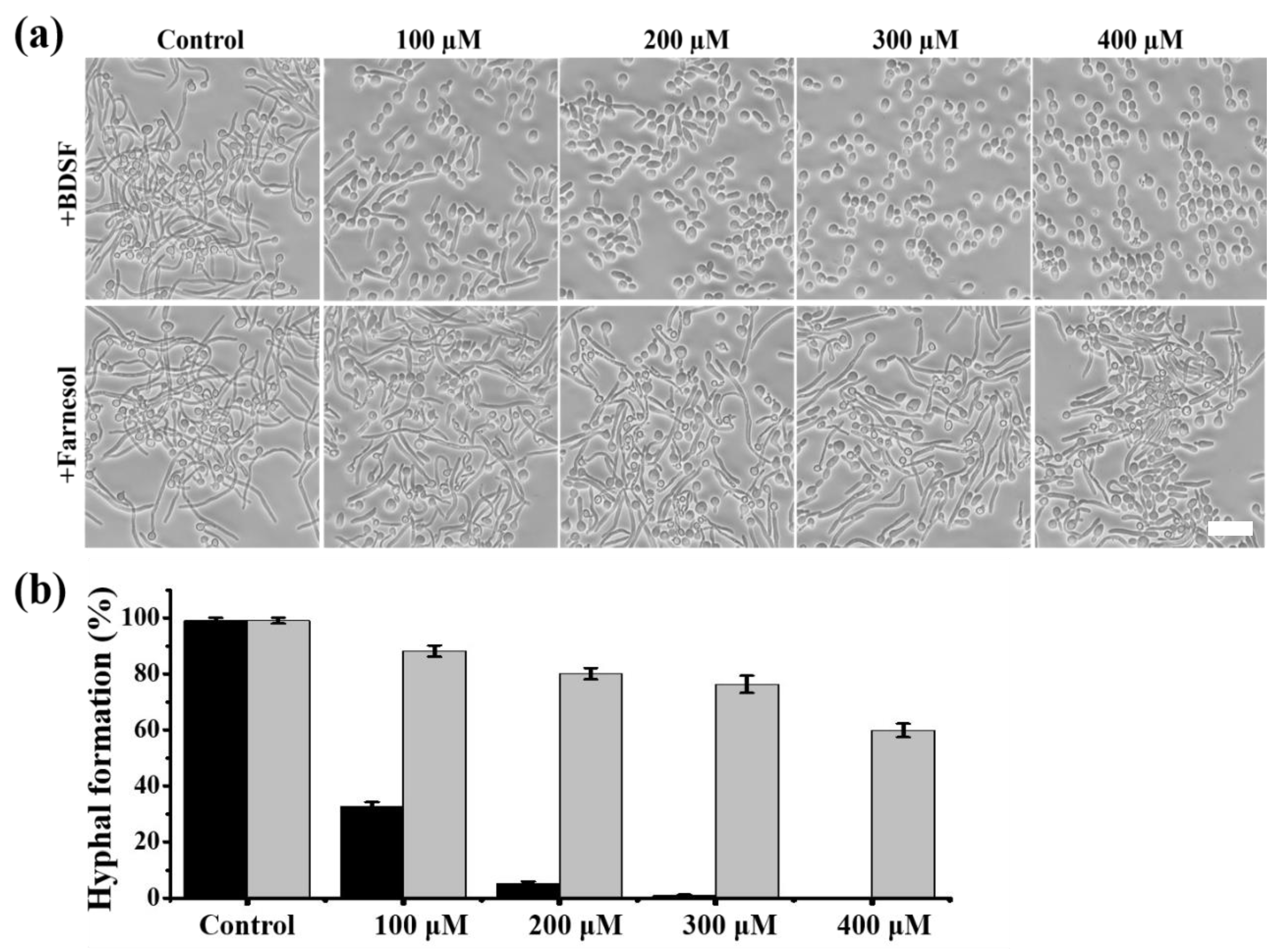
3.1. BDSF Inhibits Filament Formation in C. albicans

Our previous study indicated that C. albicans filament formation is more effectively inhibited by exogenous addition of BDSF than farnesol in glucose minimum medium (GMM) containing 20% filtered FBS or RPMI 1640 supplemented with 4-morpholinepropanesulfonic acid [32,33]. At present, C. albicans remains the main species of Candida bloodstream infections [28,34]. We therefore checked the inhibitory effect of BDSF on hyphal formation induced by the whole FBS. As shown in Figure 1, BDSF inhibits C. albicans hyphal growth in a dose-dependent manner and has a greater inhibitory activity than farnesol. The hyphal growth inhibition rate was increased from 67.3% to 94.9% when the concentration of BDSF increased from 100 μM to 200 μM. On the contrary, when the concentration of farnesol increased from 100 μM to 400 μM, the hyphal growth inhibition rate was increased from 11.8% to 40.2% (Figure 1b). These results indicate that BDSF possesses a superior ability of inhibiting hyphal growth.

3.2. Differential Gene Expression in C. albicans Treated with BDSF

To investigate the molecular mechanism by which BDSF inhibits filament formation, we next conducted cDNA microarray analysis to examine the altered gene expression profile of C. albicans treated with BDSF. The microarray data of this experiment are available from the Gene Expression Omnibus (GEO) database under accession number GSE137546. Comparison between BDSF- and ethanol-treated cells revealed that 305 genes were significantly affected on the expression level by BDSF treatment. These genes were classified according to their biological functions (http://www.candidagenome.org/). We found that these BDSF-modulated genes are associated with a variety of biological functions including hyphal formation, cell wall structure, adherence, antibiotic resistance, transportation, cell membrane permeabilization, ergosterol biosynthesis, glucose metabolism, and fatty acid metabolism. As shown in Figure 2, seven genes related to filament formation were differentially expressed. Among them, CHK2, orf19.5576, ERG3, and SFL2 are required for filamentous growth [35,36]. Downregulation of these genes may therefore block filament formation. UBI4 encodes an ubiquitin precursor and disruption of UBI4 promotes hyphae and pseudohyphae formation under certain circumstances [37,38]. SFL1 encodes a negative regulator of hyphal development. Thus, upregulation of UBI4 and SFL1 may inhibit filament formation. Currently, most commercial antifungals target the ergosterol biosynthesis pathway by, for example, blocking ergosterol synthesis or selectively binding to ergosterol to further disrupt membrane integrity [39]. ERG1, ERG3, and ERG11 encode enzymes necessary for ergosterol synthesis [40]. UPC2 encodes a Zn2-Cys6 transcript factor that upregulates ERG2 and ERG11 expression. The UPC2 disruption mutant shows reduced ergosterol level and high susceptibility to antifungals [41]. SUR2 encodes ceramide hydroxylase, which is needed for sphingolipid biosynthesis. Both ergosterol and sphingolipid are involved in membrane polarization and hyphal morphogenesis in C. albicans [42]. Therefore, downregulation of the genes of the sphingolipid and ergosterol biosynthetic pathway may result in cells maintaining in yeast form. BDSF treatment also resulted in the upregulation of several genes encoding cell cycle proteins, DNA repair enzymes, and heat shock proteins. This was also observed in C. albicans biofilm treated with farnesol [43].

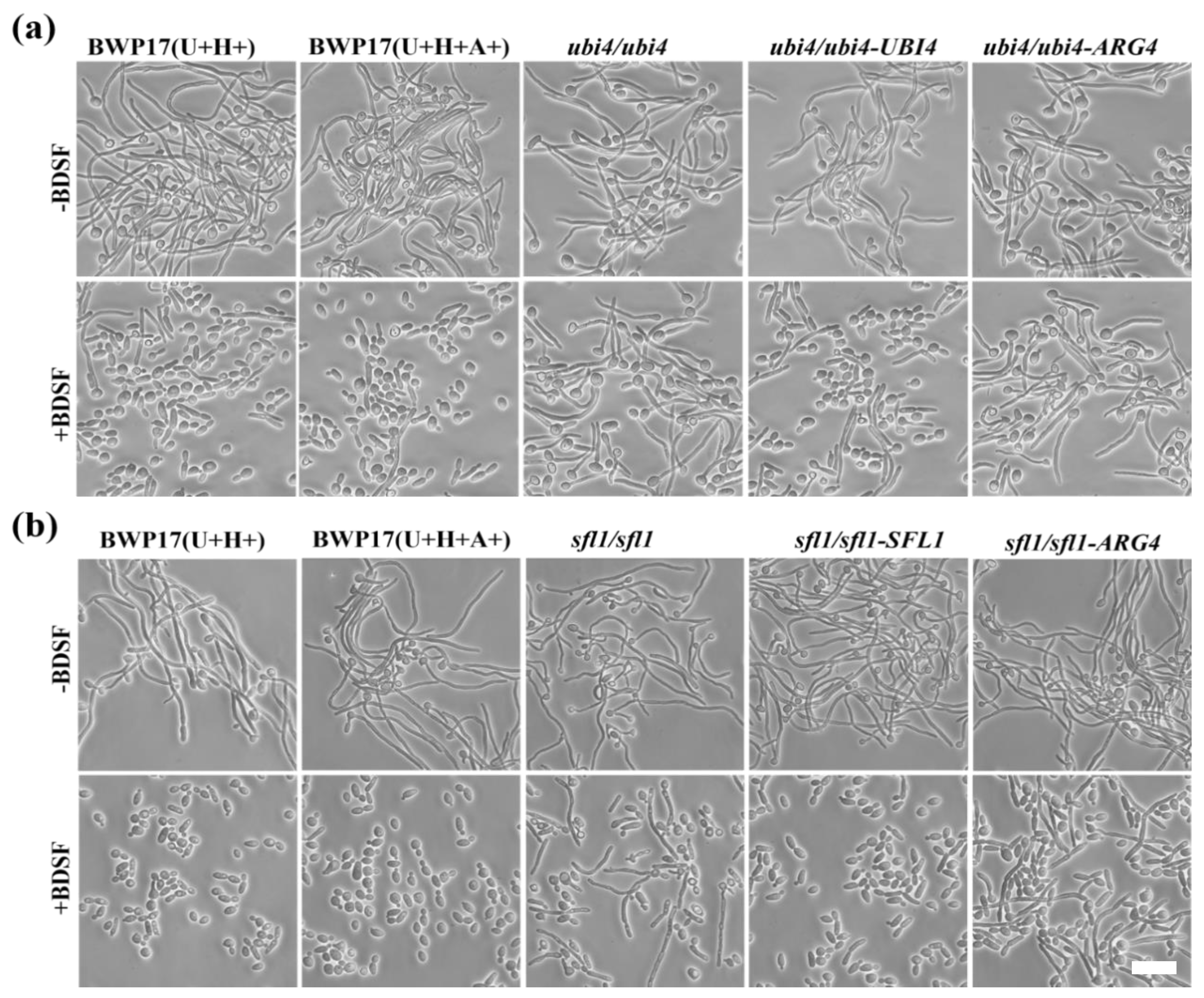
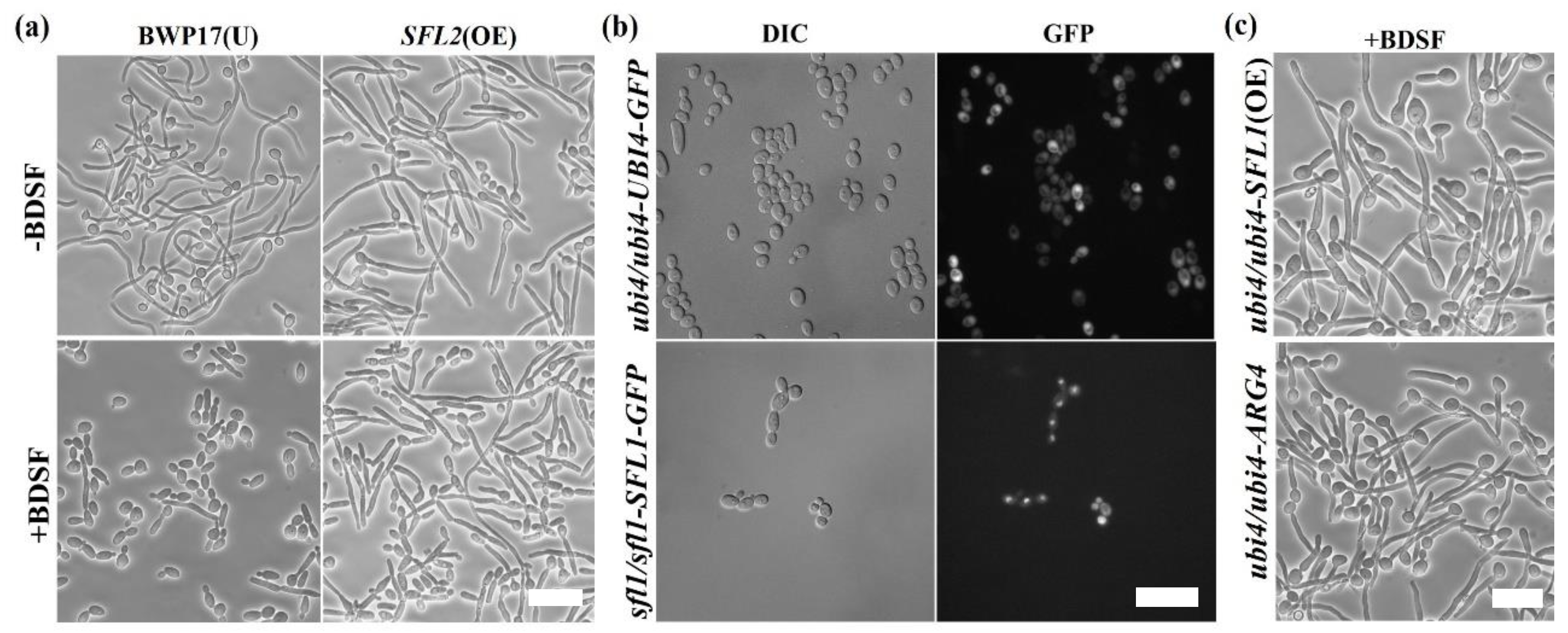
3.3. Screening for BDSF-Resistant Mutants

To verify the findings deduced from the microarray analysis, we chose genes involved in filament growth and regulation for further examination. We knocked out each of the genes to construct a mutant library. Each mutant strain was cultured in YPD medium and induced for hyphal growth with 5% FBS at 37 °C for 4 h in the presence or absence of 150 μM BDSF. Both ubi4∆/∆ and sfl1∆/∆ mutants exhibited insensitivity to BDSF and continued to produce hyphae in the presence of BDSF. In contrast, cells of wild-type control BWP17(U+H+) largely remained in yeast form under the same condition (Figure 3a,b). To further confirm that UBI4 and SFL1 are truly the genes required for BDSF resistance, wild-type UBI4 and SFL1 genes were introduced into the corresponding deletion mutants to generate rescue strains. Similar to the wild-type control BWP17(U+H+A+), both rescue strains (ubi4/ubi4-UBI4 and sfl1/sfl1-SFL1) regained the sensitivity to BDSF and failed to produce normal hyphae, while mutants transformed with the vector (ubi4/ubi4-ARG4 and sfl1/sfl1-ARG4) maintained the resistance to BDSF (Figure 3a,b).
In S. cerevisiae, both CaSFL1 and CaSFL2 can complement the loss of ScSFL1, indicating that these two genes perform similar functions in S. cerevisiae. However, CaSFL1 and CaSFL2 have antagonistic functions in C. albicans morphogenesis [21]. In support of this finding, we found that cells overexpressing SFL2 conferred resistance to BDSF similar to the SFL1 deletion cells under the same condition (Figure 4a). Previous studies indicated that Ubi4 is located in the cytoplasm and Sfl1 is located in the nucleus [20,44]. We further confirmed the results by examining the subcellular localization of GFP-tagged Ubi4 and Sfl1 (Figure 4b). Based on their subcellular localization, it is possible that SFL1 may act downstream of UBI4 to regulate filament formation. However, as shown in Figure 4c, overexpression of SFL1 in ubi4∆/∆ mutant failed to reduce the resistance of ubi4∆/∆ mutant to BDSF, suggesting that SFL1 acts in a pathway different from that of UBI4.

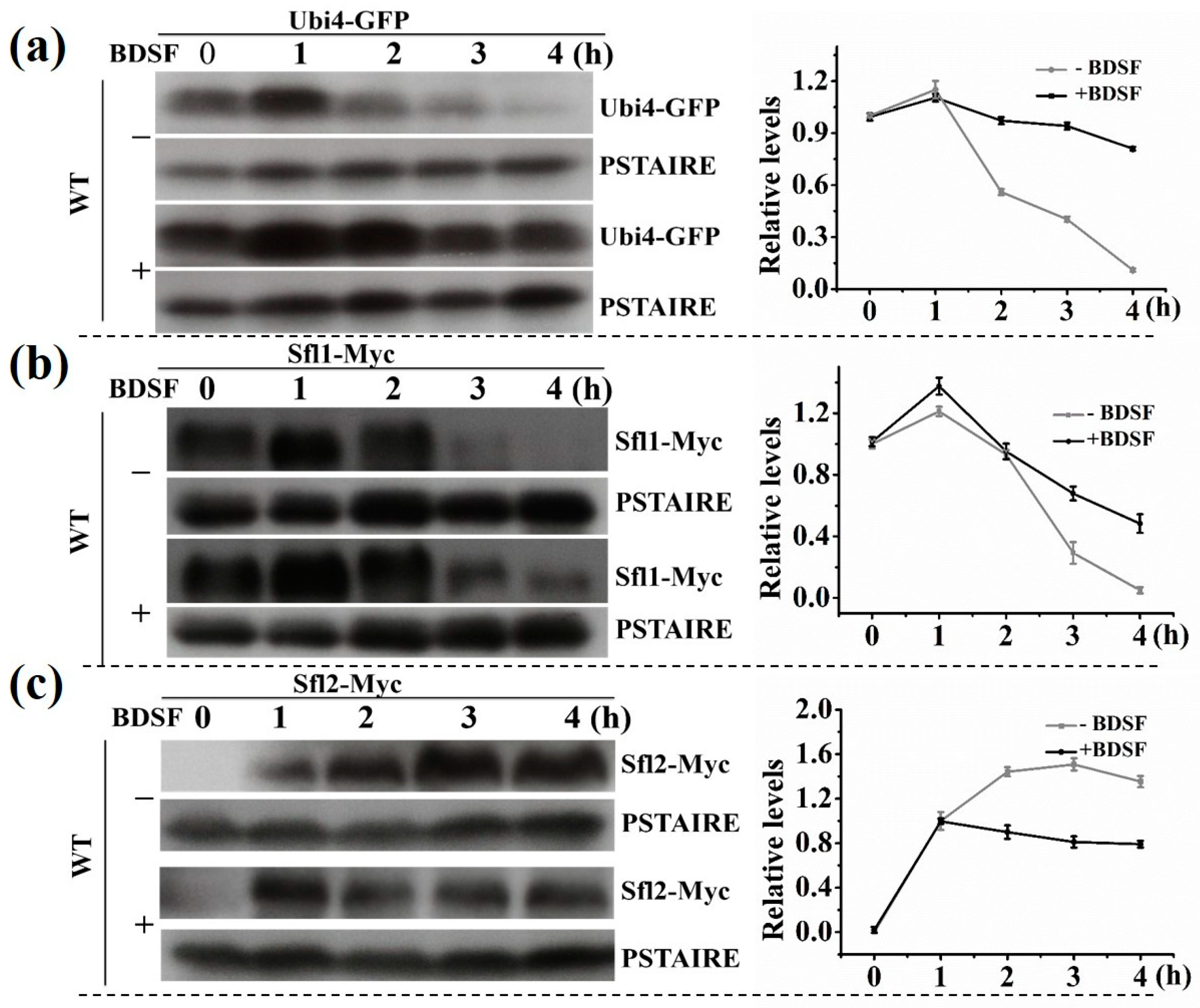
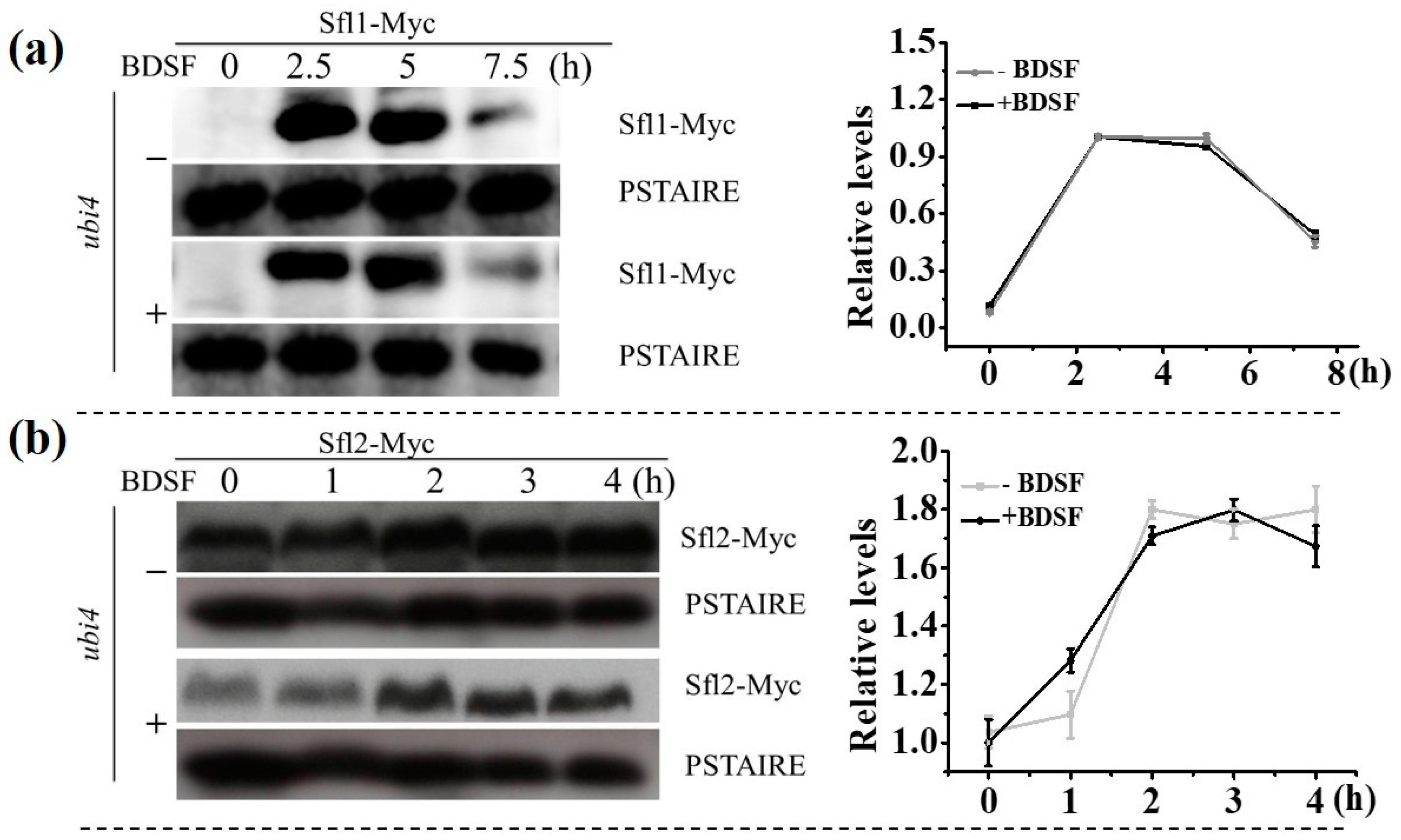
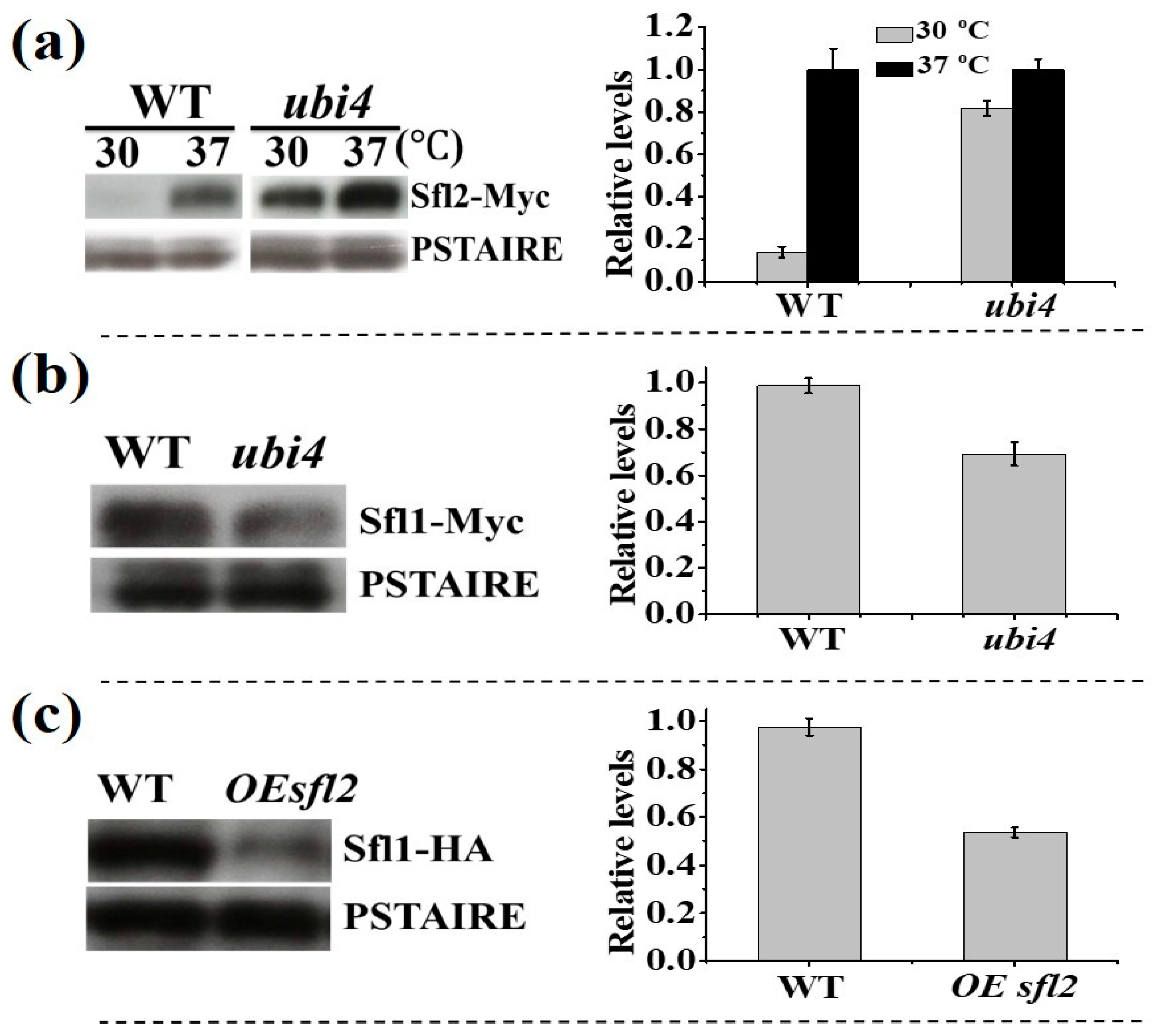
3.4. The Change in Expression Level of Proteins

According to the Candida genome database, Sfl1 is a transcription factor that represses hyphal morphogenesis. In contrast, Sfl2 is a transcription factor that is required for hyphal morphogenesis. UBI4 encodes an ubiquitin precursor that participates in the management of protein concentration [45]. To examine the protein level of Ubi4, Sfl1, and Sfl2 during hyphal growth, we tagged each of the proteins with GFP or Myc at the C terminus, respectively. Upon hyphal induction (without BDSF), the level of Ubi4 and Sfl1 slightly increased initially, then rapidly decreased following the extension of hyphal induction time (Figure 5a,b). Sfl2, as an activator of hyphal growth, was upregulated in this process (Figure 5c). In the presence of BDSF, the protein level of Ubi4 and Sfl1 decreased in a much slower rate; but the protein level of Sfl2 began to drop rapidly after an initial increase (Figure 5a–c).
In order to investigate the differential expression of SFL1 and SFL2 in ubi4∆/∆ mutant, both Sfl1 and Sfl2 in the mutant were tagged with Myc at the C terminus. Deletion of UBI4 showed very little effect on the protein expression pattern of Sfl1 (Figure 5b and Figure 6a) and Sfl2 (Figure 5c and Figure 6b) during the hyphal growth in the absence of BDSF. With the presence of BDSF, the protein level of Sfl1 decreased slightly faster in ubi4∆/∆ mutant cells (Figure 5b) than in wild-type cells (Figure 6a). However, in the presence of BDSF, the rapid decrease of Sfl2 protein amount seen in wild-type cells (Figure 5c) was mostly inhibited and remained in a high level (Figure 6b), indicating that Ubi4 may mediate the degradation of Sfl2.
To further confirm that Ubi4 is required for Sfl2 degradation, wildtype and ubi4∆/∆ mutant cells were cultured at 37 °C for 4 h to induce Sfl2 expression, then transferred to 30 °C and further cultured overnight. As shown in Figure 7a, Sfl2-Myc was detected in both strains when cells were grown at 37 °C. When the temperature was changed to 30 °C, Sfl2 protein could only be detected in the ubi4∆/∆ mutant. This result further indicated that Ubi4 is necessary for Sfl2 degradation. Christophe et al. found that Sfl1 and Sfl2 act as a morphology control center and antagonistically control C. albicans morphogenesis [21]. In contrast to Sfl2 whose protein level was higher in ubi4∆/∆ mutant cells than in wild-type cells (Figure 7a), We found that Sfl1 had lower protein level in ubi4∆/∆ mutant cells than in wild-type cells (Figure 7b). We also found that Sfl1 protein level was reduced when the cells overexpressed SFL2 (Figure 7c). Based on the above results, we hypothesized that, in wild-type cells under hyphal inducing condition, BDSF treatment helps to maintain Ubi4 and Sfl1 proteins at a high level, resulting in rapidly decrease of Sfl2 protein, hence inhibits hyphal morphogenesis.

4. Discussion

BDSF is an unsaturated fatty acid that is secreted by B. cenocepacia [32]. As a cross-kingdom signal molecule, BDSF regulates virulence of B. cenocepacia and activates the quorum-sensing system of Xanthomonas campestris. Furthermore, BDSF also suppresses the germination of C. albicans [13,32,33]. Compared to farnesol, BDSF is a more effective inhibitor of C. albicans hyphal morphogenesis. However, the mechanisms by which BDSF regulates hyphal formation remain to be elucidated. According to cDNA microarray analysis, a number of genes were differentially expressed when C. albicans cells were exposed to BDSF. We used gene knockout technology to confirm the functions of those genes that were involved in the BDSF response. We found that UBI4, SFL1, and SFL2 play a crucial role in the BDSF regulation of hyphal morphogenesis. Deletion of UBI4 completely abolished C. albicans response to BDSF, but the mutant cells regained sensitivity to BDSF when UBI4 was ectopically expressed. This phenomenon suggested that Ubi4 is required for BDSF to block hyphal formation. In support of this hypothesis, we found that Ubi4 protein level was upregulated upon BDSF treatment. Consistently, downregulation of UBI4 has been shown to promote filament formation [38]. The ubiquitin-mediated proteolytic degradation system has been known to implicate in the regulation of signal transduction networks that control filamentous growth [12]. For example, Lu et al. found that Ubr1-mediated degradation of Cup9 helps farnesol control hyphal initiation [12]. Atir-Lande and Roig et al. discovered that ubiquitin depletion results in expression level changes of genes associated with morphogenesis regulation [23,24].
Sfl1 and Sfl2 are transcription factors that act antagonistically to control morphogenesis in C. albicans [21]. SFL1 and SFL2 encode a transcriptional repressor and a transcriptional activator of hyphal formation, respectively [21]. C. albicans germ tube formation was inhibited under hyphal-inducing conditions when coincubated with B. cenocepacia, while in the sfl1 deficiency mutant the hyphal growth was normal [44]. In this work, we further confirmed that Sfl1 is needed for BDSF to inhibit C. albicans hyphal morphogenesis. In the presence of BDSF, C. albicans remained in yeast form even in the hyphal induction condition. Under yeast culture condition, Sfl1 had higher protein amount in wild-type than in the ubi4 mutant. In contrast, Sfl2 had lower protein amount in wild-type than in the ubi4 mutant. In addition, overexpression of SFL2 resulted in a reduction of Sfl1 protein level. Based on these results, we hypothesize that BDSF treatment may cause Sfl1 protein to remain at a relatively high level, and as a suppressor, Sfl1 may bind to other transcriptional activators of hyphal formation to inhibit hyphal formation.
Previous works indicated that Sfl1 and Sfl2 both localize to the nucleus in yeast and hyphal cells [16,20], while Ubi4 is found in the cytoplasm. Ubiquitin-mediated protein degradation plays a vital role in the morphological switch of C. albicans. The polyubiquitination-deficient mutant may cause an alteration in DNA arrangement and chromatin structure, further altering the expression of genes related to colony morphology, such as TUP1 and EFG1 [23]. Indeed, we found that the expression of genes involved in morphology control was changed in the ubi4 deletion mutant. In general, Sfl2 cannot be detected when cells were cultured at 30 °C [16], but it could be detected in the ubi4 mutant under the same condition. This result suggested that Ubi4 is needed for Sfl2 degradation.

5. Conclusions

In summary, DNA microarray analysis was used to identify the differentially-expressed genes between cells growing in hyphal-inducing condition in the absence or presence of BDSF. Subsequent mutant screens confirmed that three characterized genes were involved in the BDSF regulation of filament formation. With the presence of BDSF, two repressors of filamentous growth (Ubi4 and Sfl1) were maintained at a relative high level, and as a result, the morphological transition from yeast to hyphae was blocked. In addition, Ubi4 is the protein required for the degradation of Sfl2. High level of Ubi4 protein contributed to the degradation of Sfl2 (an activator of filamentous growth), thereby maintaining the C. albicans cells in the yeast form.

Author Contributions

G.Z. and L.W. conceived and designed the experiments. D.Y. and G.Z. performed the experiments. F.Y.C. provided general technique support. D.Y., G.Z., L.W., Y.H., L.W., F.Y.C, Z.Y., Q.G., Y.Z., and Y.W. analyzed the data. D.Y., G.Z. and L.W. wrote the paper. All authors have read and agreed to the published version of the manuscript.

Funding

This work was financially supported by the National Key Research and Development Program of China (2017YFA0205301, 2017YFA0205302), the Program for Changjiang Scholars and Innovative Research Team in University (IRT_15R37), the Key Research and Development Program of Jiangsu (BE2018732), the Natural Science Key Fund for Colleges and Universities in Jiangsu Province (17KJA430011), the Priority Academic Program Development of Jiangsu Higher Education Institutions (YX03002), the Natural Science Foundation of the Jiangsu Higher Education Institutions (19KJB150030, 19KJB150031), and the open research fund of Jiangsu Key Laboratory for Biosensors (51204080).

Conflicts of Interest

The authors declare no conflict of interest. The funders had no role in the design of the study; in the collection, analyses, or interpretation of data; in the writing of the manuscript, or in the decision to publish the results.

References

  1. Shapiro, R.S.; Robbins, N.; Cowen, L.E. Regulatory circuitry governing fungal development, drug resistance, and disease. Microbiol. Mol. Biol. R 2011, 75, 213–267. [Google Scholar] [CrossRef] [PubMed] [Green Version]
  2. Wang, Y. Fungal adenylyl cyclase acts as a signal sensor and integrator and plays a central role in interaction with bacteria. PLoS Pathog. 2013, 9, e1003612. [Google Scholar] [CrossRef] [PubMed] [Green Version]
  3. Mallick, E.M.; Bennett, R.J. Sensing of the microbial neighborhood by Candida albicans. PLoS Pathog. 2013, 9, e1003661. [Google Scholar] [CrossRef] [PubMed] [Green Version]
  4. Yang, D.L.; Hu, Y.L.; Yin, Z.X.; Zeng, G.S.; Li, D.; Zhang, Y.Q.; Xu, Z.H.; Guan, X.M.; Weng, L.X.; Wang, L.H. Cis-2-dodecenoic acid mediates its synergistic effect with triazoles by interfering with efflux pumps in fluconazole-resistant Candida albicans. Biomed. Environ. Sci. 2019, 32, 199–209. [Google Scholar] [CrossRef]
  5. Santolaya, M.E.; Thompson, L.; Benadof, D.; Tapia, C.; Legarraga, P.; Cortés, C.; Rabello, M.; Valenzuela, R.; Rojas, P.; Rabagliati, R.; et al. A prospective, multi-center study of Candida bloodstream infections in Chile. PLoS ONE 2019, 14, e0212924. [Google Scholar] [CrossRef] [Green Version]
  6. Pfaller, M.A.; Diekema, D.J. Epidemiology of invasive Candidiasis: A persistent public health problem. Clin. Microbiol. Rev. 2007, 20, 133–163. [Google Scholar] [CrossRef] [Green Version]
  7. Calderone, R.A.; Fonzi, W.A. Virulence factors of Candida albicans. Trends Microbiol. 2001, 9, 327–335. [Google Scholar] [CrossRef]
  8. Yang, D.; Lv, X.; Xue, L.; Yang, N.; Hu, Y.; Weng, L.; Fu, N.; Wang, L.; Dong, X. A lipase-responsive antifungal nanoplatform for synergistic photodynamic/photothermal/pharmaco-therapy of azole-resistant Candida albicans infections. Chem. Commun. 2019, 55, 15145–15148. [Google Scholar] [CrossRef]
  9. Whiteway, M.; Bachewich, C. Morphogenesis in Candida albicans. Annu. Rev. Microbiol. 2007, 61, 529–553. [Google Scholar] [CrossRef] [Green Version]
  10. Sudbery, P.E. Growth of Candida albicans hyphae. Nat. Rev. Microbiol. 2011, 9, 737–748. [Google Scholar] [CrossRef]
  11. Hogan, D.A.; Vik, Å.; Kolter, R.A. Pseudomonas aeruginosa quorum-sensing molecule influences Candida albicans morphology. Mol. Microbiol. 2004, 54, 1212–1223. [Google Scholar] [CrossRef] [PubMed]
  12. Lu, Y.; Su, C.; Unoje, O.; Liu, H. Quorum sensing controls hyphal initiation in Candida albicans through Ubr1-mediated protein degradation. Proc. Natl. Acad. Sci. USA 2014, 111, 1975–1980. [Google Scholar] [CrossRef] [Green Version]
  13. Wang, L.H.; He, Y.; Gao, Y.; Wu, J.E.; Dong, Y.H.; He, C.; Wang, S.X.; Weng, L.X.; Xu, J.L.; Tay, L.; et al. A bacterial cell-cell communication signal with cross-kingdom structural analogues. Mol. Microbiol. 2004, 51, 903–912. [Google Scholar] [CrossRef] [PubMed]
  14. Tian, J.; Weng, L.X.; Zhang, Y.Q.; Wang, L.H. BDSF inhibits Candida albicans adherence to urinary catheters. Microb. Pathog. 2013, 64, 33–38. [Google Scholar] [CrossRef] [PubMed]
  15. Yang, D.L.; Zhang, Y.Q.; Hu, Y.L.; Weng, L.X.; Zeng, G.S.; Wang, L.H. Protective effects of cis-2-dodecenoic acid in an experimental mouse model of vaginal candidiasis. Biomed. Environ. Sci. 2018, 31, 816–828. [Google Scholar] [CrossRef] [PubMed]
  16. Song, W.; Wang, H.; Chen, J. Candida albicans Sfl2, a temperature-induced transcriptional regulator, is required for virulence in a murine gastrointestinal infection model. FEMS Yeast Res. 2011, 11, 209–222. [Google Scholar] [CrossRef] [PubMed] [Green Version]
  17. Lu, Y.; Su, C.; Liu, H. Candida albicans hyphal initiation and elongation. Trends Microbiol. 2014, 22, 707–714. [Google Scholar] [CrossRef] [PubMed] [Green Version]
  18. Zhang, T.; Li, D.; Li, W.; Wang, Y.; Sang, J. CaSfl1 plays a dual role in transcriptional regulation in Candida albicans. Chin. Sci. Bull. 2008, 53, 2624–2631. [Google Scholar] [CrossRef] [Green Version]
  19. Li, Y.; Su, C.; Mao, X.; Cao, F.; Chen, J. Roles of Candida albicans Sfl1 in hyphal development. Eukaryot. Cell 2007, 6, 2112–2121. [Google Scholar] [CrossRef] [Green Version]
  20. Bauer, J.; Wendland, J. Candida albicans Sfl1 suppresses flocculation and filamentation. Eukaryot. Cell 2007, 6, 1736–1744. [Google Scholar] [CrossRef] [Green Version]
  21. Znaidi, S.; Nesseir, A.; Chauvel, M.; Rossignol, T.; d’Enfert, C. A comprehensive functional portrait of two heat shock factor-type transcriptional regulators involved in Candida albicans morphogenesis and virulence. PLoS Pathog. 2013, 9, e1003519. [Google Scholar] [CrossRef] [PubMed] [Green Version]
  22. Weathington, N.M.; Mallampalli, R.K. Emerging therapies targeting the ubiquitin proteasome system in cancer. J. Clin. Investig. 2014, 124, 6–12. [Google Scholar] [CrossRef] [PubMed] [Green Version]
  23. Roig, P.; Gozalbo, D. Depletion of polyubiquitin encoded by the UBI4 gene confers pleiotropic phenotype to Candida albicans cells. Fungal Genet. Biol. 2003, 39, 70–81. [Google Scholar] [CrossRef]
  24. Atir-Lande, A.; Gildor, T.; Kornitzer, D. Role for the SCF(CDC4) ubiquitin ligase in Candida albicans morphogenesis. Mol. Biol. Cell 2005, 16, 2772–2785. [Google Scholar] [CrossRef] [PubMed] [Green Version]
  25. Wang, Y.; Cao, Y.Y.; Jia, X.M.; Cao, Y.B.; Gao, P.H.; Fu, X.P.; Ying, K.; Chen, W.S.; Jiang, Y.Y. Cap1p is involved in multiple pathways of oxidative stress response in Candida albicans. Free Radic. Biol. Med. 2006, 40, 1201–1209. [Google Scholar] [CrossRef] [PubMed]
  26. Butler, D.K.; All, O.; Goffena, J.; Loveless, T.; Wilson, T.; Toenjes, K.A. The GRR1 gene of Candida albicans is involved in the negative control of pseudohyphal morphogenesis. Fungal Genet. Biol. 2006, 43, 573–582. [Google Scholar] [CrossRef]
  27. Wang, L.H.; Zhang, L.H. Inhibitors of Yeast Filamentous Growth and Method of Their Manufacture. U.S. Patent 7915313B2, 29 March 2011. [Google Scholar]
  28. Zeng, G.; Wang, Y.M.; Chan, F.Y.; Wang, Y. One-step targeted gene deletion in Candida albicans haploids. Nat. Protoc. 2014, 9, 464–473. [Google Scholar] [CrossRef]
  29. Gao, J.; Wang, H.; Wong, A.H.; Zeng, G.; Huang, Z.; Wang, Y.; Sang, J.; Wang, Y. Regulation of Rfa2 phosphorylation in response to genotoxic stress in Candida albicans. Mol. Microbiol. 2014, 94, 141–155. [Google Scholar] [CrossRef]
  30. Liu, Q.; Han, Q.; Wang, N.; Yao, G.; Zeng, G.; Wang, Y.; Huang, Z.; Sang, J.; Wang, Y. Tpd3-Pph21 phosphatase plays a direct role in Sep7 dephosphorylation in Candida albicans. Mol. Microbiol. 2016, 101, 109–121. [Google Scholar] [CrossRef]
  31. Zeng, G.; Wang, Y.M.; Wang, Y. Cdc28-Cln3 phosphorylation of Sla1 regulates actin patch dynamics in different modes of fungal growth. Mol. Biol. Cell 2012, 23, 3485–3497. [Google Scholar] [CrossRef]
  32. Boon, C.; Deng, Y.; Wang, L.; He, Y.; Xu, J.; Fan, Y.; Pan, S.Q.; Zhang, L. A novel DSF-like signal from Burkholderia cenocepacia interferes with Candida albicans morphological transition. ISME J. 2007, 2, 27–36. [Google Scholar] [CrossRef] [PubMed]
  33. Zhang, Y.; Cai, C.; Yang, Y.; Weng, L.; Wang, L. Blocking of Candida albicans biofilm formation by cis-2-dodecenoic acid and trans-2-dodecenoic acid. J. Med. Microbiol. 2011, 60, 1643–1650. [Google Scholar] [CrossRef] [PubMed]
  34. Moran, C.; Grussemeyer, C.A.; Spalding, J.R.; Benjamin, D.K.; Reed, S.D. Candida albicans and non-albicans bloodstream infections in adult and pediatric patients: Comparison of mortality and costs. Pediatr. Infect. Dis. J. 2009, 28, 433–435. [Google Scholar] [CrossRef] [PubMed] [Green Version]
  35. Yamada-Okabe, T.; Mio, T.; Ono, N.; Kashima, Y.; Matsui, M.; Arisawa, M.; Yamada-Okabe, H. Roles of three histidine kinase genes in hyphal development and virulence of the pathogenic fungus Candida albicans. J. Bacteriol. 1999, 181, 7243–7247. [Google Scholar] [CrossRef] [PubMed] [Green Version]
  36. Uhl, M.A.; Biery, M.; Craig, N.; Johnson, A.D. Haploinsufficiency-based large-scale forward genetic analysis of filamentous growth in the diploid human fungal pathogen C. albicans. EMBO J. 2003, 22, 2668–2678. [Google Scholar] [CrossRef] [PubMed] [Green Version]
  37. Roig, P.; Gozalbo, D. Candida albicans UBI3 and UBI4 promoter regions confer differential regulation of invertase production to Saccharomyces cerevisiae cells in response to stress. Int. Microbiol. 2002, 5, 33–36. [Google Scholar] [CrossRef] [PubMed] [Green Version]
  38. Leach, M.D.; Stead, D.A.; Argo, E.; MacCallum, D.M.; Brown, A.J.P. Molecular and proteomic analyses highlight the importance of ubiquitination for the stress resistance, metabolic adaptation, morphogenetic regulation and virulence of Candida albicans. Mol. Microbiol. 2011, 79, 1574–1593. [Google Scholar] [CrossRef] [Green Version]
  39. Pierce, C.G.; Lopez-Ribot, J.L. Candidiasis drug discovery and development: New approaches targeting virulence for discovering and identifying new drugs. Expert Opin. Drug. Discov. 2013, 8, 1117–1126. [Google Scholar] [CrossRef] [Green Version]
  40. Onyewu, C.; Blankenship, J.R.; Del Poeta, M.; Heitman, J. Ergosterol biosynthesis inhibitors become fungicidal when combined with calcineurin inhibitors against Candida albicans, Candida glabrata, and Candida krusei. Antimicrob. Agents Chemother. 2003, 47, 956–964. [Google Scholar] [CrossRef] [Green Version]
  41. Silver, P.M.; Oliver, B.G.; White, T.C. Role of Candida albicans transcription factor Upc2p in drug resistance and sterol metabolism. Eukaryot. Cell 2004, 3, 1391–1397. [Google Scholar] [CrossRef] [Green Version]
  42. Martin, S.W.; Konopka, J.B. Lipid raft polarization contributes to hyphal growth in Candida albicans. Eukaryot. Cell 2004, 3, 675–684. [Google Scholar] [CrossRef] [PubMed] [Green Version]
  43. Cao, Y.Y.; Cao, Y.B.; Xu, Z.; Ying, K.; Li, Y.; Xie, Y.; Zhu, Z.Y.; Chen, W.S.; Jiang, Y.Y. cDNA microarray analysis of differential gene expression in Candida albicans biofilm exposed to farnesol. Antimicrob. Agents Chemother. 2005, 49, 584–589. [Google Scholar] [CrossRef] [PubMed] [Green Version]
  44. Hall, R.A.; Turner, K.J.; Chaloupka, J.; Cottier, F.; De Sordi, L.; Sanglard, D.; Levin, L.R.; Buck, J.; Mühlschlegel, F.A. The quorum-sensing molecules farnesol/homoserine lactone and dodecanol operate via distinct modes of action in Candida albicans. Eukaryot. Cell 2011, 10, 1034–1042. [Google Scholar] [CrossRef] [PubMed] [Green Version]
  45. Kornitzer, D.; Ciechanover, A. Modes of regulation of ubiquitin-mediated protein degradation. J. Cell Physiol. 2000, 182, 1–11. [Google Scholar] [CrossRef]
Figure 1. (a) Hyphal formation was induced in YPD medium (1% Difco yeast extract, 2% Difco peptone, and 2% dextrose) + 5% fetal bovine serum (FBS) with different concentrations of Cis-2-dodecenoic acid (BDSF) or farnesol at 37 ℃ for 4 h. (b) Effects of different concentrations of BDSF (black) or farnesol (grey) on C. albicans hyphal formation. Bar, 15 µm.
Microorganisms 08 00075 g001
Figure 2. Functional classification of C. albicans genes regulated by BDSF (150 μM) treatment.
Microorganisms 08 00075 g002
Figure 3. (a) ubi4∆/∆ and (b) sfl1∆/∆ mutants exhibited BDSF resistance. Cells were cultured in YPD medium and induced for hyphal formation with 5% FBS at 37 °C for 4 h in the presence or absence of 150 μM BDSF. Bar, 15 µm.
Microorganisms 08 00075 g003
Figure 4. (a) Overexpression (OE) of SFL2 leads to BDSF resistance. (b) Subcellular localization of SFL1-GFP and UBI4-GFP in C. albicans cells. (c) Overexpression of SFL1 in ubi4∆/∆ mutant remained insensitive to BDSF. Hyphal induction was performed with the addition of 5% FBS and maintained at 37 °C for 4 h in the presence or absence of 150 μM BDSF. Bars, 15 µm.
Microorganisms 08 00075 g004
Figure 5. Hyphal growth of wild-type (WT) cells carrying (a) Ubi4-GFP, (b) Sfl1-Myc, and (c) Sfl2-Myc was induced with 5% FBS at 37 °C in the presence or absence of 150 μM BDSF. Samples were taken at 1 h intervals for Western blot analysis (left panels). Relative protein expression levels (right panels) were analyzed using ImageJ software. Normalizations were performed against the corresponding protein signal at 0 h (a,b) or 1 h (c).
Microorganisms 08 00075 g005
Figure 6. ubi4 mutant cells expressing (a) Sfl1-Myc or (b) Sfl2-Myc were induced for hyphal growth with 5% FBS at 37 °C in the presence or absence of 150 μM BDSF. Samples were taken for Western blot analysis at the time points as indicated (left panels). Relative protein expression levels (right panels) were analyzed using ImageJ software. Normalizations were performed against the corresponding protein signal at 2.5 h (a) and 0 h (b), respectively.
Microorganisms 08 00075 g006
Figure 7. (a) WT and ubi4 mutant cells expressing Sfl2-Myc were first grown at 37 °C in GMM medium (2% glucose and 0.67% Difco yeast nitrogen base) for 4 h, then shifted to 30 ℃ and cultured overnight. (b) WT and ubi4 mutant cells expressing Sfl1-Myc were cultured overnight in GMM at 30 °C. (c) WT and SFL2 overexpression cells expressing Sfl1-HA were cultured at 30 °C overnight. Right panels show corresponding protein relative expression level. Protein bands on Western blot (left panels) were analyzed using ImageJ software. Normalizations were performed against the corresponding protein signal at 37 °C (a) or in wildtype cells (b,c).
Microorganisms 08 00075 g007
Table 1. C. albicans strains used in this study.
StrainRelevant GenotypeSource
BWP17ura3::imm434/ura3::imm434 his1::hisG/his1::hisG arg4::hisG/arg4::hisGWY lab
BWP17(A+)ARG4WY lab
BWP17(U+)URA3WY lab
BWP17(U+H+)URA3, HIS1WY lab
BWP17(U+A+)URA3, ARG4WY lab
BWP17(H+A+)HIS1, ARG4WY lab
BWP17(U+H+A+)HIS1, ARG4, URA3WY lab
WLX 11ubi4Δ::HIS1/ubi4Δ::URA3This study
WLX 12ubi4Δ::HIS1/ubi4Δ::URA3, ARG4This study
WLX 13ubi4Δ::HIS1/ubi4Δ::URA3, UBI4-GFP-ARG4This study
WLX 19sfl1Δ::HIS1/sfl1Δ::URA3This study
WLX 20sfl1Δ::HIS1/sfl1Δ::URA3, ARG4This study
WLX 21sfl1Δ::HIS1/sfl1Δ::URA3, SFL1-GFP-ARG4This study
WLX 60SFL1-HA-ARG4, SFL2-Myc (overexpression)-URA3This study
WLX 67SFL1-HA-ARG4This study
WLX 68sfl1Δ::HIS1/SFL1-Myc-ARG4This study
WLX 70ubi4Δ::HIS1/ubi41Δ::URA3, SFL1-Myc (overexpression)-ARG4This study
WLX 71ubi4Δ::HIS1/ubi41Δ::URA3, SFL2-Myc-ARG4This study
WLX 72SFL2-Myc::ARG4This study

