Article

Methylcellulose Hydrogel with Melissa officinalis Essential Oil as a Potential Treatment for Oral Candidiasis

1
Microbiology at Interfaces, Faculty of Science and Engineering, Manchester Metropolitan University, Manchester M1 5GD, UK
2
Department of Materials, School of Natural Sciences, The University of Manchester, Manchester M13 9PL, UK
3
School of Dentistry, School of Dentistry, Cardiff University, Cardiff CF14 4XY, UK
*
Author to whom correspondence should be addressed.
Microorganisms 2020, 8(2), 215; https://doi.org/10.3390/microorganisms8020215
Submission received: 24 January 2020 / Accepted: 5 February 2020 / Published: 6 February 2020
(This article belongs to the Special Issue Oral Biofilms)

Abstract

:
Candida spp. are the most prevalent fungi of the human microbiota and are opportunistic pathogens that can cause oral candidiasis. Management of such infections is limited due to the low number of antifungal drugs available, their relatively high toxicity and the emergence of antifungal resistance. Therefore, much interest in the antimicrobial potential of natural compounds has recently been evident. The use of hydrogels in the delivery of biocides has been explored due to their biocompatibility, ease with drug encapsulation, and due to their potential to confer mechanical and structural properties similar to biological tissue. Methylcellulose hydrogels (10% (w/v)) with 1% (v/v) and 2% (v/v) Melissa officinalis oil were synthesised. The rheological properties and gelation time of the hydrogels were evaluated. Antimicrobial action, the antifungal potential and ability to displace Candida were determined. Rheological tests revealed that the hydrogel jellified in three minutes at 37 °C. Loaded hydrogels successfully inhibited Candida albicans growth as evident by zone of inhibition and time-kill assays. A significant reduction in retained C. albicans was demonstrated with the hydrogel at 2% Melissa officinalis concentration. This work demonstrated that an essential oil-loaded hydrogel had the potential to provide a novel antimicrobial therapy for the treatment of oral candidiasis.

1. Introduction

Candida albicans is a commensal fungus found in 30–50% of healthy humans [1]. However, changes to the oral environment can lead to one of the most common human fungal infection, oral candidiasis. Treatments for oral candidiasis often involves administration of topical or systemic antifungals, and denture cleansers in case of denture stomatitis [2].
Compared to antibiotics, commercially available antifungals are limited and development without toxicity is complicated due to the eukaryotic nature of Candida [2]. The rise of resistance of Candida to clinically used antifungals has led to great interest in the antifungal properties of natural compounds [3].
Essential oils are products of aromatic plants and mainly composed of terpenes and terpenoids and other molecules [4]. Being hydrophobic, they interact with the cell membrane changing its permeability and causing leaching of intracellular components (e.g., radicals, proteins, calcium ions) and the inactivation of enzymatic mechanisms [5]. Melissa officinalis is a plant already used to give fragrance to food and beverage products [6]. Furthermore, it has been utilised as a medical plant for different therapeutic effects (e.g., energizer, anticonvulsant, tranquilizer, and to aid digestion) [7]. Besides these functions, it has been demonstrated to have both antimicrobial and antioxidant properties against periodontal pathogens [8,9] and Candida both in planktonic and biofilm form [10,11].
Hydrogels are 3D-networks obtained from natural and/or synthetic polymers that once cross-linked form an insoluble structure [12]. The property of hydrogels is their ability to absorb and retain a significant amount of water (>90%) without dissolving. Swollen hydrogels have some properties in common with living tissues (e.g., consistency, low interfacial tension, water content) and biocompatibility [13]. Therefore, they have been widely used in tissue engineering and regenerative medicine, mainly as drug and/or cell delivery systems, as pre-formed or injectable cell scaffolds [12].
Methylcellulose is widely used in food packaging. Formulations of methylcellulose films containing ginja cherry extract [14], olive leaf extract [15], clove bud (Syzygium aromaticum) and oregano (Origanum vulgare) essential oils [16] that demonstrate antimicrobial properties have been synthesised. The use of methylcellulose as a drug delivery system, has potential since it is a bioadhesive polymer that shows adhesion to mucosal surfaces [17]. This characteristic has the potential to prolong the retention of the drug delivery system at the site of clinical application (oral mucosa and/or vaginal applications) [18].
The aim of this study was to develop a methylcellulose hydrogel in combination with Melissa officinalis essential oil as treatment for oral candidiasis.

2. Materials and Methods

2.1. Hydrogel Preparation

Methylcellulose (M7140, Sigma-Aldrich, Gillingham, UK) was used to prepare hydrogels (10% (w/v)) containing 1% and 2% (v/v) Melissa officinalis (Essential Oils Direct Ltd., Oldham, UK) essential oil. Melissa officinalis (Essential Oils Direct Ltd., Oldham, UK) was used as in a previous study by Serra et al. [11]. Sterile distilled water, with or without 1% or 2% (v/v) Melissa officinalis and Tween 80 in equal amounts, was held at 80 °C for 10 min. Methylcellulose (10% (w/v)) was then gently added into the solvent. Once the powder was fully wet, the temperature was reduced by placing the solution on ice to form a clear viscous solution which was vortexed for 30 s until the powder was solubilised and then incubated overnight at 4 °C shaking at 200 rpm. Prior to use, the solution was vortexed for 1 min and equilibrated at room temperature for 1h to allow air bubble dispersion. The equilibrated solution was transferred to a Petri dish and incubated at 37 °C until a stable hydrogel was formed (within 30 min). Discs of the hydrogel were cut with a sterile cork-borer at different sizes (0.75 mm, 10 mm or 11.25 mm in diameter).

2.2. Rheology

The rheological characterisation was performed on a Discovery Hybrid Rheometer DHR-2, (TA Instruments, DE) equipped with a 20 mm diameter plate and a solvent trap to minimise evaporation. Hydrogel solution (180 µL) was placed on the rheometer and the rheometer head was lowered to the desired gap height of 500 µm. Hydrogel gelation was studied under time sweep at 37 °C, while hydrogel viscoelasticity was analysed under strain and frequency sweep after keeping the hydrogel solution at 37 °C for eight minutes, with a gap size of 500 µm. Strain, frequency and time sweeps were carried out respectively using the following parameters: Equilibrium time of 8, 8 and 0 min; Frequency of 1 Hz, 0.01–100 Hz, 1 Hz; % Strain 0.1–100%, 1%, 1% and running time of 10 min for time sweep tests (n = 3) [19].

2.3. Melissa Officinalis Composition

Prior to evaluating the release of Melissa officinalis from the methylcellulose hydrogels, the composition of the essential oil was analysed by gas chromatography (Agilent Technologies 7890BUK) coupled to a mass spectrometer (Agilent Technologies 5977B, UK). The gas chromatograph was equipped with a capillary column Agilent J&W HP-5ms (30 m × 0.25 mm, 0.25 µm film thickness) (Agilent Technologies, UK). The method was adapted from that previously reported by Rajkowska et al. (2016) [20].
The initial temperature was set to 50 °C and held for 3 min, followed by a linear increase up to 300 °C (40 °C/min) and held for 2 min. A 5:1 split ratio with an injection volume of 2 μL was set. Inlet and transfer line temperatures were kept at 275 °C and 300 °C, respectively. Helium, CP grade, was used as a carrier gas at a flow rate of 1 mL/min. Quadrupole temperature was fixed to 150 °C. The mass spectra were obtained by scanning a range of masses from 45 to 550 atomic mass unit (AMU). The ion source temperature was 230 °C and the ionisation was obtained by electron impact at 70 eV. All samples were prepared in dichloromethane (DCM) and the composition of the oil was analysed by comparing the mass spectra with those of the computer library (NIST14 MS Search library).
Once the compounds were identified, abundance was determined using standard curves of each compound. Briefly, analytical standards of citronellol, citronellal, geraniol, and linalool (Sigma-Aldrich, UK) were used in double serial dilutions of each compound from 20 ppm to 2.5 ppm were prepared in DCM and analysed by GC-MS. The correlation between peak areas and concentrations was obtained and used to evaluate the abundance of each compound.

2.4. Melissa Officinalis Release

Ten percent (10% (w/v)) methylcellulose hydrogels containing 1% and 2% (v/v) Melissa officinalis essential oil were prepared. Once gellified at 37 °C, 1 g of hydrogel was cut with a 11.25 mm diameter sterile cork-borer and incubated in 20mL distilled water in a sterile glass universal at 37 °C under shaking conditions at 150 rpm (New Brunswick™ I26, Eppendorf UK Limited, Stevenage, UK). Two millilitres of liquid were collected at 1, 2, 3, 4, 5, 6, 7, 8, 24, 30 and 48 h and replaced by 2 mL of distilled water, to keep the volume constant throughout the experiment. On the same day the samples were used for GCMS analysis.
To evaluate the Melissa officinalis oil release, 1 mL of each liquid sample was added to 5 mL DCM and vortexed briefly before incubation overnight at 4 °C with shaking at 200 rpm to disperse the Melissa officinalis oil into DCM. The sample was centrifuged at 3000rpm (SIGMA 3-16 Centrifuge, Sigma Centrifuges, UK) for 5 min and the DCM, containing the essential oil, was collected in a 1 mL glass or plastic vial and analysed by GC-MS.
Controls included the contentment of the hydrogel prior incubation. To extract the oil within the hydrogel, 1 g of hydrogel was placed into 10mL DCM, vortexed briefly and incubated overnight at 4 °C with shaking at 200 rpm. After incubation, the suspension was centrifuged at 3000 rpm for 10 min and the DCM was collected in a vial and run in the GC-MS. All experiments were completed in triplicate and drug release was expressed as percentage cumulative release and milligrams released.

2.5. Candida albicans

Candida albicans 135BM2/94 was used to test the antifungal properties of the synthesised hydrogels. Candida albicans 135BM2/94 was a clinical strain from the School of Dentistry (Cardiff University, UK), which had been described as a high invader using an in vitro tissue model [21]. The strain was sub-cultured on Sabouraud Dextrose Agar (SDA) (Oxoid Ltd., UK) and grown at 37 °C for 24 h. A colony of C. albicans was inoculated into 20 mL of Sabouraud Dextrose Broth (SDB) (Oxoid Ltd., UK) and incubated aerobically with shaking (200 rpm) overnight at 37 °C.

2.6. Retention Assay

Overnight Candida cultures were washed and re-suspended to OD 1.0 at 600 nm, corresponding to concentrations of 3.7 × 10⁴ CFU/mL. Polymethylmethacrylate (PMMA) coupons (0.4 g), (0.1 cm2 × 0.4 cm) were fixed to the base of a sterilised container (70 cm3). Two replicate substrata were placed horizontally into the container and one of the 6 following preparations were added: 30 mL hydrogel containing 1% (v/v) Melissa officinalis; 30 mL hydrogel containing 2% (v/v) Melissa officinalis; 30 mL pure hydrogel without Melissa officinalis; 30 mL sterilised distilled water as a negative control; 30 mL of 2% (v/v) Melissa officinalis without the hydrogel; or 35 mL standardised Candida culture (positive control). The box with the hydrogel was placed on a heating plate (50 °C) for the hydrogel to jellify. The standardised Candida culture (35 mL of 3.7 × 10⁴ CFU/mL) was added and incubated for one hour at 37 °C aerobically.
Following incubation, the suspension was serial diluted and plated on SDA agar plates. The coupons were removed with sterile forceps, washed gently with 5 mL sterilised water and each coupon was transferred to a universal containing 2 mL phosphate buffered saline (PBS). The universals were vortexed (Hook and Tucker Rotamixer) for one minute and after that each coupon was added to a new universal with 2 mL PBS and again vortexed for one minute. The dilution of the first universal was added to the second universal, serial diluted and plated on SAD agar plates. To determine Candida growth in the hydrogel, 1–1.5 g of the hydrogel was weighed and diluted with 2 mL PBS, vortexed, serial diluted and plated on SDA plates. All concentrations were tested in quadruplicate and on three separate occasions. The method of Whitehead et al. [22] was used with minor adaptions (n = 3).

2.7. Disc Diffusion Method

The antifungal activity of 10% (w/v) methylcellulose hydrogels containing Melissa officinalis was evaluated. The method was adapted from that previously reported by Campos et al. (2014) [14]. Briefly, a 100 µL volume of an overnight C. albicans 135BM2/94 suspension was diluted to 107 CFU/mL and uniformly spread onto SDA. The prepared hydrogel discs were placed onto the agar and incubated overnight at 37 °C, the diameter of zone of inhibition was measured (n = 9).

2.8. Time-Kill Assay

The method was adapted from that previously reported by Kong et al. (2016) [23]. One hundred and fifty µL of an overnight C. albicans 135BM2/94 culture diluted to 105 CFU/mL was added to 150 µL of SDB in a sterile 1.5mL microcentrifuge tube. A disc of 0.75mm diameter (equivalent to 0.15 g) of 10% (w/v) methylcellulose hydrogels with 1% or 2% (v/v) Melissa officinalis essential oil was added to the tube. The cultures were incubated at 37 °C with shaking at 150rpm. At 2, 4, 6 and 24 h the suspension containing C. albicans 135BM2/94 was collected, and viable cell numbers counted. Briefly, samples were serially diluted in PBS and 50 µL were spread onto SDA plates using a Whitley Automated Spiral Plate system (WASP, Don Whitley Scientific Limited, UK). Agar plates were incubated overnight at 37 °C, and the resulting CFU/mL were counted. Controls included C. albicans cultured in SDB with or without 10% (w/v) methylcellulose hydrogel (n = 6).

2.9. Statistical Analysis

Statistical analysis was performed using GraphPad Prism Version 7.0 (GraphPad Software, La Jolla, CA, USA). Data were presented as arithmetic mean ± SD. The difference between treatments was statistically analyzed using one-way analysis of variance (ANOVA) followed by Tukey multiple comparisons test or T-test. Statistically significant differences were determined at p < 0.05.

3. Results

3.1. Optimisation of Hydrogel Containing Melissa Officinalis

The strain dependence of the storage (G′) and loss modulus (G″) of the hydrogel was measured with strain amplitude between 0.1% and 100% at a constant frequency of 1Hz. The storage modulus, which represents the hydrogel’s elasticity, was constant at strain amplitude below 10%, before increasing (>100 Pa), due to a re-arrangement of methylcellulose network and gel stiffening (Figure 1A,B). The frequency dependence of the storage and loss modulus was measured by stimulating the hydrogel with frequencies ranging from 0.01 Hz to 100 Hz at constant strain of 1%. At frequencies above 10 Hz, the loss modulus, which represents hydrogel’s viscous component, exceeded the storage modulus leading to gel disruption and loss of the hydrogels’ structure (Figure 1C,D). The gelation time was studied by observing G′ and G″ behaviour at 1% strain and 1Hz of frequency. In this range, the viscoelastic properties of the hydrogel were independent of the strain applied, as seen in Figure 1A,B,E,F show the storage and loss modulus as a function of time at a constant temperature of 37 °C. Initially, the solution was liquid (the loss modulus exceeded the storage modulus, G″ > G′), while over time the storage modulus increased reaching a time point called the ‘gelation time’ when storage and loss modulus intersected and the hydrogel was formed (G′ ≥ G″). Ten percent (10% (w/v)) methylcellulose hydrogel containing 1% (v/v) Melissa officinalis jellified in 167 s, whilst 10% (w/v) methylcellulose with 2% (v/v) Melissa officinalis jellified in 188 s. This difference was not significant (p = 0.054).

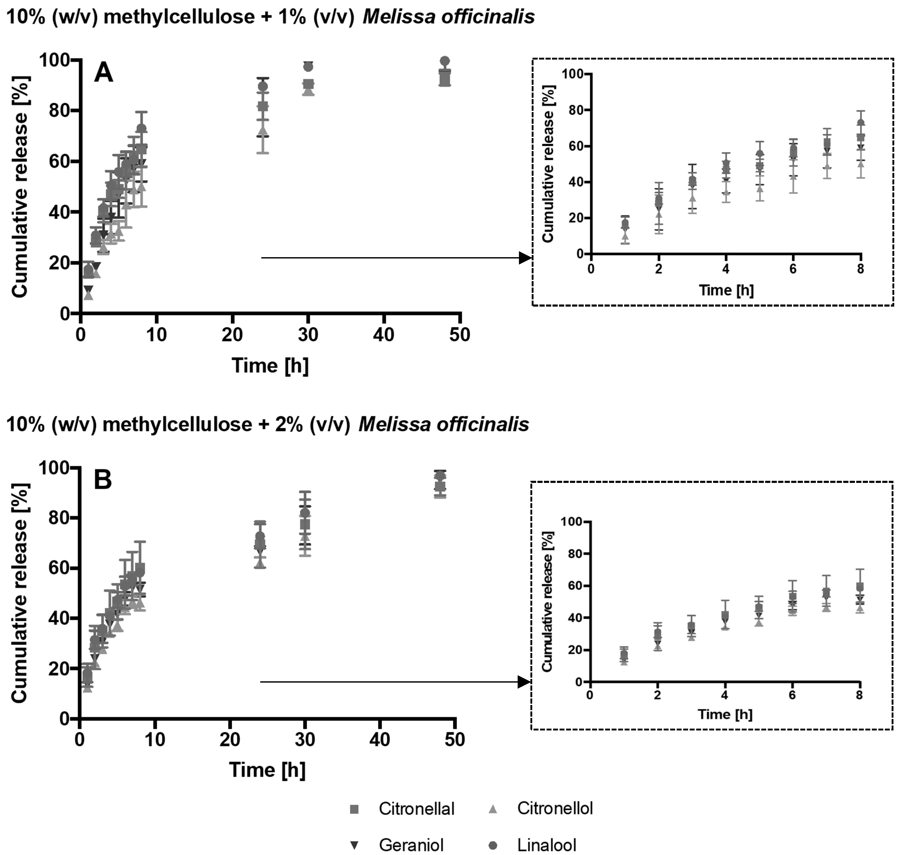
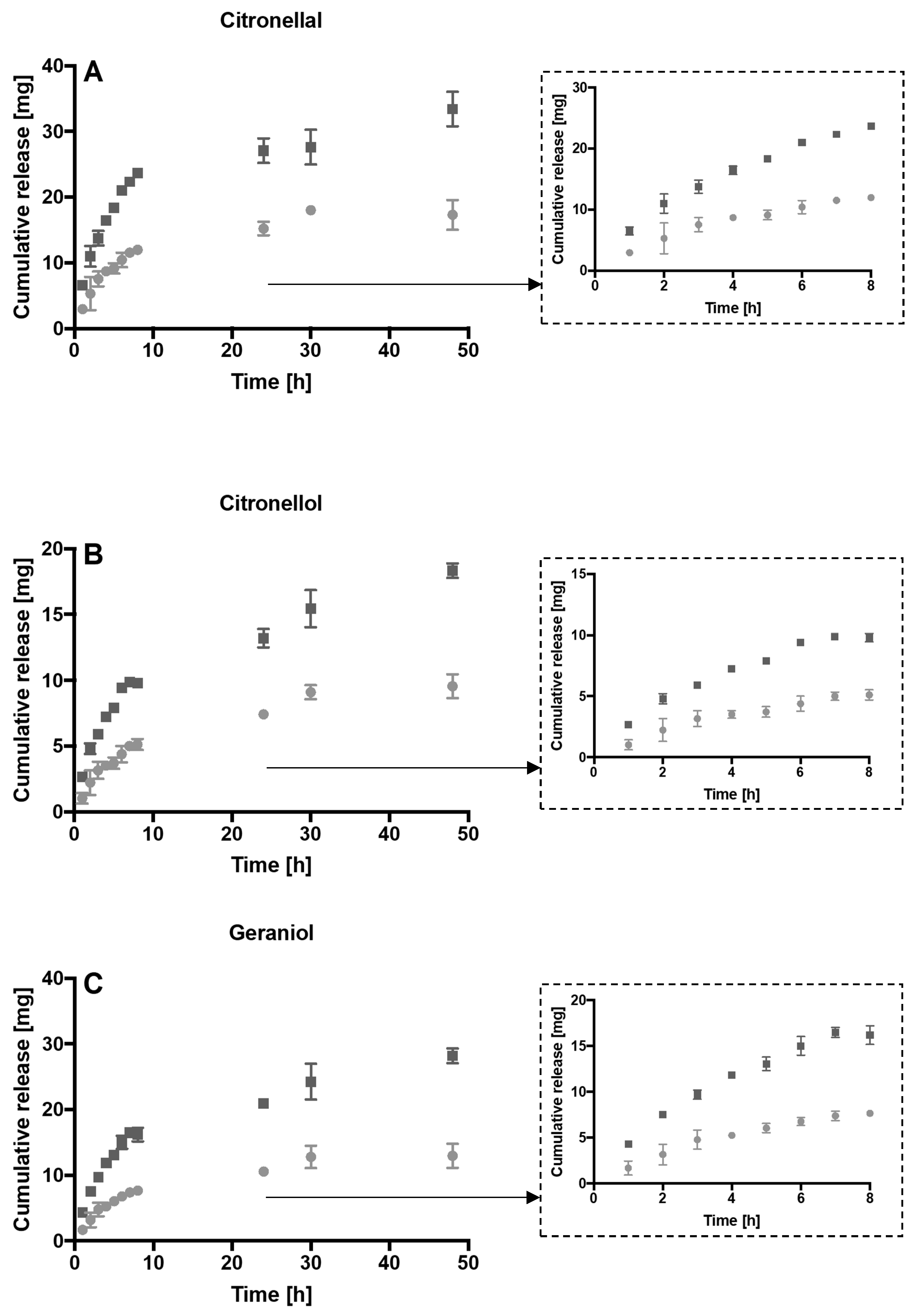
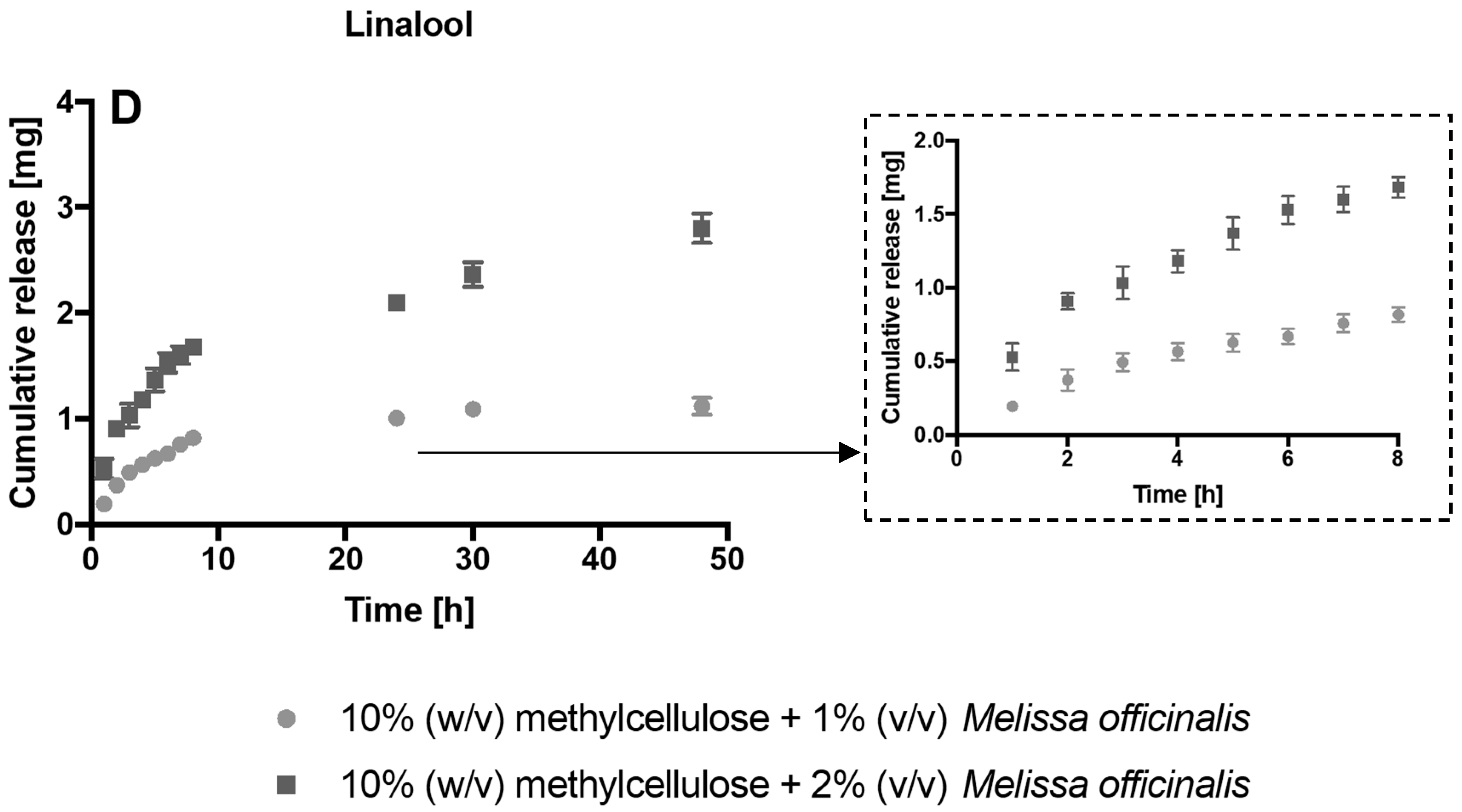
3.2. Melissa Officinalis Release

Prior to the evaluation of the release of the essential oil from the hydrogels, the composition of Melissa officinalis was analysed by GC-MS. Peaks were qualitatively identified by matching the mass spectra with those found in the NIST14 MS Search library. The quantification was carried out using a standard curve that correlated the peak areas to the concentrations. Three main compounds were identified, namely citronellal (50%), citronellol (10%) and geraniol (14%). In addition, 2.5% of linalool, a terpene with antimicrobial properties against oral pathogens, was also contained in the essential oil.
Therefore, the release from the hydrogel of citronellol, citronellal, geraniol and linalool was investigated. The drug release profile from 10% (w/v) methylcellulose hydrogels with 1 or 2% (v/v) Melissa officinalis essential oil over 48 h was demonstrated (Figure 2). Release was maintained for the duration of the experiment. After 8 h more than the 45% of the compounds was released, while within 48 h more than the 92% of each compound leached out. As expected, by increasing the initial concentration of Melissa officinalis into the hydrogel, the amount of drug release increased (Figure 3).

3.3. Retention Assay

The 10% (w/v) methylcellulose hydrogels with 2% (v/v) Melissa officinalis essential oil significantly reduced the retention of C. albicans (5.01 × 10³ CFU/mL; p = 0.03). There was also a significant difference in the reduction of the C. albicans retained on the hydrogels when loaded with either a 1% or 2% Melissa oil (p = 0.003). The lowest retention was achieved with 2% (v/v) Melissa officinalis on its own (solution), without the addition of the hydrogel (4.31 × 10 2 CFU/mL) (Figure 4).

3.4. Disk Diffusion Method

The antifungal activity of the methylcellulose hydrogels was determined by the disc diffusion method and the inhibitory zone measured (Table 1). The 10% (w/v) methylcellulose hydrogels with 1 or 2% (v/v) Melissa officinalis essential oil exhibited antifungal activity (p < 0.0001), while methylcellulose itself was not antifungal.

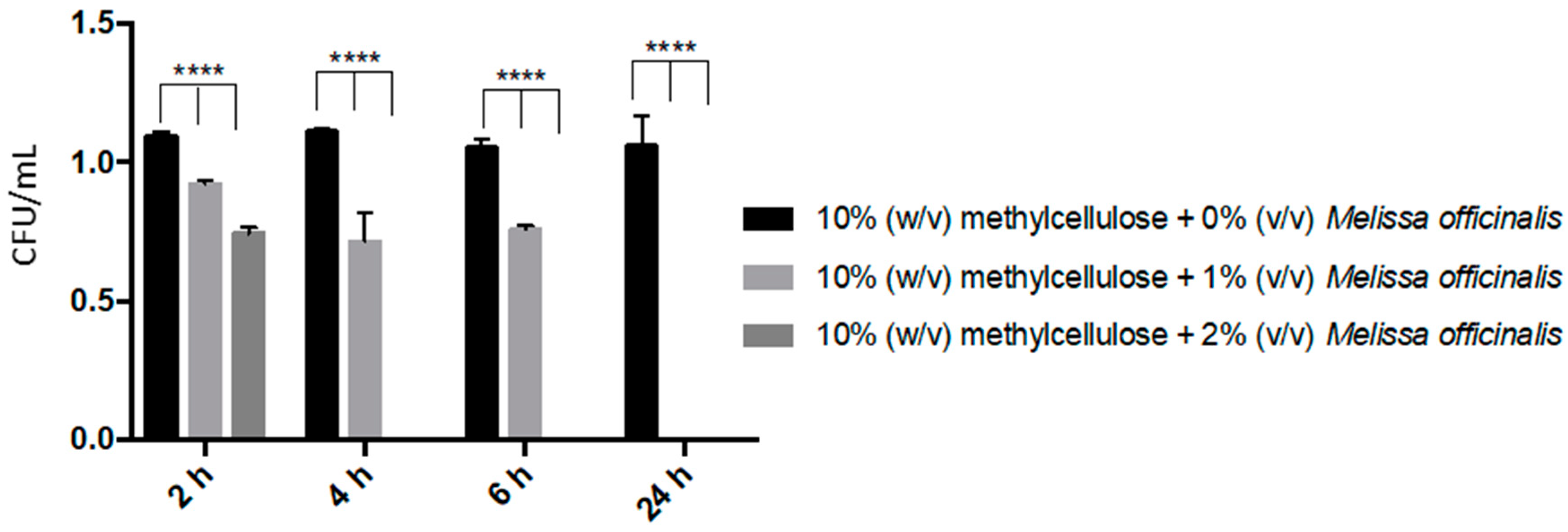
3.5. Time-Kill Assay

Antifungal activity was evaluated with a time-kill assay. The amount of C. albicans recovered within 2 h was decreased significantly (p < 0.001) by 15% and 30% in the presence of 10% (w/v) methylcellulose hydrogel with 1% (v/v) Melissa officinalis and 10% (w/v) methylcellulose hydrogel with 2% (v/v) Melissa officinalis, respectively (Figure 5). After 4 h application of 10% (w/v) methylcellulose hydrogels with 2% (v/v) Melissa officinalis, no viable cells were recovered, while it took 24 h to completely kill C. albicans when it was cultured in the presence of 10% (w/v) methylcellulose hydrogels with 1% (v/v) Melissa officinalis (Figure 5). It was observed that the presence of 10% (w/v) methylcellulose promoted C. albicans growth. This difference was significant until 6h incubation (p < 0.003), but not after 24 h incubation (p > 0.33).

4. Discussion

Candida species are commensal microorganisms of the oral cavity, mainly found on the posterior part of the tongue and on the oral mucosa. Candida is an opportunistic pathogen and can cause oral candidiasis when suitable conditions arise.
The emergence of antimicrobial resistance, defined by the World Health Organization (WHO) as a global threat to the effective treatment of infections, has led to a new interest in the antifungal properties of natural and chemical compounds [24]. Melissa officinalis has been previously reported to be active against C. albicans both in the planktonic and biofilm form at Minimum Inhibitory Concentrations (MIC) of 0.06% (v/v) and 1.5% (v/v), respectively [11]. Candida albicans is a predominately studied yeast because it significantly changes its morphology and expresses a range of virulence factors associated with biofilm formation, invasion of the host tissue and adaptation to the environment [25,26,27,28]. Being lipophilic, essential oils typically integrate into membrane structures causing increased cell permeability, leaching of intracellular components and inactivation of enzymes [5,29]; blocking the synthesis of membrane; inhibiting spore germination, proliferation of fungi and cellular respiration [28,30]. Furthermore, essential oils can also interact with the mitochondrial membrane leading to cidal effects [4]. Therefore, a hydrogel to be used as delivery vehicle to release the essential oil at the site of infection was developed. It is important to emphasize that methylcellulose is an US Food and Drug Administration (FDA)-approved material, it is approved for food industry and biomedical applications and it has been tested over the years to be not toxic for human and animal use [31,32,33].
Ten percent (10% (w/v)) methylcellulose hydrogels with 1 or 2% (v/v) Melissa officinalis essential oil were successfully synthesised. Rheological analysis was performed to evaluate the gelation time at body temperature. The essential oil content did not influence the rheological properties, and no significant differences in the loss and storage modulus were observed. The synthesised hydrogels are intended to be used as vehicles for drug delivery. Accordingly, it was important that once applied into the mouth, they undergo gelation at 37 °C within an appropriate time. For this reason, the viscoelastic properties of the hydrogel were evaluated to investigate the gelation time in the linear viscoelastic region at body temperature. It should be noted that these hydrogels were not supposed to be used to resist deformations such as in load-bearing applications (e.g., cartilage and bone regeneration). Considering the gelation time, the hydrogels were considered suitable for an oral application, being able to form a self-supporting hydrogel at 37 °C in less than three minutes.
Before investigating the essential oil release from the hydrogels, the composition of Melissa officinalis essential oil was analysed by GC-MS, since it is known that the composition and the ratio of the components depend on the plant’s origins and influence the antimicrobial properties of the essential oils [5]. The main component of the oil was citronellol (50% (v/v)) followed by geraniol (14% (v/v)) and citronellal (10% (v/v)). Two-point five percent (2.5% (v/v)) of linalool, a terpene with antimicrobial properties and generally recognized as safe by the FDA [34] was also contained in Melissa officinalis essential oil. Therefore, it was decided to evaluate the release of the three main compounds as well as of linalool.
Hydrogels are characterised by a 3D-polymeric network that allows liquid and molecules to diffuse. The diffusion of the drug depends on the mesh size: if the mesh size is larger than the drug then molecules are free to migrate through the network, while if the drug size is comparable to the mesh size then the drug is physically entrapped inside the hydrogel [35]. However, the mesh size can change over time allowing the release of the drug. Two main phenomena can lead to changes in the 3D-polymeric network, namely, degradation and swelling. Degradation, a loss of polymer mass, can be mediated by enzyme activity or by hydrolysis. Degradation results in an increase of mesh size that allows drug diffusion. Similarly, swelling due to water absorption, increases the mesh size and allows drug release [35]. In general, diffusion and drug release are affected by different parameters such as the hydrophilicity/hydrophobicity of the drug, the surface area and geometry of the hydrogel, the type and volume of solvent, the degradation of the hydrogel, the mesh size and the swelling rate [36]. Different volumes, solvents, ratios of hydrogel to solvent, flasks (e.g., universal, conical flasks), the presence of dialysis membranes and different shaking rate were found to be variable in a review of the literature [23,37,38,39,40]. In the current study, the drug release was evaluated over 48 h, the amount of compounds released were a function of the initial content of oil. Indeed, the amount released from the hydrogel containing 2% (v/v) of Melissa officinalis essential oil was significantly greater than that released from the hydrogels synthesized with 1% (v/v) of Melissa officinalis essential oil.
The antifungal activity of the hydrogels was screened against a clinical strain described previously as a high tissue invader (C. albicans 135BM2/94). The clinical strain had previously been isolated from a patient with chronic hyperplastic candidiasis and squamous cell carcinoma [21]. Candida albicans was not recovered when cultured in the presence of (10% (w/v) methylcellulose with 2% (v/v) Melissa officinalis essential oil) for 4 h with (10% (w/v) methylcellulose with 1% (v/v) Melissa officinalis essential oil for 24 h. Besides evaluating the antifungal potential of the drug released from the hydrogel, the antifungal potential of methylcellulose itself was investigated by comparing the viable cell count with that obtained culturing C. albicans in broth without the hydrogel.
The disc diffusion method demonstrated that the methylcellulose on its own did not inhibit Candida. In contrast, when the methylcellulose hydrogel contained Melissa officinalis essential oil, a zone of inhibition was observed and it was proportional to the amount of Melissa officinalis contained in the hydrogel. These results were in accordance with those of Campos et al. [14] and Ayana and Turhan [15] who observed a zone of inhibition when methylcellulose hydrogel was loaded with Ginja cherry or olive oil extracts, respectively.
The retention assay was used to observe if the methylcellulose hydrogel could be used as a direct drug delivery system to inhibit the retention of C. albicans on PMMA denture surfaces. For this reason, the hydrogel suitability for antimicrobial applications was tested on custom-made PMMA coupons. The 10% (w/v) methylcellulose hydrogels with 2% (v/v) Melissa officinalis essential oil significantly reduced the retention of C. albicans compared to the hydrogel on its own and to the 1% (v/v) Melissa officinalis hydrogel on PMMA. It is important to highlight that the lowest retention was achieved with 2% (v/v) Melissa officinalis pure oil without the application of the hydrogel. However, it is worth noting that in an in vivo situation firstly the 2% (v/v) essential oil concentration would not be directed to the target but, due to its liquid form, it would spread around on the surrounding tissues. Secondly, the 2% (v/v) concentration would be quickly diluted in the oral cavity, diminishing its effective antimicrobial potency. Finally, having the essential oil in its liquid form would make it difficult to determine what drug concentration will reach the target area. Other studies have focused on hydrogels as delivery systems for the oral cavity [41,42,43]. Kong et al. (2016) developed a hydroxypropyl methylcellulose hydrogel for use against oral candidiasis. A 4% (w/w) hydroxypropyl methylcellulose hydrogel was formulated with 2 mg/mL of Histatin-5, a peptide with antimicrobial properties. The authors tested the antifungal activity of the hydrogel against C. albicans standard strain SC5314, observing Histatin-5 release from the hydrogel within 2 h and a significant decrease in CFU/mL compared to the control (i.e., hydroxypropyl methylcellulose hydrogel without antimicrobial). In addition, the novel formulation was tested in vivo. The tongues of mice were infected with C. albicans and the hydrogel was applied for 1 h. Untreated mice developed oral candidiasis with tissue damage and C. albicans was recovered from the site. By contrast, most of the mice treated with the hydrogel showed less fungal adherence, hyphal penetration and tissue damage [23].

5. Conclusions

A 10% (w/v) methylcellulose hydrogel with 1% (v/v) and 2% (v/v) Melissa officinalis was successfully synthesised. The rheological analysis highlighted that the hydrogels jellified at body temperature in less than 3 min. The antifungal properties of the hydrogels were confirmed by both zone of inhibition method and time-kill assays and the 2% (v/v) hydrogel significantly reduced the retention of C. albicans. The novel findings of this work in future, will be expanded to include a range of Candida species as well as bacterial species associated with oral infections.

Author Contributions

E.S. performed all the experiments, analysed the data and wrote a draft of the paper. A.H.-B., A.S. and C.L. drafted the experiments related to hydrogel rheology, reviewed and revised the draft manuscript. F.S. and K.W. drafted the experiments related to the retention assay, reviewed and revised the draft manuscript. S.M., J.V. and D.W.W. drafted the work, designed the experiments related to the microbiology work, oversaw the laboratory work, reviewed and revised the draft manuscript. All authors have read and agreed to the published version of the manuscript.

Funding

This research was funded by Manchester Metropolitan University. C.L. thanks the EPSRC & MRC CDT in Regenerative Medicine for their financial support (EP/L014904/1).

Conflicts of Interest

The authors declare no conflict of interest. The funders had no role in the design of the study; in the collection, analyses, or interpretation of data; in the writing of the manuscript, or in the decision to publish the results.

References

  1. Singh, S.; Fatima, Z.; Hameed, S. Predisposing factors endorsing Candida infections. Infez. Med. 2015, 23, 211–223. [Google Scholar] [PubMed]
  2. Williams, D.; Lewis, M. Pathogenesis and treatment of oral candidosis. J. Oral Microbiol. 2011, 3. [Google Scholar] [CrossRef] [PubMed] [Green Version]
  3. Vandeputte, P.; Ferrari, S.; Coste, A.T. Antifungal resistance and new strategies to control fungal infections. Int. J. Microbiol. 2012, 2012, 713687. [Google Scholar] [CrossRef] [PubMed]
  4. Nazzaro, F.; Fratianni, F.; de Martino, L.; Coppola, R.; de Feo, V. Effect of essential oils on pathogenic bacteria. Pharmaceuticals 2013, 6, 1451–1474. [Google Scholar] [CrossRef]
  5. Bakkali, F.; Averbeck, S.; Averbeck, D.; Idaomar, M. Biological effects of essential oils—A review. Food Chem. Toxicol. 2008, 46, 446–475. [Google Scholar] [CrossRef]
  6. Da Silva, S.; Moreira, C.B.; Esquibel, M.A.; da Silva San Gil, A.; da Silva Riehl, C.A.; Sato, A. Effect of salicylic acid on essential oil compounds of Melissa officinalis in vitro plants. Agropecuária Técnica 2014, 35, 178–184. [Google Scholar]
  7. Babpour, E.; Angaji, S.A.; Angaji, S.M. Antimicrobial effects of four medicinal plants on dental plaque. J. Med. Plants Res. 2009, 3, 132–137. [Google Scholar]
  8. Iauk, L.; Bue, A.M.L.; Milazzo, I.; Rapisarda, A.; Blandino, G. Antibacterial activity of medicinal plant extracts against periodontopathic bacteria. Phytother. Res. 2003, 17, 599–604. [Google Scholar] [CrossRef]
  9. Abdellatif, F.; Boudjella, H.; Zitouni, A.; Hassani, A. Chemical composition and antimicrobial activity of the essential oil from leaves of algerian. EXCLI J. 2014, 13, 772–781. [Google Scholar]
  10. Mimica-Dukic, N.; Bozin, B.; Sokovic, M.; Simin, N. Antimicrobial and antioxidant activities of Melissa officinalis L. (Lamiaceae) essential oil. J. Agric. Food Chem. 2004, 52, 2485–2489. [Google Scholar] [CrossRef]
  11. Serra, E.; Hidalgo-bastida, L.A.; Verran, J.; Williams, D.; Malic, S. Antifungal activity of commercial essential oils and biocides against Candida albicans. Pathogens 2018, 7, 15. [Google Scholar] [CrossRef] [PubMed] [Green Version]
  12. Slaughter, B.V.; Khurshid, S.S.; Fisher, O.Z.; Khademhosseini, A.; Peppas, N.A. Hydrogels in regenerative medicine. Adv. Mater. 2009, 21, 3307–3329. [Google Scholar] [CrossRef] [PubMed] [Green Version]
  13. Bhattarai, N.; Gunn, J.; Zhang, M. Chitosan-based hydrogels for controlled, localized drug delivery. Adv. Drug Deliv. Rev. 2010, 62, 83–99. [Google Scholar] [CrossRef] [PubMed]
  14. Campos, D.; Piccirillo, C.; Pullar, R.C.; Castro, M.L.; Pintado, M.M.E. Characterization and antimicrobial properties of food packaging methylcellulose films containing stem extract of Ginja cherry. J. Sci. Food Agric. 2014, 94, 2097–2103. [Google Scholar] [CrossRef]
  15. Ayana, B.B.; Turhan, K.N. Use of antimicrobial methylcellulose films to control Staphylococcus aureus during storage of Kasar cheese and science, Packag. Technol. Sci. 2009, 22, 461–469. [Google Scholar]
  16. Otoni, C.G.; Pontes, S.F.O.; Medeiros, E.A.A.; Soares, N.F. Edible films from methylcellulose and nanoemulsions of clove bud (Syzygium aromaticum) and oregano (Origanum vulgare) essential oils as shelf life extenders for sliced bread. J. Agric. Food Chem. 2014, 62, 5214–5219. [Google Scholar] [CrossRef]
  17. Klouda, L. Thermoresponsive hydrogels in biomedical applications: A seven-year update. Eur. J. Pharm. Biopharm. 2015, 97, 338–349. [Google Scholar] [CrossRef]
  18. Gafitanu, C.A.; Filip, D.; Cernatescu, C.; Rusu, D.; Tuchilus, C.G.; Macocinschi, D.; Zaltariov, M.-F. Design preparation and evaluation of HPMC-based PAA or SA freeze-dried scaffolds for vaginal delivery of fluconazole. Pharm. Res. 2017, 34, 2185–2196. [Google Scholar] [CrossRef]
  19. Zuidema, J.M.; Rivet, C.J.; Gilbert, R.J.; Morrison, F.A. A protocol for rheological characterization of hydrogels for tissue engineering strategies. J. Biomed. Mater. Res. B Appl. Biomater. 2014, 102, 1063–1073. [Google Scholar] [CrossRef]
  20. Rajkowska, K.; Nowak, A.; Kunicka-Styczy, A.; Siadura, A. Biologicaleffects of various chemically characterized essential oils: Investigation of the mode of action against Candida albicans and HeLa cells. RSC Adv. 2016, 6, 97199–97207. [Google Scholar] [CrossRef] [Green Version]
  21. Malic, S.; Hill, K.E.; Ralphs, J.R.; Hayes, A.; Thomas, D.W.; Potts, A.J.; Williams, D.W. Characterization of Candida albicans infection of an in vitro oral epithelial model using confocal laser scanning microscopy. Oral Microbiol. Immunol. 2007, 22, 188–194. [Google Scholar] [CrossRef] [PubMed]
  22. KWhitehead, A.; Colligon, J.; Verran, J. Retention of microbial cells in substratum surface features of micrometer and sub-micrometer dimensions. Colloids Surf. B Biointerfaces 2005, 41, 129–138. [Google Scholar] [CrossRef] [PubMed]
  23. Kong, E.F.; Tsui, C.; Boyce, H.; Ibrahim, A.; Hoag, S.W.; Karlsson, A.J.; Meiller, T.F.; Jabra-Rizk, M.A. Development and in vivo evaluation of a novel Histatin-5 bioadhesive hydrogel formulation against oral candidiasis. Antimicrob. Agents Chemother. 2016, 60, 881–889. [Google Scholar] [CrossRef] [PubMed] [Green Version]
  24. World Health Organization. Antimicrobial Resistance: Global Report on Surveillance 2014; World Health Organization: Geneva, Switzerland, 2014. [Google Scholar]
  25. Henriques, M.; Azeredo, J.; Oliveira, R. Adhesion of Candida albicans and Candida dubliniensis to acrylic and hydroxyapatite. Biointerfaces 2004, 33, 235–241. [Google Scholar] [CrossRef] [Green Version]
  26. Ramage, G.; Saville, S.P.; Thomas, D.P.; López-Ribot, J.L. Candida biofilms: An update. Eukaryot. Cell 2005, 4, 633–638. [Google Scholar] [CrossRef] [Green Version]
  27. Thein, Z.M.; Samaranayake, Y.H.; Samaranayake, L.P. Effect of oral bacteria on growth and survival of Candida albicans biofilms. Arch. Oral Biol. 2006, 51, 672–680. [Google Scholar] [CrossRef] [PubMed]
  28. de Castro, G.M.F.A.; Santos, B.V.O.; Costa, E.M.M.D.; Cavancanti, A.L.; Trindade, L.A.; Lima, E.D.; de Castro, R.D. Chemical and antifungal analysis of essential oils and phytochemicals against Candida albicans and Candida tropicalis. Clin. Microbiol. Res. 2018. [Google Scholar] [CrossRef]
  29. Sikkematb, J.; Bontt, J.A.M.D. Interactions of cyclic hydrocarbons with biological membranes. J. Biol. Chem. 1994, 269, 8022–8028. [Google Scholar]
  30. Harris, R. Progress with superficial mycoses using essential oils. Int. J. Aromather. 2002, 12, 83–91. [Google Scholar] [CrossRef]
  31. Sannino, A.; Demitri, C.; Madaghiele, M. Biodegradable Cellulose-based Hydrogels: Design and Applications. Materials 2009, 2, 353–373. [Google Scholar] [CrossRef]
  32. Stalling, S.S.; Akintoye, S.O.; Nicoll, S.B. Development of photocrosslinked methylcellulose hydrogels for soft tissue reconstruction. Acta Biomater. 2009, 5, 1911–1918. [Google Scholar] [CrossRef] [PubMed]
  33. Nasatto, P.L.; Pignon, F.; Silveira, J.L.M.; Duarte, M.E.R.; Noseda, M.D.; Rinaudo, M. Methylcellulose, a Cellulose Derivative with Original Physical Properties and Extended Applications. Polymers 2015, 7, 777–803. [Google Scholar] [CrossRef] [Green Version]
  34. Hsu, C.-C.; Lai, W.-L.; Chuang, K.-C.; Lee, M.-H.; Tsai, Y.-C. The inhibitory activity of linalool against the filamentous growth and biofilm formation in Candida albicans. Med. Mycol. 2013, 51, 473–482. [Google Scholar] [CrossRef] [PubMed] [Green Version]
  35. Li, J.; Mooney, D.J. Designing hydrogels for controlled drug delivery. Nat. Rev. Mater. 2016, 1, 1–18. [Google Scholar] [CrossRef]
  36. Zarzycki, R.; Modrzejewska, Z.; Nawrotek, K.; Lek, U. Drug release from hydrogel matrices. Ecol. Chem. Eng. S 2010, 17, 117–136. [Google Scholar]
  37. Senel, S.; Ikinci, G.; Kas, S.; Yousefi-Rad, A.; Sargon, M.F.; Hincal, A.A. Chitosan films and hydrogels of chlorhexidine gluconate for oral mucosal delivery. Int. J. Pharm. 2000, 193, 197–203. [Google Scholar] [CrossRef]
  38. Ji, Q.X.; Zhao, Q.S.; Deng, J.; Lü, R. A novel injectable chlorhexidine thermosensitive hydrogel for periodontal application: Preparation, antibacterial activity and toxicity evaluation. J. Mater. Sci. Mater. Med. 2010, 21, 2435–2442. [Google Scholar] [CrossRef]
  39. Kodadova, A.; Vitkova, Z.; Herdova, P.; Oremusova, J.; Kodadova, A.; Mikus, P. Formulation of sage essential oil (Salvia officinalis, L.) monoterpenes into chitosan hydrogels and permeation study with GC-MS analysis. Drug Dev. Ind. Pharm. 2014, 41, 1080–1088. [Google Scholar] [CrossRef]
  40. Low, W.L.; Kenward, M.a.K.; Cairul, M.; Mohd, I.; Martin, C. Ionically crosslinked chitosan hydrogels for the controlled release of antimicrobial essential oils and metal ions for wound management applications. Medicines 2016, 3, 8. [Google Scholar] [CrossRef] [Green Version]
  41. Perchyonoka, V.T.; Zhang, S.; Oberholze, T. Novel Melatonin-chitosan Hydrogels as Suitable Oral Bio-drug Delivery Systems to Fight Oral Mucositis: Synergy of Antioxidants and Bioactives in Action. Curr. Org. Chem. 2012, 16, 2430–2436. [Google Scholar] [CrossRef]
  42. Echazú, M.I.A.; Olivetti, C.E.; Anesini, C.; Perez, C.J.; Alvarez, G.S.; Desimone, M.F. Development and evaluation of thymol-chitosan hydrogels with antimicrobial-antioxidant activity for oral local delivery. Mater. Sci. Eng. C 2017, 81, 588–596. [Google Scholar] [CrossRef] [PubMed]
  43. Galler, K.M.; Hartgerink, J.D.; Cavender, A.C.; Schmalz, G.; D’Souza, R.N. A Customized Self-Assembling Peptide Hydrogel for Dental Pulp Tissue Engineering. Tissue Eng. Part A 2012, 18, 176–184. [Google Scholar] [CrossRef] [PubMed]
Figure 1. Rheological characterization of the synthesized methylcellulose hydrogels. (A,B) Strain sweep for 10% (w/v) methylcellulose hydrogel with 1% (v/v) Melissa officinalis (A) and 2% (v/v) Melissa officinalis (B). (C,D) Frequency sweep for 10% (w/v) methylcellulose hydrogel with 1% (v/v) Melissa officinalis (C) and 2% (v/v) Melissa officinalis (D). (E,F) Time sweep for 10% (w/v) methylcellulose hydrogel with 1% (v/v) Melissa officinalis (E) and 2% (v/v) Melissa officinalis (F) determined at an amplitude strain of 1% and angular frequency of 1 Hz. G′ (black circle) storage modulus and G″ (grey square) loss modulus.
Microorganisms 08 00215 g001
Figure 2. Percentage cumulative release of citronellal (square), citronellol (triangle), geraniol (inverted triange), and linalool (circle) of 10% (w/v) methylcellulose and 1% (v/v) Melissa officinalis (A), and 10% (w/v) methylcellulose and 2% (v/v) Melissa officinalis (B). A zoom of the percentage cumulative release between 0 and 8 h is also shown in the dashed rectangles.
Microorganisms 08 00215 g002
Figure 3. Cumulative release in milligrams of citronellal (A), citronellol (B), geraniol (C) and linalool (D) from 10% (w/v) methylcellulose with 1% (v/v) Melissa officinalis (circle), 10% (w/v) methylcellulose with 2% (v/v) Melissa officinalis (square). A zoom of the cumulative release between 0 and 8 h is also shown in the dashed rectangles.
Microorganisms 08 00215 g003aMicroorganisms 08 00215 g003b
Figure 4. Retention assay after 1 h of exposure of C. albicans to 10% (w/v) methylcellulose hydrogels with or without 1 or 2% (v/v) Melissa officinalis.
Microorganisms 08 00215 g004
Figure 5. In vitro time-kill assay after 2, 4, 6 and 24 h of exposure of C. albicans to 10% (w/v) methylcellulose hydrogels with or without 1 or 2% (v/v) Melissa officinalis. **** = p < 0.0001.
Microorganisms 08 00215 g005
Table 1. Inhibitory zone diameters of 10% (w/v) methylcellulose hydrogels with or without 1 or 2% (v/v) Melissa officinalis. **** = p < 0.0001.
Inhibitory Zone Diameter (mm)
10% (w/v) methylcellulose0
10% (w/v) methylcellulose +
1% (v/v) Melissa officinalis
10.2 ± 0.4
10% (w/v) methylcellulose +
2% (v/v) Melissa officinalis
17.5 ± 2.6 (****)

