Communication

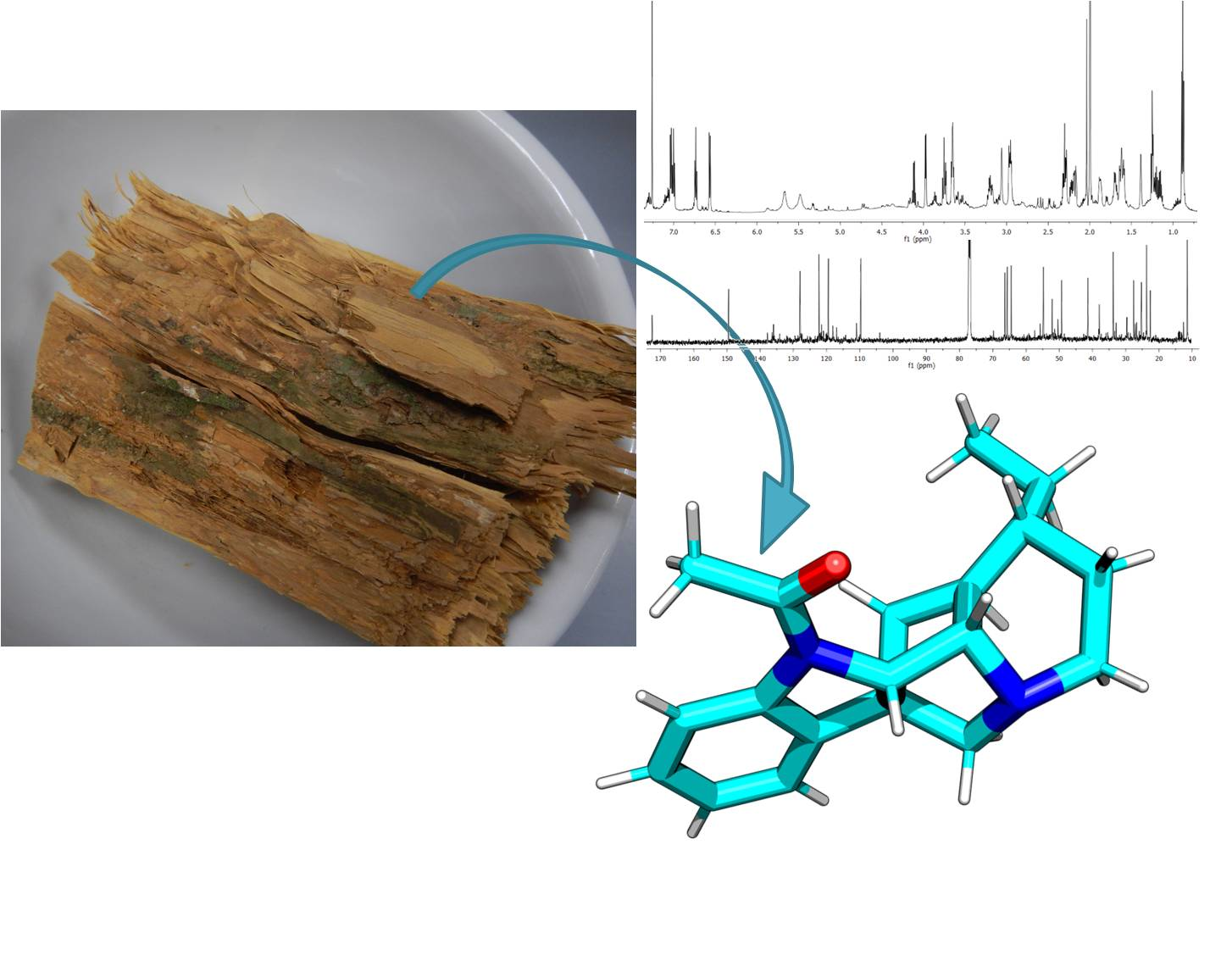
Geissospermiculatine, a New Alkaloid from Geissospermum reticulatum Bark

by
Joanna J. Sajkowska-Kozielewicz
1,*,
Paweł Kozielewicz
2,3,
Katerina Makarova
1,
Marcin Stocki
4,
Nicholas M. Barnes
3 and
Katarzyna Paradowska
1
1
Department of Physical Chemistry, Faculty of Pharmacy with Laboratory Medicine Division, Medical University of Warsaw, 02-097 Warsaw, Poland
2
Department of Physiology and Pharmacology, Karolinska Institutet, 17165 Stockholm, Sweden
3
Institute of Clinical Sciences, College of Medical and Dental Sciences, University of Birmingham, Birmingham B152TT, UK
4
Institute of Forest Sciences, Faculty of Civil Engineering and Environmental Sciences, Bialystok University of Technology, 15-351 Bialystok, Poland
*
Author to whom correspondence should be addressed.
Molecules 2021, 26(1), 143; https://doi.org/10.3390/molecules26010143
Submission received: 29 November 2020 / Revised: 16 December 2020 / Accepted: 28 December 2020 / Published: 31 December 2020
(This article belongs to the Section Natural Products Chemistry)

Abstract

:
A new alkaloid, geissospermiculatine was characterized in Geissospermum reticulatum A. H. Gentry bark (Apocynaceae). Here, following a simplified isolation protocol, the structure of the alkaloid was elucidated through GC-MS, LC-MS/MS, 1D, and 2D NMR (COSY, ROESY, HSQC, HMBC, 1H-15N HMBC). Cytotoxic properties were evaluated in vitro on malignant THP-1 cells, and the results demonstrated that the cytotoxicity of the alkaloid (30  μg/mL) was comparable with staurosporine (10  μM). Additionally, the toxicity was tested on zebrafish (Danio rerio) embryos in vivo by monitoring their development (0–72 h); toxicity was not evident at 30  μg/mL.

Graphical Abstract

1. Introduction

Geissospermum species (Apocynaceae) grow in the Amazonian rainforest [1,2]. For many years they have proven popular as therapeutic plants used in traditional medicines as remedies for malarial, tumors, bacterial infections, and pain relief, and as anti-inflammatory agents [1,3,4]. First investigations of the chemical composition of these trees date back to the end of the 19th century [1]. However, in recent decades, little progress has been made in the characterization of the compounds from the Geissospermum genus. In fact, among 12 species, only six (G. argenteum, G. fuscum, G. leave, G. reticulatum, G. sericeum, and G. urceolatum) have been studied phytochemically [1,3,5,6,7,8,9,10,11,12,13,14]. Specifically, it has been reported that these trees are a rich source of indole alkaloids [1,3]. Nevertheless, the phytochemical profile of Geissospermum reticulatum A. H. Gentry bark has received minimal attention with only one published study that described the presence of three alkaloids: 11-methoxygeissospermidine, flavopereirine, and geissosreticulatine [3].
As a part of our investigation into the composition and properties of plants from the Amazon region, we now document discovery of a new indole alkaloid from G. reticulatum bark—geissospermiculatine. Utilizing LC-MS/MS and GC-MS, the presence in the alkaloidal fraction was identified after a short isolation procedure. Moreover, the alkaloid structure was determined using 1D and 2D NMR. Finally, to assess biological properties of the compound, potential toxic effects on THP-1 cells and Danio rerio embryos were evaluated.

2. Results and Discussion

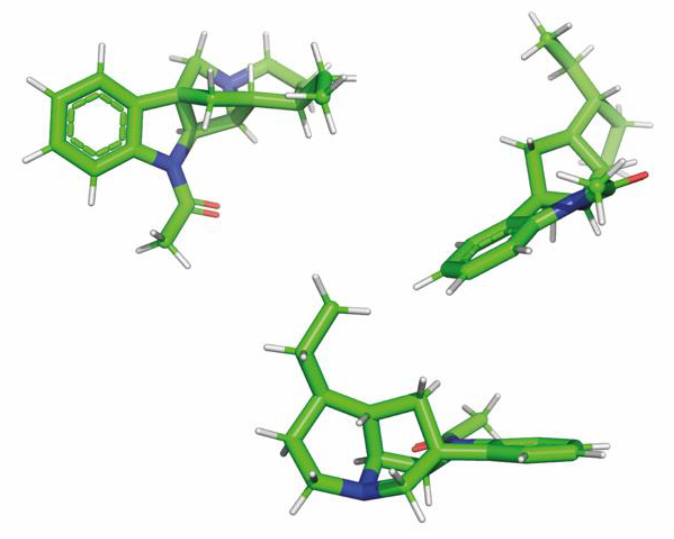
2.1. Structural Determination by 1D and 2D NMR Spectroscopy

Using our isolation procedure, approximately 1.8 g of the alkaloid was obtained from 100 g of G. reticulatum bark following. Next, we analyzed the chemical properties utilizing a range of analytical techniques. The mass spectrum of the alkaloidal fraction demonstrated that one alkaloid with a molecular ion at m/z 368 constituted 86% of the total composition (Figures S1 and S2 in Supplementary Materials). Since it was the silylated fraction, it was predicted that the molecular weight of this alkaloid was 296 g/mol. Next, the molecular formula of this alkaloid was established to be C19H24N2O by LC-MS/MS.
The 1H-NMR spectrum (in CDCl3) demonstrated the presence of an ethyl side chain (methyl group at δH 0.89 ppm, t, H-19, and methylene protons at δH 1.21 and 1.24 ppm, m, H-18). The 1H-NMR spectrum (Figure S4) also revealed four signals at δH 6.57 (d, J = 7.7 Hz, H-9), δH 6.74 (t, J = 7.4 Hz, H-10), δH 7.02 (t, J = 7.6 Hz, H-11), and δH 7.04 (d, J = 7.4 Hz, H-12) which were characteristic for aromatic moieties. The 13C-NMR spectrum (Figure S5), complemented by DEPT-135 and DEPT-90 experiments (Figure S6), displayed 19 signals from the alkaloid, including two methyl groups, five methylenes, eight methines, and four quaternary carbons, at δC 52.14 (C-7), δC 135.97 (C-8), δC 149.51 (C-13), and δC 172.51 (C-20).
Additionally, a range of 2D NMR spectroscopic techniques (COSY, ROESY, HSQC, HMBC, 1H-15N HMBC, Figures S7–S11) was applied to identify the structure of geissospermiculatine. The HMBC correlation networks of H-9/C-10, C-8, and H-11/C-12, C-13 indicated the linkages of C-8–C-9–C-10 and C-11–C-12–C-13, respectively. Next, HMBC analysis also showed correlations from H-2 to C-7. Moreover, the presence of a quaternary carbon at δC 172.51 (C-20) with one strong correlation to δH 2.00 (m, H-21) suggested that they formed a substituted indole ring. Correspondingly, a chemical shift of δC 172.51 (characteristic for R1CONR2 groups) and a chemical shift of δH 2.00 typical for methyl protons near a carbonyl group (CH3-C=O) revealed the presence of an indole ring substituted by CH3-C=O. In the 1H-15N HMBC spectrum, there was a correlation from H-21 (δH 2.00) and a nitrogen signal (δN 103.34), specific for primary amides, which confirmed the structure of substituent. Additionally, COSY NMR spectra displayed correlations between H-18–H-19, H-14–H-15, and H-16–H-17. The above analysis helped to determine the presence of octahydroindolizine moiety in the structure. Importantly, the presence of the bridge between C-6 and C-14 was concluded from COSY cross-peaks supported with ROESY analysis. The structure of this indole alkaloid (geissospermiculatine, Figure 1, Table 1) was, therefore, elucidated as 1-(7-ethyl-6,7,8,9-tetrahydro-6,11a-methanoindolizino[1,2-b]indol-5(5aH,5bH,11H)-yl)ethanone, which we named geissospermiculatine. Additionally, its three-dimensional conformation was predicted using in silico calculations.

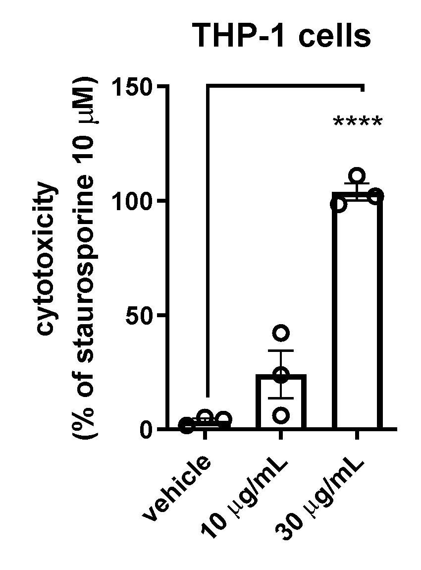
2.2. Cytotoxic Activity on In Vitro Cultured Cells

We have previously assessed and reported cytotoxic properties of the extracts from G. reticulatum using THP-1 cell line [4]. This model has also been employed by others to study cytotoxic properties of plant substances, including indole alkaloids [15,16]. Our results show that the alkaloid fraction (30  μg/mL with 48 h of exposure) induced significant cell death, comparable to staurosporine (10 μM) (Figure 2). Importantly, the vehicle (0.12% ethanol) had no effect on the cell viability in this assay. Additionally, the treatment of the cells with the lower concentration (10 μg/mL) of the fraction also resulted in a decrease in the number of live cells. However, the more modest reduction did not reach statistical significance but demonstrated a concentration-dependent effect of geissospermiculatine (Figure 2). Our results suggest that the alkaloid, geissospermiculatine, could be further evaluated for the treatment of leukemia.

2.3. Effect on Zebrafish (Danio rerio)

The introduction of a new compound onto the market requires performing various analyses, including animal tests of toxicity. Aside from the ethical concerns raised by the public, the industry is also interested in alternative testing methods that are less time and space consuming. Fish embryos represent an attractive model for toxicological assays since they offer the possibility to perform small-scale, high-throughput analyses. Moreover, as a toxicology model, zebrafish has the potential to reveal the pathways of developmental toxicity due to their similarity with those present in mammals. Therefore, zebrafish are often considered a useful model to screen as a part of a Target Product Profile to support selection of relatively safe lead candidates early in the drug discovery process [17,18].
To test the toxicity of the fraction in vivo, we administered the alkaloid fraction to zebrafish embryos and subsequently monitored their development (0–72 h) (Table 2 and Table 3). We did not observe any evident toxic effect at 30 μg/mL—that is the concentration that caused significant THP-1 cell death in our assays (Figure 2). However, the embryos did display growth retardation upon exposure to higher concentrations of the alkaloid (100 and 300 μg/mL) (Table 3).

3. Materials and Methods

3.1. General Experimental Procedures

GC-MS analysis was performed with an Agilent 7890A gas chromatograph equipped with an Agilent 5975C mass selective detector. The injection of a 1 µL sample (10 mg of extract dissolved with 1 mL of pyridine and 100 μL BSTFA was added, after which the sample was heated for 30 min at 60 °C for silylation) was performed with the aid of Agilent 7693A autosampler. The separation was performed on an HP-5MS (30 m × 0.25 mm × 0.25 µm film thickness) fused silica column at a helium flow rate of 1 mL/min. The ion source and quadrupole temperatures were 230 °C and 150 °C, respectively. The electron ionization mass spectra (EIMS) were obtained at ionization energy 70 eV. LC-MS/MS analysis was carried out by liquid chromatography (Agilent 1260 Infinity, Agilent Technologies, Santa Clara, CA, USA) coupled with a hybrid triple quadrupole/linear ion trap mass spectrometer (QTRAP 4000; AB SCIEX, Framingham, MA, USA). The 1D (1H, 13C, DEPT-135, and DEPT-90) and 2D (COSY, ROESY, HSQC, HMBC, and 1H-15N HMBC) NMR spectra were acquired on a Bruker Avance III 600-MHz spectrometer (Bruker Co., Ettlingen, Germany). The experiments were performed in a 5 mm, three-channel probe (TXI-inverse) at 295 K. Impulse sequences that came from the Bruker standard library of programs were used for the measurements. Chemical shifts for the spectra were calibrated relative to the shift of the 1H and 13C signals of the solvent (CDCl3). The spectra were analyzed using the MestReNova program (version 14.1.2, Mestrelab Research S.L., Santiago de Compostela, Spain) and TopSpinTM (Bruker). The 3D conformation (Figure 3) was predicted of the alkaloid using the PM3 semi-empirical calculations followed by the HF/6-31G quantum mechanical calculations.

3.2. Plant Material

The sample of dried bark of Geissospermum reticulatum was collected in the Amazon rainforest and stored at National Agrarian University—La Molina (Lima, Peru) in June 2013. The identity of the plant was confirmed at the same University by Engr. Santos Jaimes Serkovic. A voucher specimen (GDMD 21760) is deposited at the Herbarium, Medical University of Gdańsk, Poland.

3.3. Extraction and Isolation

The process of extraction was based on that proposed by Pilarski et al. [19], with some modifications made by our team. Moreover, in comparison with Reina et al. [3], in the present study, we employed a quicker and potentially cheaper extraction protocol. Twenty mL MeOH(aq) (Chempur) was added to 1 g of minced bark and sonicated for 45 min in an ultrasonic bath (Polsonic type sonic-2) set to 45 °C. Then, 10 mL MeOH(aq) was added, and the mixture was sonicated again: for 15 min at 45 °C, and then for 45 min with switched-off ultrasound. The mixture was then filtered and evaporated to dryness. The residue was dissolved in the mixture of 10 mL of 2% H2SO4(aq) (POCH) and 10 mL of EtOAc(aq) (POCH) and sonicated for 15 min in an ultrasonic bath. The aqueous phase was separated and extracted with 10 mL of EtOAc(aq). The aqueous phase was collected and stored at 4 °C for 24 h. Then the solution was decanted from the sediment. The aqueous phase was adjusted to pH 10 with 10% NH4OH(aq) (Chempur), 10 mL of EtOAc(aq) was added, and the mixture was shaken (Dragon Lab shaker, Beijing, China) for 10 min. The organic phase separated, while the aqueous one was extracted again with 10 mL of EtOAc(aq). The organic fractions of alkaloids were combined and evaporated to dryness.

3.4. Cell Culture

THP-1 (cell line typifying human monocytic leukemia; source ATCC TIB-202TM) cells were cultured in RPMI-1640 medium (Sigma-Aldrich, St. Louis, MI, USA) supplemented with 1% l-glutamine (Life Technologies, Carlsbad, CA, USA), 1% penicillin/streptomycin (Sigma-Aldrich, 10,000 units penicillin and 10 mg streptomycin per mL) and 10% heat-inactivated fetal bovine serum. Cells were subcultured at a density of 1–1.5 × 10⁶ cells/mL.

3.5. Cytotoxic Activity Test with 7-AAD

Cells were stimulated for 24 h and, following the incubation, they were washed with PBS by centrifugation at 400× g for 5 min. The supernatant was discarded, and the cells were resuspended in 7-AAD solution (50 μg/mL, eBioscience, San Diego, CA, USA) and incubated for 20 min at 4 °C. Cells were kept on ice and immediately analyzed on an ADP Cyan flow cytometer (Beckman Coulter, Brea, CA, USA). The flow cytometry data were analyzed using FlowJo V10 (Tree Star, Ashland, OR, USA).

3.6. Zebrafish (Danio rerio) Embryo Toxicity Test

The zebrafish embryo toxicity test was performed according to published guidelines. It should be noted that according to the EU Directive 2010/63/EU on the protection of animals used for scientific purposes, the earliest life stages of animals are not defined as protected and, therefore, do not fall into the regulatory frameworks dealing with animal experimentation. Zebrafish (Danio rerio) used in this experiment were of the wild-type AbxTL strain. The fish were housed in a circulating system that continuously aerates and filters the water to maintain it in the right quality. The temperature in the room and tanks was maintained between 26.0–28.5 °C (pH 6.8–7.5) on a 14 h light:10 h dark cycle. For all experiments, E3 medium (5 mM NaCl, 0.17 mM KCl, 0.33 mM CaCl2, and 0.33 mM MgSO4) was used. Twenty fertilized eggs of 4 h postfertilization (hpf) were used for each experiment. The selected eggs were exposed to 5 mL of alkaloid fraction of 100 µg/mL and 300 µg/mL Geissospermum reticulatum dissolved in E3 or E3 alone as a control.
All experiments were performed in duplicates. E3 controls were performed with each set of experiments. The samples were incubated at 28 °C for 24, 48, and 72 h and embryonic development was observed with a Leica M165 microscope after exposure to the studied environment. The observations were made at room temperature (20 °C). The delay in the development, the number of spontaneous movements (in 20 s), heartbeats, length of each fish, development of ears, size of the pericardium, tail deformation, and mortality were evaluated. The delay in the development was estimated based on the percent of embryos not developed to the same stage as those treated with E3. To measure the heart rate, individual embryos were placed under the microscope, and then a 10-s lapse video was recorded. The number of heartbeats was recalculated to the beats per minute. To measure the length of fish or the size of the pericardium, a photo was taken while individual embryos were placed under the microscope. Other observations were appraised visually. The figures were analyzed using the ImageJ program.

4. Conclusions

In comparison with the previous report, here, we employed an optimized (faster and potentially more cost efficient) protocol that enabled us to extract a novel indole alkaloid (geissospermiculatine). The compound exhibited cytotoxic properties upon malignant THP-1 cells in vitro, yet did not cause substantial defects of normal zebrafish embryos at a THP-1 cell cytotoxic concentration.

Supplementary Materials

The following are available online. Figure S1: GC-MS chromatogram of studied fraction after the silylation process. Figure S2: Mass spectrum of silylated geissospermiculatine (Rt = 58.124 min.) from GC-MS analysis. Figure S3: Significant HMBC (in black) and COSY (in blue) correlations. Figure S4: 1H-NMR spectrum of geissospermiculatine. Figure S5: 13C-NMR spectrum of geissospermiculatine. Figure S6: DEPT-135 (A) and DEPT-90 (B) NMR spectra of geissospermiculatine. Figure S7: COSY spectrum of geissospermiculatine. Figure S8: ROESY spectrum of geissospermiculatine. Figure S9: HSQC spectrum of geissospermiculatine. Figure S10: HMBC spectrum of geissospermiculatine. Figure S11: 1H-15N HMBC spectrum of geissospermiculatine.

Author Contributions

Conceptualization, J.J.S.-K. and K.P.; methodology, J.J.S.-K., P.K., M.S. and K.M.; software, J.J.S.-K., P.K., M.S. and K.M.; validation, J.J.S.-K., P.K., M.S. and K.M.; formal analysis, J.J.S.-K., P.K., M.S. and K.M.; investigation: J.J.S.-K., P.K., M.S. and K.M.; resources, J.J.S.-K., P.K., M.S., K.M. and N.M.B.; data curation, J.J.S.-K., P.K., M.S. and K.M.; writing—original draft preparation, J.J.S.-K.; writing—review and editing, J.J.S.-K., P.K., M.S., K.M., K.P. and N.M.B.; visualization, J.J.S.-K. and M.S.; supervision, K.P. and N.M.B.; funding acquisition, J.J.S.-K., P.K. and N.M.B. All authors have read and agreed to the published version of the manuscript.

Funding

This project was financially supported by the Medical University of Warsaw research fellowships for Young Scientists (No. FW28/PM2/16), Erasmus + (2015) and Karolinska Institutet (No. 2020-01426).

Data Availability Statement

The data presented in this study are available on request from the corresponding author.

Acknowledgments

The bark of G. reticulatum was a kind gift from National Agrarian University-La Molina (Lima, Peru). The authors acknowledge The FishMed facility at The International Institute of Molecular and Cell Biology in Warsaw (IIMCB) for cooperation and providing equipment for zebrafish embryo toxicity tests. The authors would also like to thank J. Giebułtowicz for her assistance in LC-MS/MS experiments. Sajkowska-Kozielewicz thanks I. Wawer for inspiring discussions.

Conflicts of Interest

Nicholas M. Barnes is the Principal Founder and a shareholder in the pharmaceutical company, Celentyx Ltd., although the company has no ownership of the present project. Otherwise, the authors declare no conflict of interest.

Sample Availability

Bark samples are available from the authors.

References

  1. Camargo, M.R.M.; das Neves Amorim, R.C.; Rocha, L.F.; Carneiro, A.L.B.; Vital, M.J.S.; Pohlit, A.M. Chemical composition, ethnopharmacology and biological activity of Geissospermum Allemao species (Apocynaceae Juss.). Rev. Fitos 2013, 8, 137–146. [Google Scholar]
  2. Gentry, A.H. New species and combinations in Apocynaceae from Peru and adjacent Amazonia. Ann. Missouri Bot. Gard. 1984, 71, 1075–1081. [Google Scholar] [CrossRef]
  3. Reina, M.; Ruiz-Mesia, W.; Rodriguez, M.L.; Mesia, L.R.; Coloma, A.G.; Diaz, R.M. Indole alkaloids from Geissospermum reticulatum. J. Nat. Prod. 2012, 75, 928–934. [Google Scholar] [CrossRef] [PubMed]
  4. Kozielewicz, J.J.S.; Kozielewicz, P.; Barnes, N.M.; Wawer, I.; Paradowska, K. Antioxidant, cytotoxic and anti-proliferative activities and total polyphenol contents of the extracts of Geissospermum reticulatum bark. Oxid. Med. Cell Longev. 2016, 2016, 2573580. [Google Scholar]
  5. Liu, L.; Cao, J.X.; Yao, Y.C.; Xu, S.P. Progress of pharmacological studies on alkaloids from Apocynaceae. J. Asian Nat. Prod. Res. 2013, 15, 166–184. [Google Scholar] [CrossRef] [PubMed]
  6. Bemis, D.L.; Capodice, J.L.; Desai, M.; Katz, A.E.; Buttyan, R. β-Carboline Alkaloid-Enriched Extract from the Amazonian Rain Forest Tree Pao Pereira Suppresses Prostate Cancer Cells. J. Soc. Integr. Oncol. 2009, 7, 59–65. [Google Scholar]
  7. Werner, J.A.; Oliveira, S.M.; Martins, D.F.; Mazzardo, L.; de Dias, F.; Lordello, A.L.; Miguel, O.G.; Royes, L.F.; Ferreira, J.; Santos, A.R. Evidence for a role of 5-HT1A receptor on antinociceptive action from Geissospermum vellosii. J. Ethnopharmacol. 2009, 125, 163–169. [Google Scholar] [CrossRef]
  8. Lima, J.A.; Costa, R.S.; Epifanio, R.A.; Castro, N.G.; Rocha, M.S.; Pinto, A.C. Geissospermum vellosii stembark: Anticholinesterase activity and improvement of scopolamine-induced memory deficits. Pharmacol. Biochem. Behav. 2009, 92, 508–513. [Google Scholar] [CrossRef] [PubMed]
  9. Correia, A.F.; Segovia, J.F.; Goncalves, M.C.; de Oliveira, V.L.; Silveira, D.; Carvalho, J.C.; Kanzaki, L.I. Amazonian plant crude extract screening for activity against multidrug-resistant bacteria. Eur. Rev. Med. Pharmacol. Sci. 2008, 12, 369–380. [Google Scholar]
  10. Sajkowska-Kozielewicz, J.J.; Gulik, K.; Makarova, K.; Paradowska, K. Antioxidant Properties and Stability of Geissospermum Reticulatum Tinctures: Lag Phase ESR and Chemometric Analysis. Acta Phys. Pol. A 2017, 132, 68–72. [Google Scholar] [CrossRef]
  11. de Andrade-Neto, V.F.; Pohlit, A.M.; Pinto, A.C.; Silva, E.C.; Nogueira, K.L.; Melo, M.R.; Henrique, M.C.; Amorim, R.C.; Silva, L.F.; Costa, M.R.; et al. In vitro inhibition of Plasmodium falciparum by substances isolated from Amazonian antimalarial plants. Mem. Inst. Oswaldo Cruz 2007, 102, 359–365. [Google Scholar] [CrossRef] [PubMed] [Green Version]
  12. Munoz, V.; Sauvain, M.; Bourdy, G.; Callapa, J.; Bergeron, S.; Rojas, I.; Bravo, J.A.; Balderrama, L.; Ortiz, B.; Gimenez, A.; et al. A search for natural bioactive compounds in Bolivia through a multidisciplinary approach. Part I. Evaluation of the antimalarial activity of plants used by the Chacobo Indians. J. Ethnopharmacol. 2000, 69, 127–137. [Google Scholar] [CrossRef]
  13. Steele, J.C.; Veitch, N.C.; Kite, G.C.; Simmonds, M.S.; Warhurst, D.C.J. Indole and ß-Carboline Alkaloids from Geissospermum sericeum. J. Nat. Prod. 2002, 65, 85–88. [Google Scholar] [CrossRef] [PubMed]
  14. Mbeunkui, F.; Grace, M.H.; Lategan, C.; Smith, P.J.; Raskin, I.; Lila, M.A. In vitro antiplasmodial activity of indole alkaloids from the stem bark of Geissospermum vellosii. J. Ethnopharmacol. 2012, 139, 471–477. [Google Scholar] [CrossRef] [PubMed]
  15. Ayesh, B.M.; Abed, A.A.; Faris, D.M. In vitro inhibition of human leukemia THP-1 cells by Origanum syriacum L. and Thymus vulgaris L. extracts. BMC Res. Notes 2014, 7, 612–617. [Google Scholar] [CrossRef] [PubMed] [Green Version]
  16. Pérez, F.F.; Almagro, L.; Pedreño, M.A.; Ros, L.V.G. Synergistic and cytotoxic action of indole alkaloids produced from elicited cell cultures of Catharanthus roseus. Pharm. Biol. 2013, 51, 304–310. [Google Scholar] [CrossRef] [PubMed]
  17. Caballero, M.V.; Cadiracii, M. Zebrafish as screening model for detecting toxicity and drug efficacy. J. Unexplored Med. Data 2018, 3, 4. [Google Scholar] [CrossRef] [Green Version]
  18. Scholz, S.; Fischer, S.; Gündel, U.; Küster, E.; Luckenbasch, T.; Voelker, D. The zebrafish embryo model in environmental risk assessment—Applications beyond acute toxicity testing. Environ. Sci. Pollut. Res. 2008, 15, 394–404. [Google Scholar] [CrossRef] [PubMed]
  19. Pilarski, R.; Zielinski, H.; Ciesiolka, D.; Gulewicz, K. Antioxidant activity of ethanolic and aqueous extracts of Uncaria tomentosa (Willd.) DC. J. Ethnopharmacol. 2006, 104, 18–23. [Google Scholar] [CrossRef] [PubMed]
Figure 1. Chemical structure of geissospermiculatine.
Molecules 26 00143 g001
Figure 2. Cytotoxic properties on THP-1 cells. The data are presented as mean ± SEM from n = 3 independent experiments. Data were analyzed for differences by one-way analysis of variance (ANOVA). Significance level is given as **** p  <  0.0001.
Molecules 26 00143 g002
Figure 3. The lowest energy conformation of geissospermiculatine (optimized using HF/6-31G level of theory). Three different views of this conformation are shown.
Molecules 26 00143 g003
Table 1. NMR data and HMBC correlations for geissospermiculatine (δ in ppm) a.
Compound
PositionδH (mult., J in HZ)δCHMBC b
23.97 (d, 5.5)64.446, 7, 14
31.91 (br s)33.76
52.99 (m) and 3.47 (m)54.757, 16
62.17 (ddd, 11.6, 11.1, 3.4) and 2.37 (m)37.932, 5, 7
7 52.14
8 135.97
96.57 (d, 7.7)109.758, 10
106.74 (d, 7.4)119.478, 9
117.02 (d, 7.6)128.0112, 13
127.04 (d, 7.4)122.3011, 13
13 149.51
143.38 (m)66.35
151.66 (dt, 9.9, 14.7)41.38
162.40 (m) and 3.04 (s)49.2814, 15
171.69 (dt, 9.9, 14.7) and 2.21 (m)25.27
181.21 and 1.24 (m)23.7015, 16
190.89 (t)11.4515, 18
20 172.51
212.00 (m)22.5920
a Measured at 600 (1H) and 150 (13C) MHz in CDCl3; b HMBC correlations are from proton(s) stated to the indicated carbon.
Table 2. Zebrafish embryo mortality.
SubstanceE3 [%]Alkaloidal Fraction
Time100 µg/mL [%]300 µg/mL [%]
24 h22.550
48 h000
72 h00100
Table 3. Toxicological effects during 72 h embryonic development of Danio rerio.
SubstanceE3 [%]Alkaloidal Fraction
Development Features100 µg/mL [%]300 µg/mL [%]
24 h n = 8n = 39n = 20
length [%]not hatchednot hatchednot hatched
heartbeats [/min]132 ± 3139 ± 2 *125 ± 2
pericardial edema [%]nonenonenone
48 h n = 8n = 7, 32 not hatchedn = 6, 14 not hatched
length [%]reference (100%)↓ 5% *↓ 12% *
heartbeats [/min]154 ± 5133 ± 3 *44 ± 2 *
pericardial edema [%]reference (100%)↑ 8%↑ 46%
72 h n = 8n = 8, 1 not hatchedn = 8
length [%]reference (100%)↓ 16% *death
heartbeats [/min]not measurednot measureddeath
pericardial edema [%]reference (100%)↑ 33%death
* Significantly different values; h, hours; ↓ decrease compared with E3; ↑ increase compared with E3.
Publisher’s Note: MDPI stays neutral with regard to jurisdictional claims in published maps and institutional affiliations.

