
Synthetic Transition from Thiourea-Based Compounds to Tetrazole Derivatives: Structure and Biological Evaluation of Synthesized New N-(Furan-2-ylmethyl)-1H-tetrazol-5-amine Derivatives

 , , , ,
Abstract
:1. Introduction
2. Results and Discussion
2.1. In Silico Structure-based Pharmacological Prediction
2.1.1. Antibacterial Activity
2.1.2. Toxicological Parameters
2.2. Chemistry
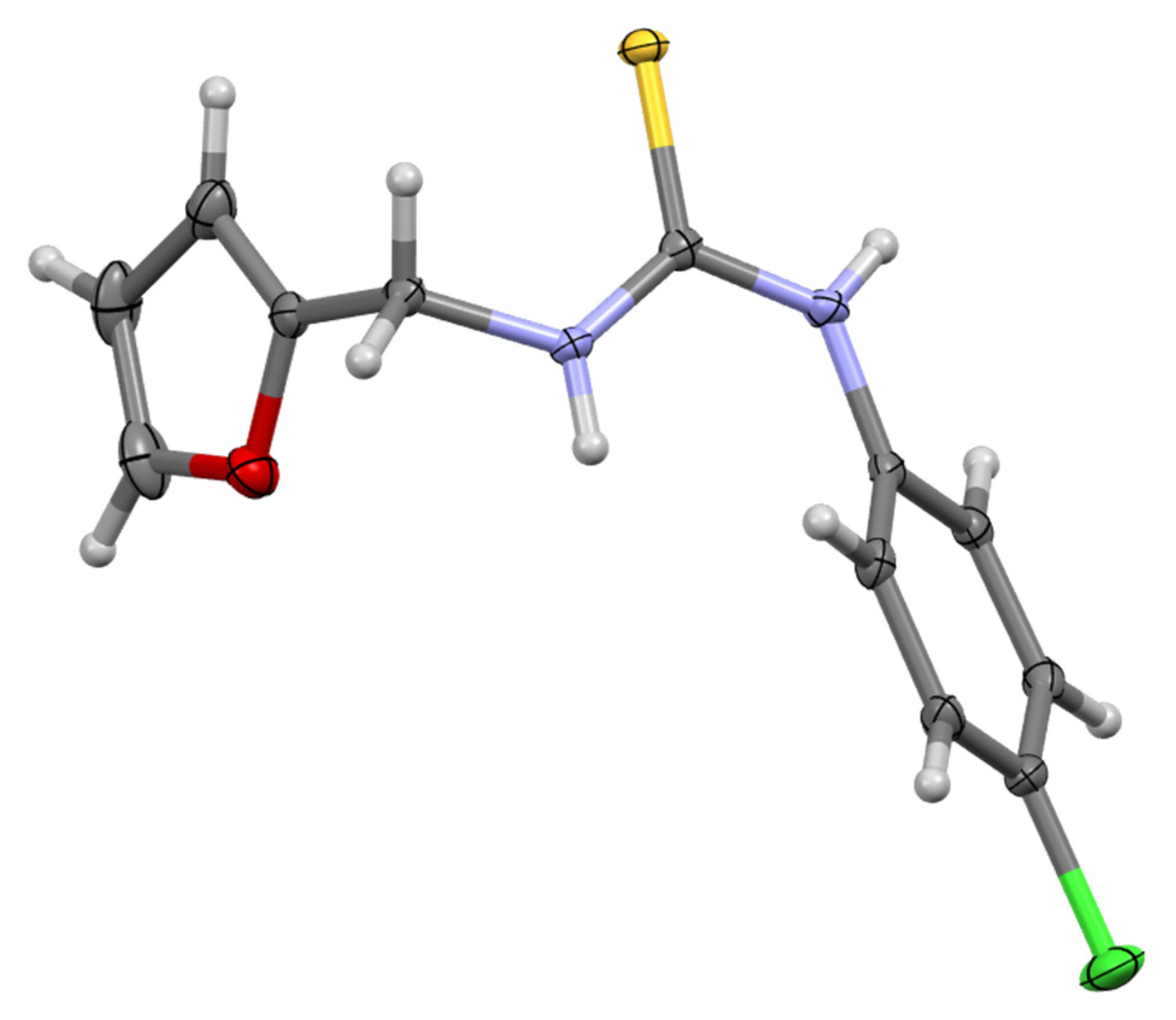
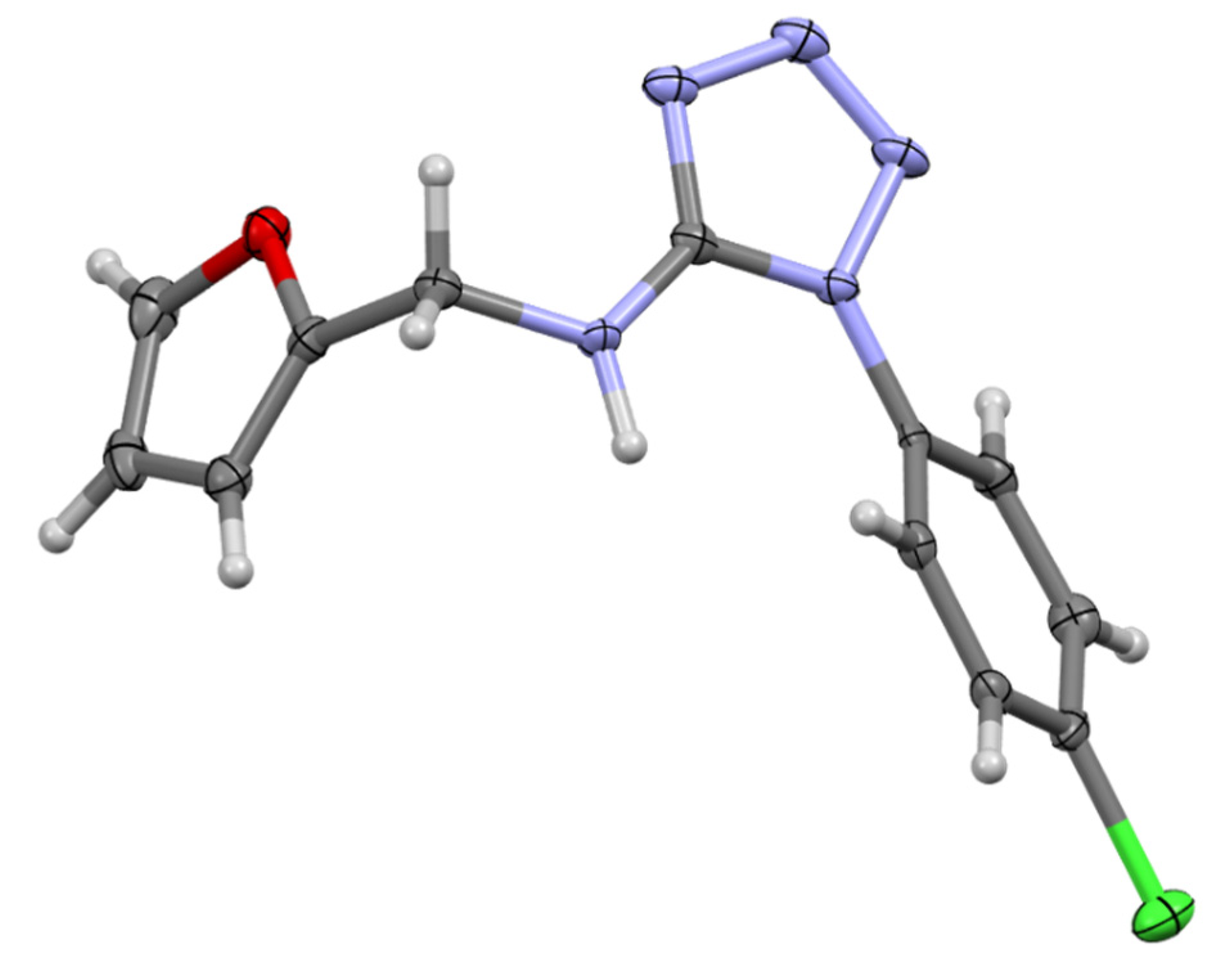
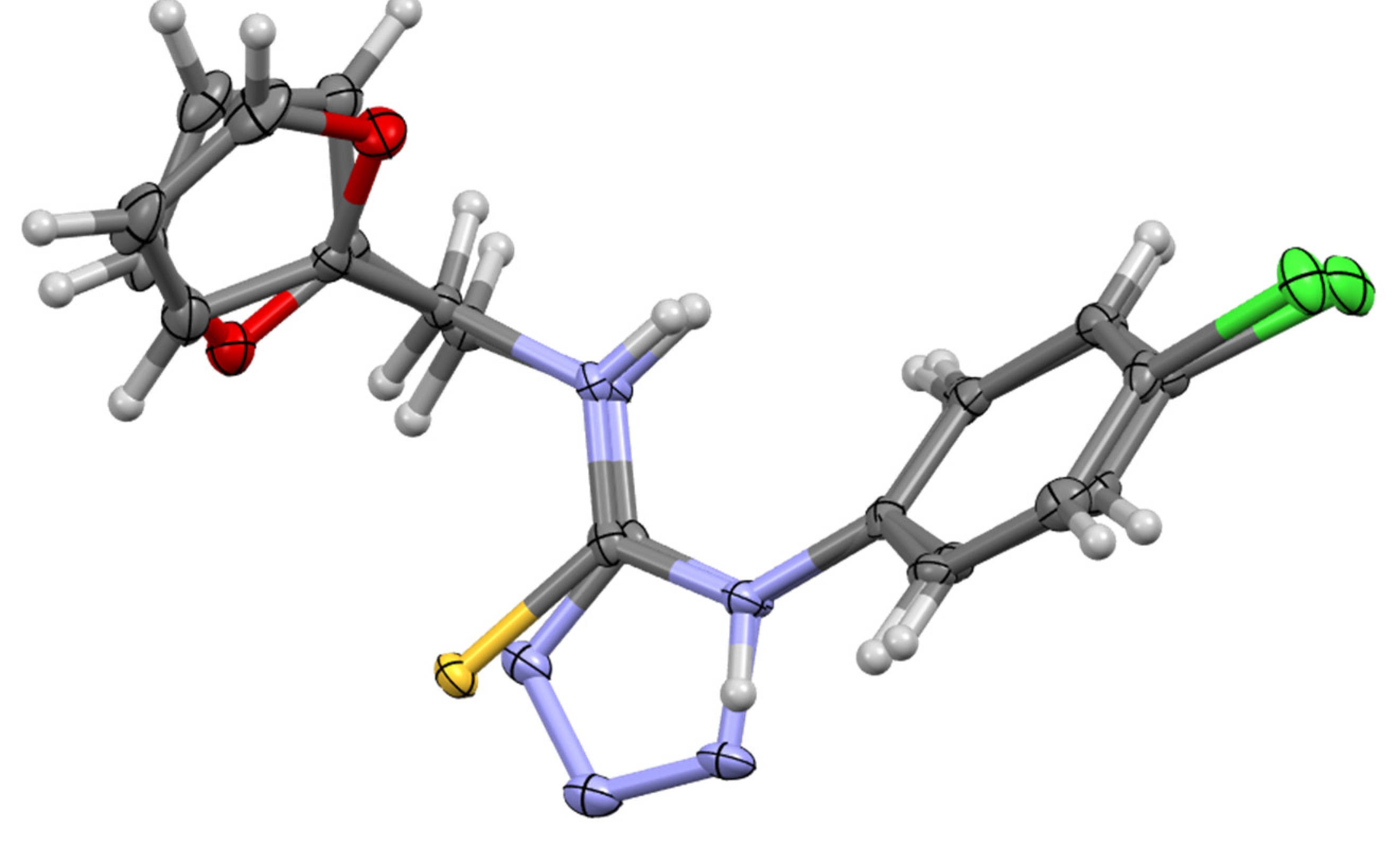
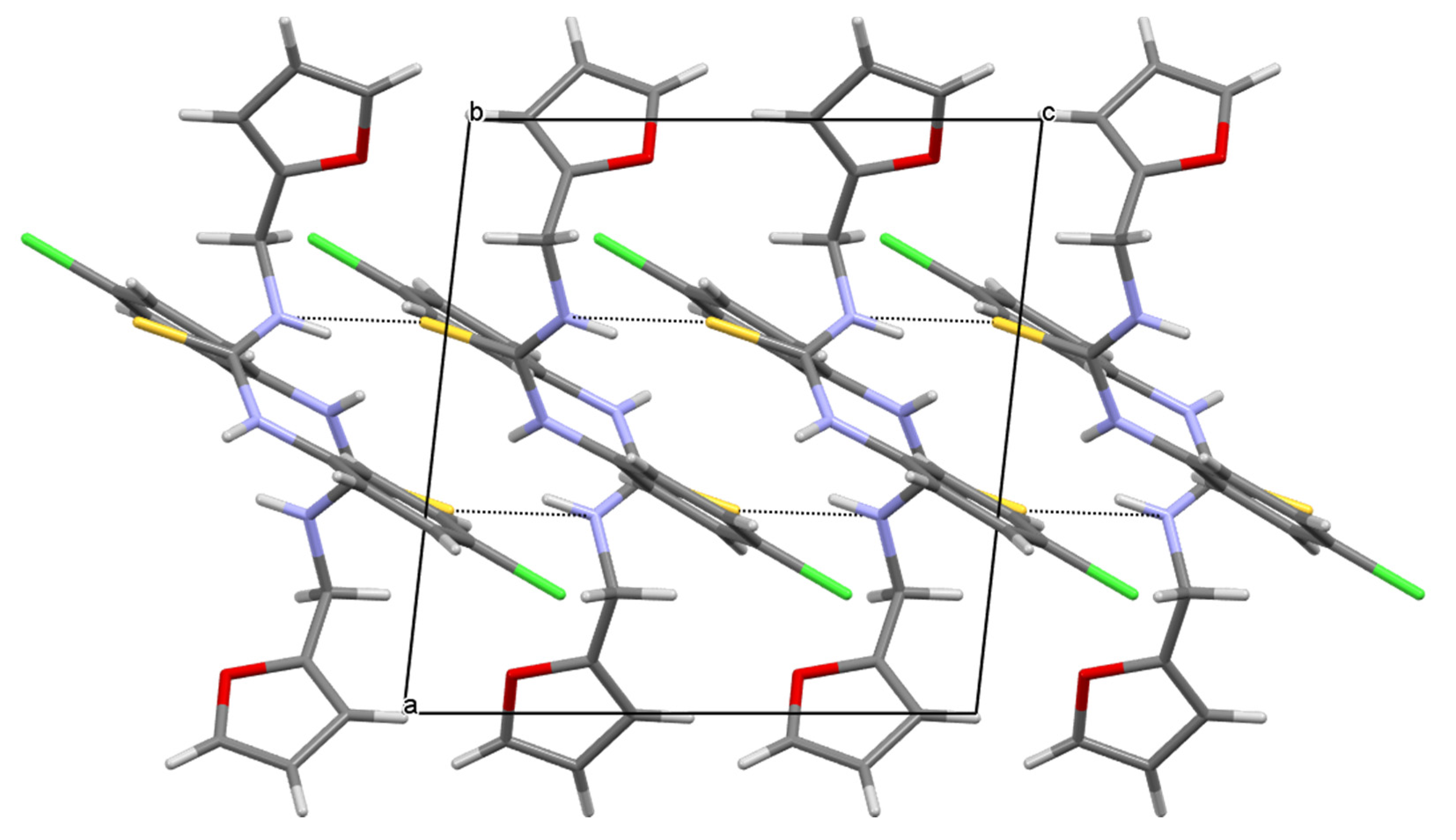
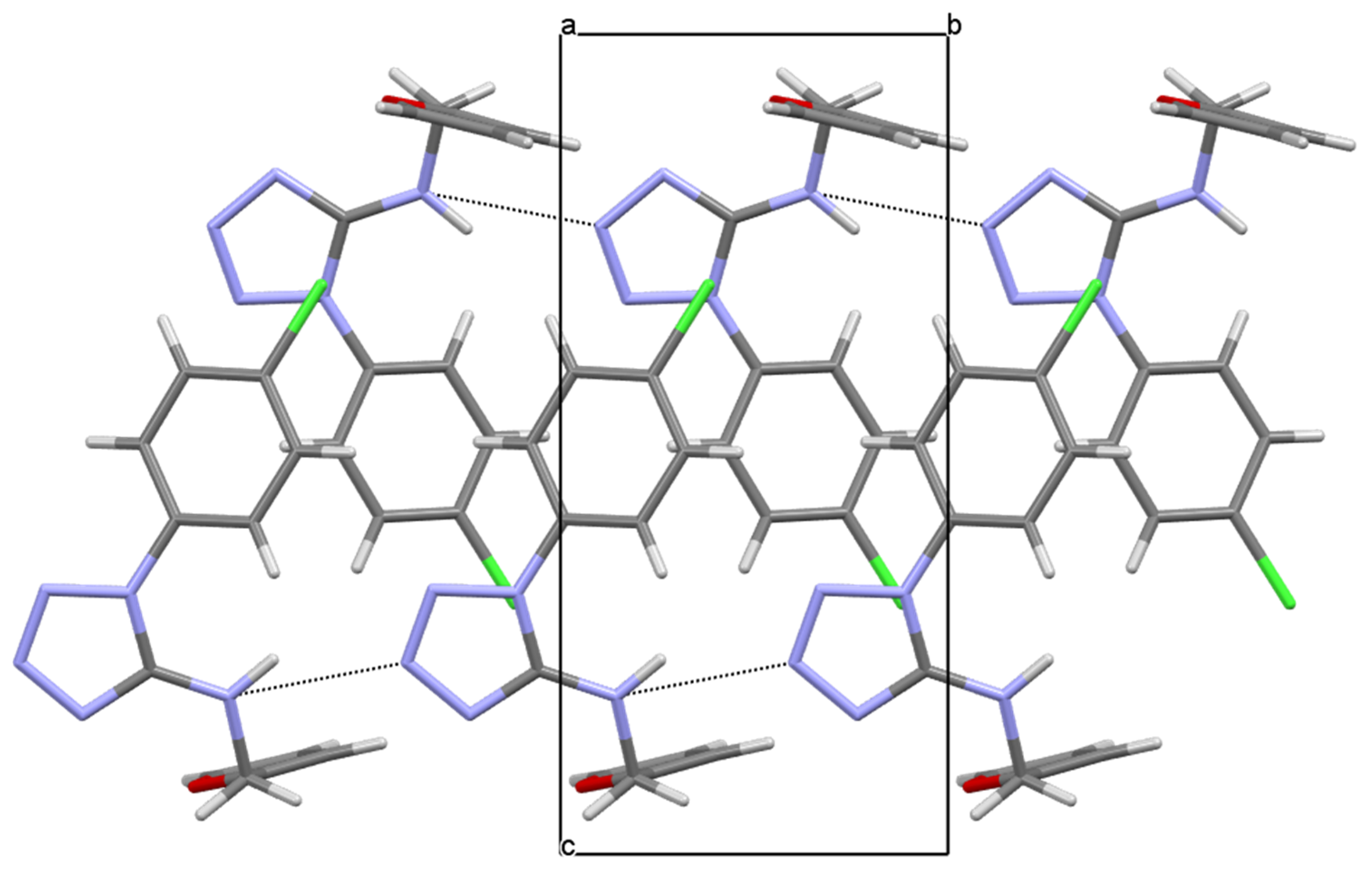
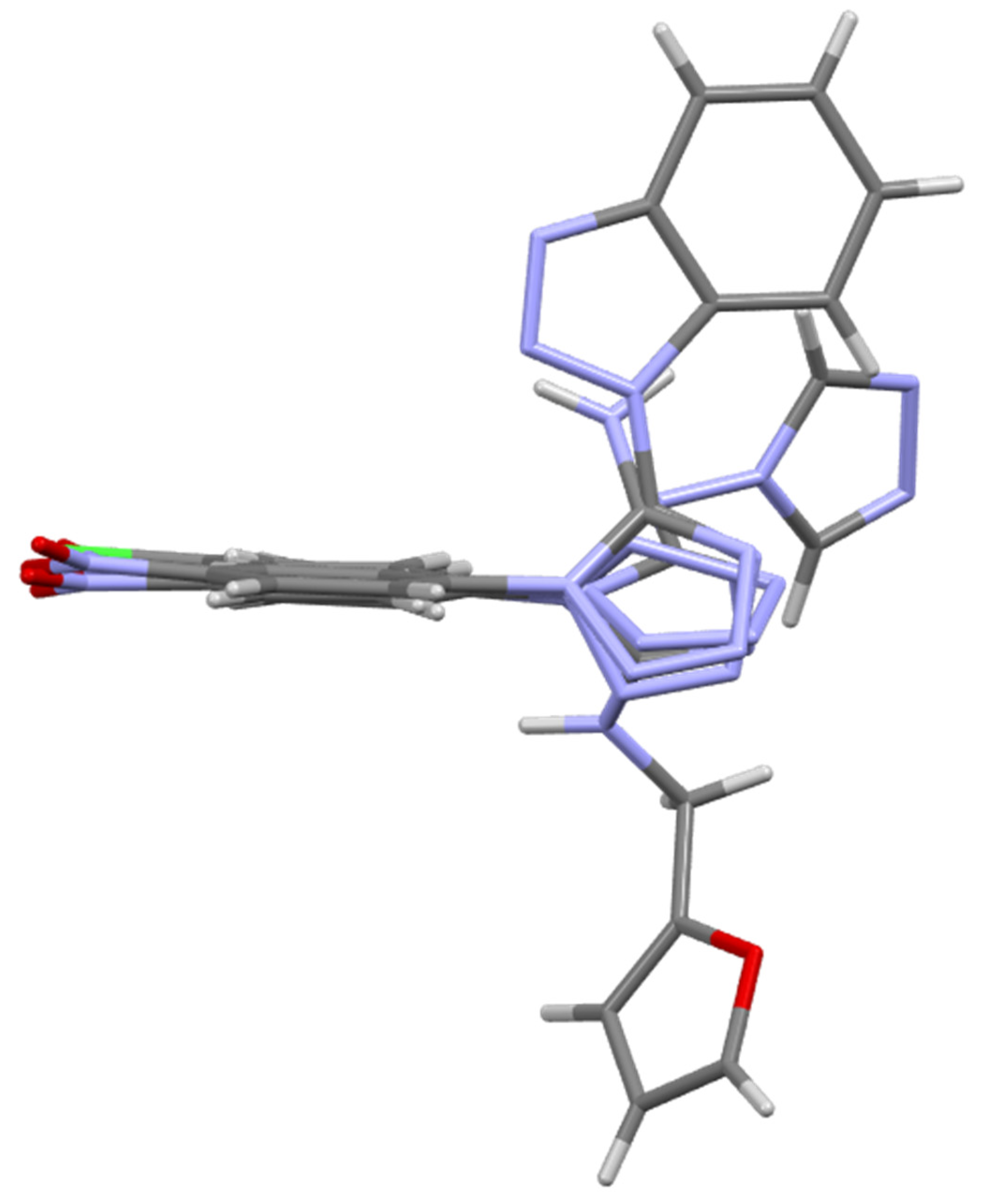
2.3. X-ray Studies
2.4. Biological Studies
2.4.1. In Vitro Antibacterial Activity Studies
2.4.2. In Vitro Antimycobacterial Activity Studies
2.4.3. Cytotoxic Studies
2.4.4. Structure-activity Relationship Studies (SAR)
3. Materials and Methods
3.1. Apparatus, Materials, and Analysis
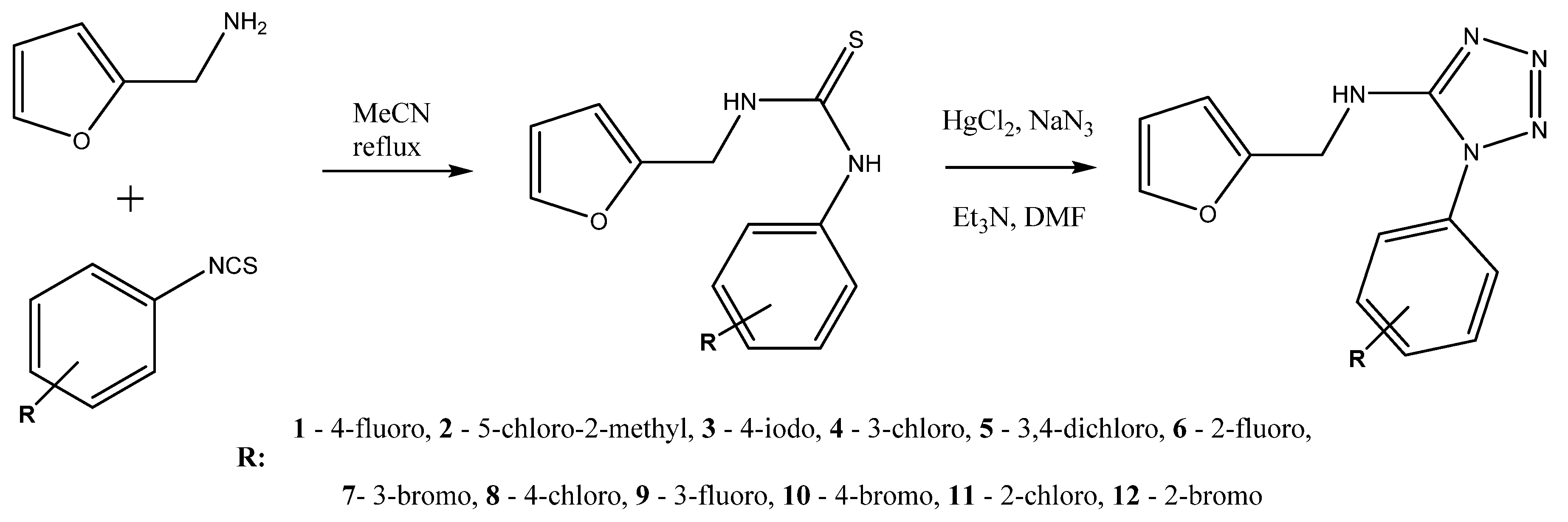
3.2. Derivatives of N-(furan-2-ylmethyl)-1H-tetrazol-5-amine
3.3. Biological Assays
3.3.1. Cytotoxic Studies (MTT Assay)
3.3.2. Antimicrobial Activity
4. Conclusions
Supplementary Materials
Author Contributions
Funding
Data Availability Statement
Acknowledgments
Conflicts of Interest
Sample Availability
References
- Bielenica, A.; Stefańska, J.; Stępień, K.; Napiórkowska, A.; Augustynowicz-Kopeć, E.; Sanna, G.; Madeddu, S.; Boi, S.; Giliberti, G.; Wrzosek, M.; et al. Synthesis, cytotoxicity and antimicrobial activity of thiourea derivatives incorporating 3-(trifluoromethyl)phenyl moiety. Eur. J. Med. Chem. 2015, 101, 111–125. [Google Scholar] [CrossRef] [PubMed]
- Abbas, S.Y.; El-Sharief, M.A.M.S.; Basyouni, W.M.; Fakhr, I.M.I.; El-Gammal, E.W. Thiourea derivatives incorporating a hippuric acid moiety: Synthesis and evaluation of antibacterial and antifungal activities. Eur. J. Med. Chem. 2013, 64, 111–120. [Google Scholar] [CrossRef] [PubMed]
- Suresha, G.P.; Suhas, R.; Kapfo, W.; Channe Gowda, D. Urea/thiourea derivatives of quinazolinone–lysine conjugates: Synthesis and structure–activity relationships of a new series of antimicrobials. Eur. J. Med. Chem. 2011, 46, 2530–2540. [Google Scholar] [CrossRef] [PubMed]
- Saeed, S.; Rashid, N.; Jones, P.G.; Ali, M.; Hussain, R. Synthesis, characterization and biological evaluation of some thiourea derivatives bearing benzothiazole moiety as potential antimicrobial and anticancer agents. Eur. J. Med. Chem. 2010, 45, 1323–1331. [Google Scholar] [CrossRef] [PubMed]
- Vega-Pérez, J.M.; Periñán, I.; Argandoña, M.; Vega-Holm, M.; Palo-Nieto, C.; Burgos-Morón, E.; López-Lázaro, M.; Vargas, C.; Nieto, J.J.; Iglesias-Guerra, F. Isoprenyl-thiourea and urea derivatives as new farnesyl diphosphate analogues: Synthesis and in vitro antimicrobial and cytotoxic activities. Eur. J. Med. Chem. 2012, 58, 591–612. [Google Scholar] [CrossRef]
- Stefanska, J.; Szulczyk, D.; Koziol, A.E.; Miroslaw, B.; Kedzierska, E.; Fidecka, S.; Busonera, B.; Sanna, G.; Giliberti, G.; La Colla, P.; et al. Disubstituted thiourea derivatives and their activity on CNS: Synthesis and biological evaluation. Eur. J. Med. Chem. 2012, 55, 205–213. [Google Scholar] [CrossRef]
- Dobrikov, G.M.; Valcheva, V.; Nikolova, Y.; Ugrinova, I.; Pasheva, E.; Dimitrov, V. Efficient synthesis of new (R)-2-amino-1-butanol derived ureas, thioureas and acylthioureas and in vitro evaluation of their antimycobacterial activity. Eur. J. Med. Chem. 2013, 63, 468–473. [Google Scholar] [CrossRef]
- Joshi, S.D.; Dixit, S.R.; Kirankumar, M.N.; Aminabhavi, T.M.; Raju, K.V.S.N.; Narayan, R.; Lherbet, C.; Yang, K.S. Synthesis, antimycobacterial screening and ligand-based molecular docking studies on novel pyrrole derivatives bearing pyrazoline, isoxazole and phenyl thiourea moieties. Eur. J. Med. Chem. 2016, 107, 133–152. [Google Scholar] [CrossRef]
- Dişli, A.; Mercan, S.; Yavuz, S. Synthesis and Antimicrobial Activity of New Pyrimidine Derivatives Incorporating 1H-Tetrazol-5-ylthio Moiety. J. Het. Chem. 2013, 50, 1446–1450. [Google Scholar] [CrossRef]
- Naresh, E.; Bhaskar, K.; Linga goud, G. Synthesis of Some New 5-(1-Phenyl-1H-1,2,3-Triazol-4-Yl)-1H-Tetrazoles and Evaluation of Their Antimicrobial Activity. Russ. J. Gen. Chem. 2016, 86, 2862–2864. [Google Scholar] [CrossRef]
- Chauhan, K.; Singh, P.; Kumar, V.; Shukla, P.K.; Siddiqi, M.I.; Chauhan, P.M.S. Investigation of Ugi-4CC Derived 1H-Tetrazol-5-Yl-(Aryl) Methyl Piperazinyl-6-Fluoro-4-Oxo-1,4-Dihydroquinoline-3-Carboxylic Acid: Synthesis, Biology and 3D-QSAR Analysis. Eur. J. Med. Chem. 2014, 78, 442–454. [Google Scholar] [CrossRef] [PubMed]
- Gao, F.; Xiao, J.; Huang, G. Current Scenario of Tetrazole Hybrids for Antibacterial Activity. Eur. J. Med. Chem. 2019, 184, 111744. [Google Scholar] [CrossRef] [PubMed]
- Bielenica, A.; Szulczyk, D.; Olejarz, W.; Madeddu, S.; Giliberti, G.; Materek, I.B.; Koziol, A.E.; Struga, M. 1H-Tetrazol-5-Amine and 1,3-Thiazolidin-4-One Derivatives Containing 3-(Trifluoromethyl)Phenyl Scaffold: Synthesis, Cytotoxic and Anti-HIV Studies. Biomed. Pharmacother. 2017, 94, 804–812. [Google Scholar] [CrossRef] [PubMed]
- Szulczyk, D.; Bielenica, A.; Głogowska, A.; Augustynowicz-Kopeć, E.; Dobrowolski, M.; Roszkowski, P.; Stępień, K.; Chrzanowska, A.; Struga, M. Development of (4-Methoxyphenyl)-1H-Tetrazol-5-Amine Regioisomers as a New Class of Selective Antitubercular Agents. Eur. J. Med. Chem. 2020, 186, 111882. [Google Scholar] [CrossRef] [PubMed]
- Szulczyk, D.; Bielenica, A.; Roszkowski, P.; Dobrowolski, M.A.; Olejarz, W.; Napiórkowska, M.; Struga, M. Cytotoxicity Evaluation of Novel Bis(2-aminoethyl)amine Derivatives. Molecules 2020, 25, 2861. [Google Scholar] [CrossRef]
- Stefanska, J.; Nowicka, G.; Struga, M.; Szulczyk, D.; Koziol, A.E.; Augustynowicz-Kopec, E.; Napiorkowska, A.; Bielenica, A.; Filipowski, W.; Filipowska, A.; et al. Antimicrobial and Anti-Biofilm Activity of Thiourea Derivatives Incorporating a 2-Aminothiazole Scaffold. Chem. Pharm. Bull. 2015, 63, 225–236. [Google Scholar] [CrossRef] [Green Version]
- Stefanska, J.; Stepien, K.; Bielenica, A.; Szulczyk, D.; Miroslaw, B.; Koziol, A.E.; Sanna, G.; Iuliano, F.; Madeddu, S.; Jozwiak, M.; et al. Antimicrobial and Anti-Biofilm Activity of Thiourea Derivatives Bearing 3-Amino-1H-1,2,4-Triazole Scaffold. Med. Chem. 2016, 12, 478–488. [Google Scholar] [CrossRef]
- Dobrowolski, M.A.; Roszkowski, P.; Struga, M.; Szulczyk, D. The Unexpected Product of Diels-Alder Reaction between “Indanocyclon” and Maleimide. J. Mol. Struct. 2017, 1130, 573–578. [Google Scholar] [CrossRef]
- Szulczyk, D.; Tomaszewski, P.; Jóźwiak, M.; Kozioł, A.E.; Lis, T.; Collu, D.; Iuliano, F.; Struga, M. Synthesis and Biological Activities of Ethyl 2-(2-Pyridylacetate) Derivatives Containing Thiourea, 1,2,4-Triazole, Thiadiazole and Oxadiazole Moieties. Molecules 2017, 22, 409. [Google Scholar] [CrossRef] [Green Version]
- Szulczyk, D.; Dobrowolski, M.A.; Roszkowski, P.; Bielenica, A.; Stefańska, J.; Koliński, M.; Kmiecik, S.; Jóźwiak, M.; Wrzosek, M.; Olejarz, W.; et al. Design and Synthesis of Novel 1H-Tetrazol-5-Amine Based Potent Antimicrobial Agents: DNA Topoisomerase IV and Gyrase Affinity Evaluation Supported by Molecular Docking Studies. Eur. J. Med. Chem. 2018, 156, 631–640. [Google Scholar] [CrossRef]
- Pogodin, P.V.; Lagunin, A.A.; Rudik, A.V.; Druzhilovskiy, D.S.; Filimonov, D.A.; Poroikov, V.V. AntiBac-Pred: A Web Application for Predicting Antibacterial Activity of Chemical Compounds. J. Chem. Info. Mod. 2019, 59, 4513–4518. [Google Scholar] [CrossRef] [PubMed]
- Pires, D.E.V.; Blundell, T.L.; Ascher, D.B. PkCSM: Predicting Small-Molecule Pharmacokinetic and Toxicity Properties Using Graph-Based Signatures. J. Med. Chem. 2015, 58, 4066–4072. [Google Scholar] [CrossRef] [PubMed]
- Bielenica, A.; Stepien, K.; Sawczenko, A.; Lis, T.; Koziol, A.E.; Madeddu, S.; Collu, D.; Iuliano, F.; Kosmider, A.; Struga, M. Synthesis, Structural Studies and Biological Evaluation of Halogen Derivative of 1,3-Disubstituted Thiourea. Lett. Drug Des. Discov. 2017, 14, 636–646. [Google Scholar] [CrossRef]
- Herschhorn, A.; Hizi, A. Virtual screening, identification, and biochemical characterization of novel inhibitors of the reverse transcriptase of human immunodeficiency virus type-1. J. Med. Chem. 2008, 51, 5702–5713. [Google Scholar] [CrossRef]
- Groom, C.R.; Bruno, I.J.; Lightfoot, M.P.; Ward, S.C. The Cambridge Structural Database. Acta Cryst. 2016, B72, 171–179. [Google Scholar] [CrossRef]
- Lyakhov, A.S.; Vorobiov, A.N.; Ivashkevich, L.S.; Gaponik, P.N. Two derivatives of 1,5-disubstituted tetrazoles: 1-(4-nitrophenyl)-1H-tetrazol-5-amine and {(E)-[1-(4-ethoxyphenyl)-1H-tetrazol-5-yl]iminomethyl}dimethylamine. Acta Cryst. 2008, 64, o414–o416. [Google Scholar]
- Lyakhov, A.S.; Egorova, N.G.; Artamonova, T.V.; Gaponik, P.N.; Koldobskii, G.I. 1-[1-(4-Nitrophenyl)-1H-tetrazol-5-yl]-1H-1,2,3-benzotriazole. Acta Cryst. 2007, 63, o2486–o2487. [Google Scholar] [CrossRef]
- Clinical and Laboratory Standards Institute Methods for Dilution Antimicrobial Susceptibility Tests for Bacteria That Grow Aerobically; Approved Standard M7-A7; Clinical and Laboratory Standards Institute: Wayne, PA, USA, 2006.
- Stroet, M.; Caron, B.; Visscher, K.M.; Geerke, D.P.; Malde, A.K.; Mark, A.E. Automated Topology Builder Version 3.0: Prediction of Solvation Free Enthalpies in Water and Hexane. J. Chem. Theory. Comput. 2018, 14, 5834–5845. [Google Scholar] [CrossRef]
- Schneidman-Duhovny, D.; Dror, O.; Inbar, Y.; Nussinov, R.; Wolfson, H.J. PharmaGist: A webserver for ligand-based pharmacophore detection. Nucleic Acids Res. 2008, 36, W223–W228. [Google Scholar] [CrossRef] [Green Version]
- APEX2 (Version 2009.1); Bruker AXS Inc.: Madison, WI, USA.
- SAINT (Version 4.050); Bruker AXS Inc.: Madison, WI, USA.
- SADABS (Version 2004/1); Bruker AXS Inc.: Madison, WI, USA.
- Sheldrick, G.M. Crystal structure refinement with SHELXL. Acta Crystallogr. C 2015, 71, 3–8. [Google Scholar] [CrossRef]
- Wilson, A.J.C.; Geist, V. International Tables for Crystallography; BKluwer Academic Publishers: Dordrecht, The Netherlands, 1993; Volume C, p. 883. [Google Scholar]
- Macrae, C.F.; Edgington, P.R.; McCabe, P.; Pidcock, E.; Shields, G.P.; Taylor, R.; Towler, M.; van de Streek, J. Mercury Visualization and Analysis of Crystal Structures. J. Appl. Crystallogr. 2006, 39, 453–457. [Google Scholar] [CrossRef] [Green Version]
- Chrzanowska, A.; Roszkowski, P.; Bielenica, A.; Olejarz, W.; Stępień, K.; Struga, M. Anticancer and Antimicrobial Effects of Novel Ciprofloxacin Fatty Acids Conjugates. Eur. J. Med. Chem. 2020, 185, 111810. [Google Scholar] [CrossRef] [PubMed]


| Toxicological Test | Unit | 1 | 2 | 3 | 4 | 5 | 6 | 7 | 8 | 9 | 10 | 11 | 12 |
|---|---|---|---|---|---|---|---|---|---|---|---|---|---|
| AMES toxicity | (Yes/No) | No | No | No | No | No | No | No | No | No | No | No | No |
| Max. tolerated dose (human) | (log mg/kg/day) | 0.056 | 0.14 | 0.016 | 0.038 | 0.074 | 0.045 | 0.031 | 0.039 | 0.055 | 0.032 | 0.028 | 0.021 |
| hERG I inhibitor | (Yes/No) | No | No | No | No | No | No | No | No | No | No | No | No |
| hERG II inhibitor | (Yes/No) | No | No | No | No | No | No | No | No | No | No | No | No |
| Oral Rat Acute Toxicity (LD50) | (mol/kg) | 2.069 | 2.211 | 2.148 | 2.145 | 2.254 | 2.052 | 2.154 | 2.151 | 2.064 | 2.16 | 2.133 | 2.141 |
| Oral Rat Chronic Toxicity (LOAEL) | (log mg/kg_bw/day) | 0.99 | 1.001 | 0.966 | 0.947 | 0.898 | 0.977 | 0.947 | 0.953 | 0.983 | 0.954 | 0.941 | 0.941 |
| Hepatotoxicity | (Yes/No) | Yes | Yes | No | Yes | No | Yes | Yes | Yes | Yes | No | Yes | Yes |
| Skin Sensitisation | (Yes/No) | No | No | No | No | No | No | No | No | No | No | No | No |
| T.Pyriformis toxicity | (log ug/L) | 0.34 | 0.341 | 0.342 | 0.34 | 0.346 | 0.337 | 0.341 | 0.342 | 0.338 | 0.343 | 0.34 | 0.34 |
| Minnow toxicity | (log mM) | 1.698 | 1.426 | 1.079 | 1.435 | 1.177 | 1.749 | 1.289 | 1.365 | 1.768 | 1.219 | 1.416 | 1.27 |
| Strain | Compound | Ciprofloxacin [µg/mL] | |||||||
|---|---|---|---|---|---|---|---|---|---|
| 1 | 2 | 3 | 4 | 6 | 8 | Ref 1 | Ref 2 | ||
| S. aureus NCTC 4163 | 64 | 64 | 64 | 256 | 8 | 128 | 4 | 2 | 0.125 |
| S. aureus ATCC 29213 | 64 | 64 | 64 | 256 | 8 | 128 | - | - | 0.5 |
| S. epidermidis ATCC 12228 | 256 | 256 | 128 | 512 | 32 | > | 2 | 2 | 0,25 |
| E. hirae ATCC 10541 | 128/64 | 64 | 128 | 512 | 16 | > | 8 | 4 | 2 |
| E. coli ATCC 25922 | 128 | 128 | 128 | > | 16 | > | - | - | 0.015 |
| P. aeruginosa ATCC 27853 | > | > | > | > | > | > | 4 | 2 | 0.25 |
| E. coli NCTC 8196 | 128 | 128 | 128 | 512 | 512 | > | - | - | 0.015 |
| P. aeruginosa ATCC 15442 | > | > | 512 | > | > | > | 32 | 64 | 0.125 |
| Strain | Compound 6 [µg/mL] | Ciprofloxacin [µg/mL] |
|---|---|---|
| S. epidermidis KR 4047 825/19 | 8 | 4 |
| S. epidermidis KR 4243 829/19 | 4 | 0.015 |
| S. epidermidis KR 4268 830/19 | 8 | 0.03 |
| S. epidermidis KR 4313 834/19 | 4 | 64 |
| S. epidermidis KR 4358/2 840/19 | 4 | 0.25 |
| S. epidermidis T 5253 845/19 | 8 | 64 |
| S. epidermidis T 5399 848/19 | 4 | 0.015 |
| S. epidermidis T 5501 851/19 | 2 | 0.03 |
| S. aureus T5384/48 | 8 | 0.2 |
| S. aureus 203/19 | 8 | 0.1 |
| S. aureus T5403/51 | 8 | 0.2 |
| S. aureus 176/13 | 8 | 0.5 |
| S. aureus T5241/46 | 8 | 0.1 |
| S. aureus 155/6 | 16 | 0.1 |
| S. aureus T5591/68 | 8 | 0.1 |
| S. aureus T5595/69 | 8 | 0.1 |
Publisher’s Note: MDPI stays neutral with regard to jurisdictional claims in published maps and institutional affiliations. |
© 2021 by the authors. Licensee MDPI, Basel, Switzerland. This article is an open access article distributed under the terms and conditions of the Creative Commons Attribution (CC BY) license (http://creativecommons.org/licenses/by/4.0/).
Share and Cite
Szulczyk, D.; Bielenica, A.; Roszkowski, P.; Dobrowolski, M.A.; Olejarz, W.; Kmiecik, S.; Podsiad, M.; Struga, M. Synthetic Transition from Thiourea-Based Compounds to Tetrazole Derivatives: Structure and Biological Evaluation of Synthesized New N-(Furan-2-ylmethyl)-1H-tetrazol-5-amine Derivatives. Molecules 2021, 26, 323. https://doi.org/10.3390/molecules26020323
Szulczyk D, Bielenica A, Roszkowski P, Dobrowolski MA, Olejarz W, Kmiecik S, Podsiad M, Struga M. Synthetic Transition from Thiourea-Based Compounds to Tetrazole Derivatives: Structure and Biological Evaluation of Synthesized New N-(Furan-2-ylmethyl)-1H-tetrazol-5-amine Derivatives. Molecules. 2021; 26(2):323. https://doi.org/10.3390/molecules26020323
Chicago/Turabian StyleSzulczyk, Daniel, Anna Bielenica, Piotr Roszkowski, Michał A. Dobrowolski, Wioletta Olejarz, Sebastian Kmiecik, Małgorzata Podsiad, and Marta Struga. 2021. "Synthetic Transition from Thiourea-Based Compounds to Tetrazole Derivatives: Structure and Biological Evaluation of Synthesized New N-(Furan-2-ylmethyl)-1H-tetrazol-5-amine Derivatives" Molecules 26, no. 2: 323. https://doi.org/10.3390/molecules26020323