
Bioimaging of Lysosomes with a BODIPY pH-Dependent Fluorescent Probe
 , , , and
Abstract
:1. Introduction
2. Results and Discussion
2.1. Synthesis and Characterization of BODIPY Derivative 3
2.2. pH-Dependent Absorption and Fluorescence Spectra
2.3. Cell Viability
2.4. Live-Imaging
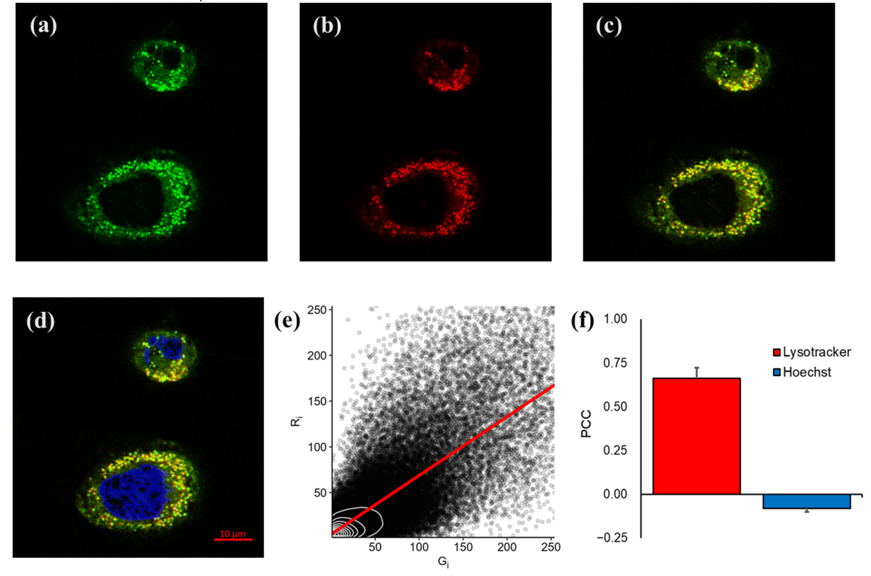
Lysosome Colocalization Assay
3. Materials and Methods
3.1. General
3.2. Synthesis
3.3. pH-Dependent Absorption and Fluorescence Spectra
3.4. Cell Culture
3.5. Cell Viability Assay
3.6. Live-Imaging
3.6.1. Lysosome Colocalization Assay
3.6.2. Quantitative Analysis of Colocalization
4. Conclusions
Supplementary Materials
Author Contributions
Funding
Institutional Review Board Statement
Informed Consent Statement
Data Availability Statement
Conflicts of Interest
Sample Availability
References
- Kobayashi, H.; Ogawa, M.; Alford, R.; Choyke, P.L.; Urano, Y. New Strategies for Fluorescent Probe Design in Medical Diagnostic Imaging. Chem. Rev. 2010, 110, 2620–2640. [Google Scholar] [CrossRef] [PubMed] [Green Version]
- Alamudi, S.H.; Chang, Y.-T. Advances in the Design of Cell-Permeable Fluorescent Probes for Applications in Live Cell Imaging. Chem. Commun. 2018, 54, 13641–13653. [Google Scholar] [CrossRef]
- Yang, X.; Lovell, J.F.; Murthy, N.; Zhang, Y. Organic Fluorescent Probes for Diagnostics and Bio-Imaging. In Fluorescent Imaging in Medicinal Chemistry; Cheng, Z., Ed.; Topics in Medicinal Chemistry; Springer International Publishing: Cham, Switzerland, 2019; Volume 34, pp. 33–53. ISBN 978-3-030-46706-7. [Google Scholar]
- Mizukami, S. Targetable Fluorescent Sensors for Advanced Cell Function Analysis. J. Photochem. Photobiol. C Photochem. Rev. 2017, 30, 24–35. [Google Scholar] [CrossRef]
- Zheng, X.; Cheng, W.; Ji, C.; Zhang, J.; Yin, M. Detection of Metal Ions in Biological Systems: A Review. Rev. Anal. Chem. 2020, 39, 231–246. [Google Scholar] [CrossRef]
- Zhang, J.; Li, J.; Chen, B.; Kan, J.; Jiang, T.; Zhang, W.; Yue, J.; Zhou, J. An Off-on Fluorescent Probe for Real-Time Sensing the Fluctuations of Intracellular PH Values in Biological Processes. Dyes Pigm. 2019, 170, 107620. [Google Scholar] [CrossRef]
- Hande, P.E.; Shelke, Y.G.; Datta, A.; Gharpure, S.J. Recent Advances in Small Molecule-Based Intracellular PH Probes. ChemBioChem 2021, 22, 1–19. [Google Scholar] [CrossRef] [PubMed]
- Kaur, P.; Singh, K. Recent Advances in the Application of BODIPY in Bioimaging and Chemosensing. J. Mater. Chem. C 2019, 7, 11361–11405. [Google Scholar] [CrossRef]
- Kowada, T.; Maeda, H.; Kikuchi, K. BODIPY-Based Probes for the Fluorescence Imaging of Biomolecules in Living Cells. Chem. Soc. Rev. 2015, 44, 4953–4972. [Google Scholar] [CrossRef]
- Bañuelos, J. BODIPY Dye, the Most Versatile Fluorophore Ever? Chem. Rec. 2016, 16, 335–348. [Google Scholar] [CrossRef]
- Ulrich, G.; Ziessel, R.; Harriman, A. The Chemistry of Fluorescent Bodipy Dyes: Versatility Unsurpassed. Angew. Chem. Int. Ed. 2008, 47, 1184–1201. [Google Scholar] [CrossRef]
- Loudet, A.; Burgess, K. BODIPY Dyes and Their Derivatives: Syntheses and Spectroscopic Properties. Chem. Rev. 2007, 107, 4891–4932. [Google Scholar] [CrossRef]
- Terai, T.; Nagano, T. Small-Molecule Fluorophores and Fluorescent Probes for Bioimaging. Pflugers Arch. - Eur J. Physiol 2013, 465, 347–359. [Google Scholar] [CrossRef] [PubMed]
- Wu, X.; Shi, W.; Li, X.; Ma, H. Recognition Moieties of Small Molecular Fluorescent Probes for Bioimaging of Enzymes. Acc. Chem. Res. 2019, 52, 1892–1904. [Google Scholar] [CrossRef]
- Choi, N.-E.; Lee, J.-Y.; Park, E.-C.; Lee, J.-H.; Lee, J. Recent Advances in Organelle-Targeted Fluorescent Probes. Molecules 2021, 26, 217. [Google Scholar] [CrossRef] [PubMed]
- Ballabio, A.; Bonifacino, J.S. Lysosomes as Dynamic Regulators of Cell and Organismal Homeostasis. Nat. Rev. Mol. Cell Biol 2020, 21, 101–118. [Google Scholar] [CrossRef] [PubMed]
- Schmid, D.; Dengjel, J.; Schoor, O.; Stevanovic, S.; Münz, C. Autophagy in Innate and Adaptive Immunity against Intracellular Pathogens. J. Mol. Med. 2006, 84, 194–202. [Google Scholar] [CrossRef]
- Savini, M.; Zhao, Q.; Wang, M.C. Lysosomes: Signaling Hubs for Metabolic Sensing and Longevity. Trends Cell Biol. 2019, 29, 876–887. [Google Scholar] [CrossRef]
- Mindell, J.A. Lysosomal Acidification Mechanisms. Annu. Rev. Physiol. 2012, 74, 69–86. [Google Scholar] [CrossRef] [PubMed] [Green Version]
- Webb, B.A.; Aloisio, F.M.; Charafeddine, R.A.; Cook, J.; Wittmann, T.; Barber, D.L. PHLARE: A New Biosensor Reveals Decreased Lysosome PH in Cancer Cells. Mol. Biol. Cell 2021, 32, 131–142. [Google Scholar] [CrossRef]
- Chen, R.; Jäättelä, M.; Liu, B. Lysosome as a Central Hub for Rewiring PH Homeostasis in Tumors. Cancers 2020, 12, 2437. [Google Scholar] [CrossRef]
- Wang, Q.; Zhou, L.; Qiu, L.; Lu, D.; Wu, Y.; Zhang, X.-B. An Efficient Ratiometric Fluorescent Probe for Tracking Dynamic Changes in Lysosomal PH. Analyst 2015, 140, 5563–5569. [Google Scholar] [CrossRef]
- Wang, X.; Fan, L.; Wang, Y.; Zhang, C.; Liang, W.; Shuang, S.; Dong, C. Visual Monitoring of the Lysosomal PH Changes during Autophagy with a Red-Emission Fluorescent Probe. J. Mater. Chem. B 2020, 8, 1466–1471. [Google Scholar] [CrossRef] [PubMed]
- Horak, E.; Kassal, P.; Murković Steinberg, I. Benzimidazole as a Structural Unit in Fluorescent Chemical Sensors: The Hidden Properties of a Multifunctional Heterocyclic Scaffold. Supramol. Chem. 2018, 30, 838–857. [Google Scholar] [CrossRef]
- Gong, T.; Li, R.; Yuan, Y.; Yu, B.; Zhao, H.; Liu, Z.; Guo, R.; Su, D.; Liang, W.; Dong, C. A Benzimidazole-Based Highly Selective Colorimetric and Far-Red Fluorometric PH Sensor for Intracellular Imaging. New J. Chem. 2018, 42, 12954–12959. [Google Scholar] [CrossRef]
- Pinto, S.C.S.; Gonçalves, R.C.R.; Costa, S.P.G.; Raposo, M.M.M. Synthesis and Characterization of a Meso-Anthracene-BODIPY Derivative for Colorimetric Recognition of Cu2+ and Fe3+. Chem. Proc. 2021; 3, p. 79. [Google Scholar]
- Gonçalves, R.C.R.; Pinto, S.C.S.; Costa, S.P.G.; Raposo, M.M.M. Anion Colorimetric Chemosensor Based on a Benzimidazole-Functionalized BODIPY Derivative. Chem. Proc. 2022; 8, p. 90. [Google Scholar]
- Sunahara, H.; Urano, Y.; Kojima, H.; Nagano, T. Design and Synthesis of a Library of BODIPY-Based Environmental Polarity Sensors Utilizing Photoinduced Electron-Transfer-Controlled Fluorescence ON/OFF Switching. J. Am. Chem. Soc. 2007, 129, 5597–5604. [Google Scholar] [CrossRef]
- Jia, Y.; Pan, Y.; Wang, H.; Chen, R.; Wang, H.; Cheng, X. Highly Selective and Sensitive Polymers with Fluorescent Side Groups for the Detection of Hg 2+ Ion. Mater. Chem. Phys. 2017, 196, 262–269. [Google Scholar] [CrossRef]
- Wagner, R.W.; Lindsey, J.S. Boron-Dipyrromethene Dyes for Incorporation in Synthetic Multi-Pigment Light-Harvesting Arrays. Pure Appl. Chem. 1996, 68, 1373–1380. [Google Scholar] [CrossRef]
- Gonçalves, R.C.R.; Pina, J.; Costa, S.P.G.; Raposo, M.M.M. Synthesis and Characterization of Aryl-Substituted BODIPY Dyes Displaying Distinct Solvatochromic Singlet Oxygen Photosensitization Efficiencies. Dyes Pigm. 2021, 196, 109784. [Google Scholar] [CrossRef]
- Lo Presti, M.; Martínez-Máñez, R.; Ros-Lis, J.V.; Batista, R.M.F.; Costa, S.P.G.; Raposo, M.M.M.; Sancenón, F. A Dual Channel Sulphur-Containing a Macrocycle Functionalised BODIPY Probe for the Detection of Hg( ii ) in a Mixed Aqueous Solution. New J. Chem. 2018, 42, 7863–7868. [Google Scholar] [CrossRef] [Green Version]
- Collado, D.; Casado, J.; González, S.R.; Navarrete, J.T.L.; Suau, R.; Perez-Inestrosa, E.; Pappenfus, T.M.; Raposo, M.M.M. Enhanced Functionality for Donor–Acceptor Oligothiophenes by Means of Inclusion of BODIPY: Synthesis, Electrochemistry, Photophysics, and Model Chemistry. Chem. Eur. J. 2011, 17, 498–507. [Google Scholar] [CrossRef]
- Li, Z.; Li, L.-J.; Sun, T.; Liu, L.; Xie, Z. Benzimidazole-BODIPY as Optical and Fluorometric PH Sensor. Dyes Pigm. 2016, 128, 165–169. [Google Scholar] [CrossRef]
- Hu, W.; Liu, M.; Zhang, X.-F.; Wang, Y.; Wang, Y.; Lan, H.; Zhao, H. Can BODIPY-Electron Acceptor Conjugates Act As Heavy Atom-Free Excited Triplet State and Singlet Oxygen Photosensitizers via Photoinduced Charge Separation-Charge Recombination Mechanism? J. Phys. Chem. C 2019, 123, 15944–15955. [Google Scholar] [CrossRef]
- Lipka, E.; Folly-Klan, M.; Charton, J.; Vaccher, M.-P.; Bonte, J.-P.; Vaccher, C. Determination of PKa Values of Benzimidazole Derivatives from Mobility Obtained by Capillary Electrophoresis. J. Pharm. Biomed. Anal. 2010, 53, 1267–1271. [Google Scholar] [CrossRef] [PubMed]
- Li, S.; Song, X.; Hu, Z.; Feng, G. A Carbon Dots-Based Ratiometric Fluorescence Probe for Monitoring Intracellular PH and Bioimaging. J. Photochem. Photobiol. A: Chem. 2021, 409, 113129. [Google Scholar] [CrossRef]
- Crosby, G.A.; Demas, J.N. Measurement of Photoluminescence Quantum Yields. Review. J. Phys. Chem. 1971, 75, 991–1024. [Google Scholar] [CrossRef]
- Montalti, M.; Credi, A.; Prodi, L.; Gandolfi, M.T. Handbook of Photochemistry, 3rd ed.; CRC Press: Boca Raton, FL, USA, 2006; ISBN 978-0-429-11538-7. [Google Scholar]
- Seixas de Melo, J.; Pina, J.; Dias, F.B.; Maçanita, A.L. Experimental Techniques for Excited State Characterisation; Evans, R., Douglas, P., Burrows, H.D., Eds.; Springer: Dordrecht, The Netherlands, 2013; ISBN 978-90-481-3829-6. [Google Scholar]
- Flors, C.; Nonell, S. On the Phosphorescence of 1H-Phenalen-1-One. Helv. Chim. Acta 2001, 84, 2533–2539. [Google Scholar] [CrossRef]
- Redmond, R.W.; Gamlin, J.N. A Compilation of Singlet Oxygen Yields from Biologically Relevant Molecules. Photochem. Photobiol. 1999, 70, 391–475. [Google Scholar] [CrossRef]
- Pineiro, M.; Carvalho, A.L.; Pereira, M.M.; d’A. Rocha Gonsalves, A.M.; Arnaut, L.G.; Formosinho, S.J. Photoacoustic Measurements of Porphyrin Triplet-State Quantum Yields and Singlet-Oxygen Efficiencies. Chem. Eur. J. 1998, 4, 2299–2307. [Google Scholar] [CrossRef]
- Belmonte-Reche, E.; Martínez-García, M.; Peñalver, P.; Gómez-Pérez, V.; Lucas, R.; Gamarro, F.; Pérez-Victoria, J.M.; Morales, J.C. Tyrosol and Hydroxytyrosol Derivatives as Antitrypanosomal and Antileishmanial Agents. Eur. J. Med. Chem. 2016, 119, 132–140. [Google Scholar] [CrossRef]
- Larson, E.M.; Doughman, D.J.; Gregerson, D.S.; Obritsch, W.F. A New, Simple, Nonradioactive, Nontoxic in Vitro Assay to Monitor Corneal Endothelial Cell Viability. Investig. Ophthalmol. Vis. Sci. 1997, 38, 1929–1933. [Google Scholar]
- Schneider, C.A.; Rasband, W.S.; Eliceiri, K.W. NIH Image to ImageJ: 25 Years of Image Analysis. Nat. Methods 2012, 9, 671–675. [Google Scholar] [CrossRef] [PubMed]
- Bolte, S.; Cordelières, F.P. A Guided Tour into Subcellular Colocalization Analysis in Light Microscopy. J. Microsc. 2006, 224, 213–232. [Google Scholar] [CrossRef] [PubMed]
- Costes, S.V.; Daelemans, D.; Cho, E.H.; Dobbin, Z.; Pavlakis, G.; Lockett, S. Automatic and Quantitative Measurement of Protein-Protein Colocalization in Live Cells. Biophys. J. 2004, 86, 3993–4003. [Google Scholar] [CrossRef] [PubMed]


| Compound | Yield (%) | λabs | λfluo | ΔSS | ϕF | ϕΔ |
|---|---|---|---|---|---|---|
| (nm) | (nm) | (cm−1) | ||||
| 1 | 74 | 505 | 515 | 385 | 0.43 | 0.29 |
| (502) | (515) | (0.03) | ||||
| 2 | 47 | 502 | 525 | 873 | 0.02 | 0.36 |
| (497) | (509) | (0.01) | ||||
| 3 | 30 | 521 | 585 | 2100 | 0.35 | 0.04 |
| (515) | (600) | (0.10) |
Publisher’s Note: MDPI stays neutral with regard to jurisdictional claims in published maps and institutional affiliations. |
© 2022 by the authors. Licensee MDPI, Basel, Switzerland. This article is an open access article distributed under the terms and conditions of the Creative Commons Attribution (CC BY) license (https://creativecommons.org/licenses/by/4.0/).
Share and Cite
Gonçalves, R.C.R.; Belmonte-Reche, E.; Pina, J.; Costa da Silva, M.; Pinto, S.C.S.; Gallo, J.; Costa, S.P.G.; Raposo, M.M.M. Bioimaging of Lysosomes with a BODIPY pH-Dependent Fluorescent Probe. Molecules 2022, 27, 8065. https://doi.org/10.3390/molecules27228065
Gonçalves RCR, Belmonte-Reche E, Pina J, Costa da Silva M, Pinto SCS, Gallo J, Costa SPG, Raposo MMM. Bioimaging of Lysosomes with a BODIPY pH-Dependent Fluorescent Probe. Molecules. 2022; 27(22):8065. https://doi.org/10.3390/molecules27228065
Chicago/Turabian StyleGonçalves, Raquel C. R., Efres Belmonte-Reche, João Pina, Milene Costa da Silva, Sónia C. S. Pinto, Juan Gallo, Susana P. G. Costa, and M. Manuela M. Raposo. 2022. "Bioimaging of Lysosomes with a BODIPY pH-Dependent Fluorescent Probe" Molecules 27, no. 22: 8065. https://doi.org/10.3390/molecules27228065