Article

Two New Cytotoxic Sesquiterpene-Amino Acid Conjugates and a Coumarin-Glucoside from Crossostephium chinense

1
Graduate School of Biomedical & Health Sciences, Hiroshima University, 1-2-3 Kasumi, Minami-ku, Hiroshima 734-8553, Japan
2
Department of Protozoology, Institute of Tropical Medicine (NEKKEN), Nagasaki University, 1-12-4 Sakamoto, Nagasaki 852-8523, Japan
3
National Research Center for Protozoan Diseases, Obihiro University of Agriculture and Veterinary Medicine, Inada, Obihiro 080-8555, Japan
4
Graduate School of Pharmacy, Yasuda Women’s University, Hiroshima 731-0153, Japan
*
Author to whom correspondence should be addressed.
Molecules 2023, 28(12), 4696; https://doi.org/10.3390/molecules28124696
Submission received: 23 May 2023 / Revised: 8 June 2023 / Accepted: 9 June 2023 / Published: 11 June 2023

Abstract

:
The Asteraceae family is a promising source of bioactive compounds, such as the famous Asteraceae plants Tanacetum cinerariifolium (pyrethrin) and Artemisia annua (artemisinin). As a result of our series of phytochemical studies of the subtropical plants, two novel sesquiterpenes, named crossoseamines A and B in this study (1 and 2, respectively), one undescribed coumarin-glucoside (3), and eighteen known compounds (421) were isolated from the aerial part of Crossostephium chinense (Asteraceae). The structures of isolated compounds were elucidated by spectroscopic methods, including 1D and 2D NMR experiments (1H, 13C, DEPT, COSY, HSQC, HMBC, and NOESY), IR spectrum, circular dichroism spectrum (CD), and high-resolution electrospray ionization–mass spectrometry (HR-ESI–MS). All isolated compounds were evaluated for their cytotoxic activities against Leishmania major, Plasmodium falciparum, Trypanosoma brucei (gambiense and rhodesiense), and human lung cancer cell line A549 because of the high demand for the discovery of new drug leads to overcome the present side effects and emerging drug-resistant strains. As a result, the new compounds (1 and 2) showed significant activities against A549 (IC50, 1: 3.3 ± 0.3; 2: 12.3 ± 1.0 μg/mL), L. major (IC50, 1: 6.9 ± 0.6; 2: 24.9 ± 2.2 μg/mL), and P. falciparum (IC50, 1: 12.1 ± 1.1; 2: 15.6 ± 1.2 μg/mL).

Graphical Abstract

1. Introduction

Crossostephium chinense (Asteraceae) is an evergreen shrub distributed in Japan, China, and other Asian countries. The shrub naturally grows on coastal raised coral reefs and can reach 10–40 cm in height. From November to December, the racemes are borne on the terminal branches and bloom tiny yellow flowers. C. chinense is widely planted as an ornamental shrub in China because of the high rate of survival for artificial cultivation [1]. The whole plants of C. chinense are traditionally used for the treatment of diabetes and arthritis in southward China [2]. The extract has been reported to have various activities, such as preventing the progression of restenosis [3], inhibiting differentiation, formation and bone resorptive abilities of osteoclasts [4], and larvicidal activity [5]. Flavonoids [6,7], coumarin [2,8], sesquiterpene [2], and triterpenoid [9] have previously been isolated from C. chinense; however, the phytochemical analysis is still insufficient, and a detailed evaluation of biological activities, especially for anticancer and antiparasitic activities, has yet to be conducted.
Cancer is a leading cause of death, especially in industrialized countries, and lung cancer causes the most deaths worldwide [10]. A549 is a cell line derived from human lung cancer and is frequently used in anticancer drug screening.
Leishmaniasis is a disease caused by protozoan parasites which are transmitted by the bite of infected phlebotomine sandflies. Some 1.5–2 million new cases occur annually [11], and emerging drug-resistant strains attract much attention for developing new chemical agents. Leishmania major is associated with cutaneous leishmaniasis, the most common form of leishmaniasis.
Trypanosoma brucei gambiense and T. brucei rhodesiense are the pathogens responsible for West and East African trypanosomiasis, also called sleeping sickness, respectively. Severe side effects and emerging resistant strains for the present clinical drugs have been reported [12,13].
Plasmodium falciparum is a unicellular protozoan parasite of humans and causes the most dangerous malaria, with a high risk of complication and mortality. According to the WHO malaria report of 2021, there were 247 million cases of malaria worldwide in 2021, resulting in an estimated 619,000 deaths. Artemisinin is a famous discovery from Artemisia annua, which belongs to the Asteraceae family and has saved millions of lives. However, artemisinin-resistant strains have been reported from southeastern Asia and Africa [14,15].
Our group focuses on the discovery of new bioactive compounds against these intractable diseases from natural resources. Asteraceae plants were one of the rich sources of bioactive compounds, such as pyrethrin from Tanacetum cinerariifolium and artemisinin from A. anua. Therefore, we are interested in an Asteraceae plant, C. chinense, to discover anticancer and antiparasitic constituents in this study.
The ethyl acetate soluble fraction from the aerial part of C. chinense was phytochemically studied, and all isolated compounds were investigated for their cytotoxic activities against human protozoan pathogens, L. major, P. falciparum, and T. brucei (gambiense and rhodesiense) and also against a human lung cancer cell line, A549. As a result, two novel sesquiterpene-amino acid conjugates (1 and 2) and one previously undescribed coumarin-glucoside (3) were discovered, of which two sesquiterpene-amino acid conjugates showed significant cytotoxic activity. Among the known compounds identified (421), the anti-Trypanosoma activity of 14 and anti-Plasmodium activity of compounds 1, 2, 11, and 14 are reported for the first time. Although the anti-Trypanosoma activity of compounds 69 and 18 and the anti-Plasmodium activity of compounds 69, 12, 13, 15, and 18 have been previously reported, we tested these compounds using our experimental conditions to allow a direct comparison with the activities of 1 and 2 [16,17,18,19,20,21,22,23,24,25,26,27].

2. Results and Discussion

Extraction of C. chinense followed by liquid–liquid partitioning, column chromatography, and HPLC resulted in the isolation of two novel compounds (1, 0.00102% and 2, 0.00175%) and a previously undescribed coumarin glucoside (3, 0.00125%) (Figure 1), together with the following 18 known compounds, i.e., which isolated and elucidated the structure previously, two coumarin derivatives (scopoletin (4, 0.0614%) [28] and scopolin (5, 0.00359%) [29,30]), five flavonoids and flavonoid glucosides (chrysosplenol D (6, 0.0354%) [31], 3-methylquercetin (7, 0.0166%) [32], luteolin (8, 0.0104%) [33], cosmosiine (9, 0.00168%) [34], quercetin-3-O-(6″-O-α-rhamnosyl)-β-glucoside (10, 0.00761%) [35]), seven caffeic acid derivatives (4,5-di-O-caffeoylquinic acid butyl ester (11, 0.0107%) [36], 4,5-di-O-caffeoylquinic acid methyl ester (12, 0.0136%) [37], 3,5-di-O-caffeoylquinic acid methyl ester (13, 0.0154%) [38], 3,4,5-tri-O-caffeoylqunic acid methyl ester (14, 0.00876%) [39], chlorogenic acid methyl ester (15, 0.0111%) [40], 2,6-dimethoxy-4-hydroxymethylphenol-1-O-(6-O-caffeoyl)-β-D-glucopyranoside (16, 0.00497%) [41], and caffeic acid (17, 0.00333%) [42]), 3,4-dihydroxy benzoic acid (18, 0.00639%) [43], one lignan (tetrecentronside B (19, 0.00477%) [44]), and two fatty acids (tianshic acid (20, 0.0241%) [45] and tianshic acid methyl ester (21, 0.00283%) [45]) (Figure 2). Known compounds were identified by comparison of their physicochemical data with those reported data. The structures of novel compounds were elucidated by 1D, 2D NMR, IR, CD, and HR-ESI-MS spectrometry and some chemical reactions.

2.1. Structure Determination of Crossoseamines A and B (1 and 2) and a Novel Coumarin Glucoside (3)

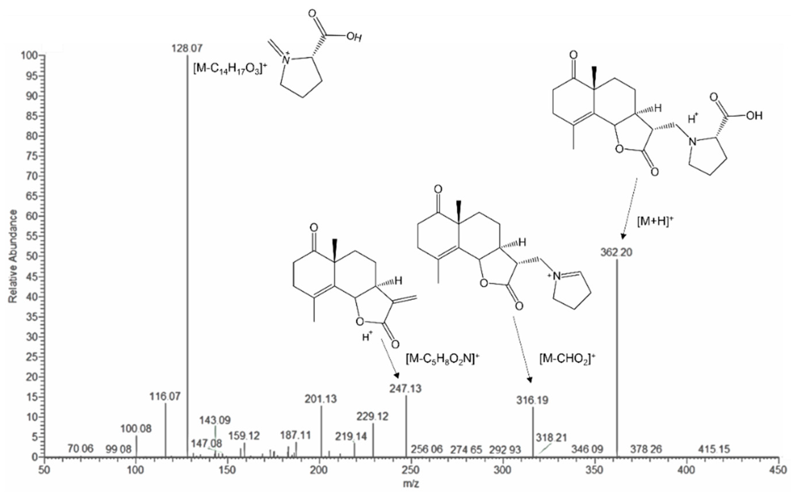
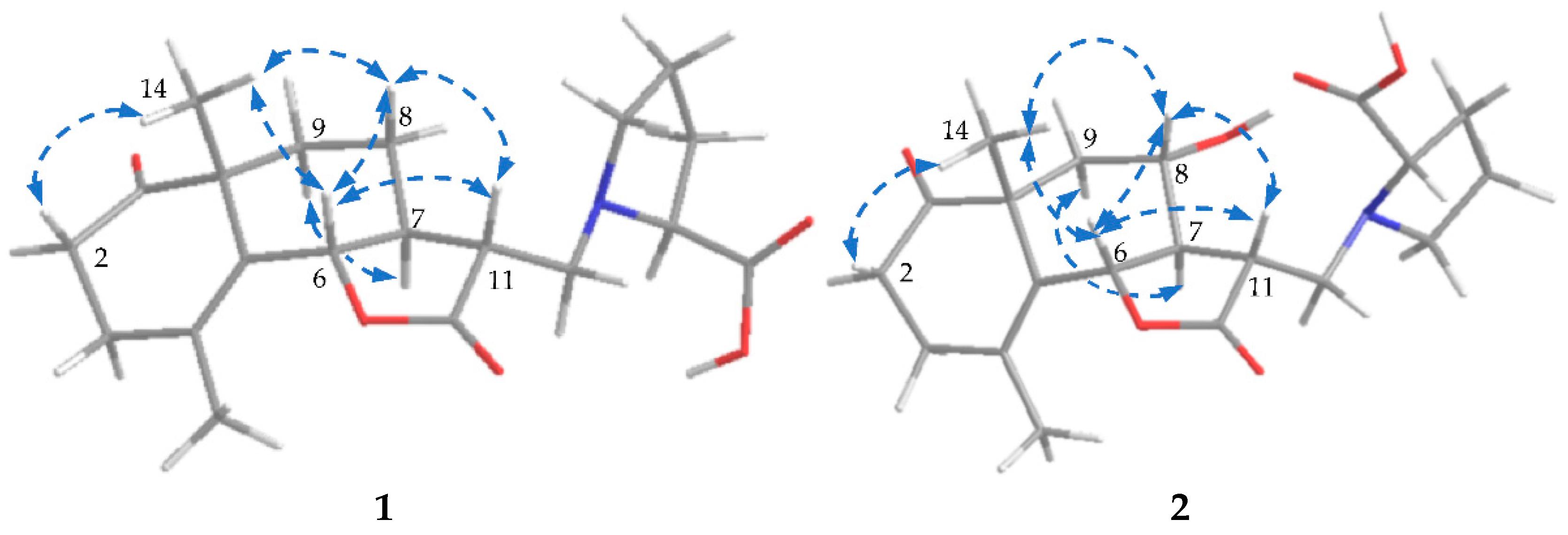
2.1.1. Structure of Crossoseamine A (1)

Crossoseamine A (1) was isolated as a colorless amorphous powder with a specific optical rotation (   [ α ] D 27 97 ,   c   0.2 ,   MeOH ) and was found to have a nitrogen function based on TLC examination using Dragendorff’s reagent. The molecular formula was determined as C20H27O5N by HR-ESI-MS data which revealed a protonated molecule [M + H]+ at m/z 362.1962 (calcd for C20H28O5N: 362.1961). The IR spectrum showed the absorption signals at 3411, 1762, and 1710 cm−1 due to hydroxy and carbonyl functions, of which 1762 cm−1 suggested a γ-lactone functional group. 13C NMR showed that of the eight indices of hydrogen deficiency, four were ascribed to one double bond (δC 125.2 and 130.0) and three carbonyl carbons (δC 212.9, 177.0, and 173.9) (Table 1). In addition, the spectra also displayed one oxymethine (δC 80.8), one aminomethine (δC 66.6), two methines (δC 48.5 and 44.2), one quaternary carbon (δC 48.4), and two methyl carbons (δC 19.4 and 23.0). The 1H-NMR spectrum (Table 1) showed a tertiary methyl, δH 1.25 (3H, s, H3-14), and a vinyl methyl, δH 1.86 (3H, br s, H3-15). The 1D NMR and HSQC spectrum displayed an oxymethine proton signal at δH 4.80 (1H, dquint-like, J = 11.2, 1.3 Hz, H-6) long-range coupled with the vinyl methyl group (H3-15) and one of the methylene protons of H2-3. The COSY correlations between H-5′ and methylene proton H-4′, H-4′ and H-3′, and H-3′ and methine proton H-2′ (Figure 3) and the 13C signals at δC 173.9 (C-6′), 66.6 (C-2′), 28.6 (C-3′), 23.4 (C-4′), and 53.2 (C-5′) suggested a proline moiety [46]. The remaining three hydrogen deficiencies and the COSY spin–spin coupling network from H-13 to H-9 and H-6 implied a eudesmane-type sesquiterpene lactone, eudesmanolide.
The HMBC correlations (Figure 3) from H-2′ (δH 3.30, dd, J = 8.7, 4.6 Hz) to C-13, C-3′, and C-5′; from H-5′ to C-13 and C-2′; and from H-13 to C-12 and C-11 indicated the attachment of proline moiety at C-13 with N. The lower-shifted chemical shift value of C-13 (δC 51.2) was also consistent with this result. Further analysis on HMBC correlations from H-14 to C-10, C-9, C-5, and C-1; from H-2 (δH 2.55, ddd, J = 13.6, 7.0, 6.2 Hz, β; δH 2.43, ddd, J = 13.6, 7.8, 6.6 Hz, α) to C-3 and C-1; and from H-3 (δH 2.35, br dt, J = 17.0, 6.2 Hz, α; δH 2.26, br dt, J = 17.0, 6.9 Hz, β) to C-1, C-2, C-4, and C-5 suggested the position of the carbonyl group at C-1. The ESI-MS/MS spectrum (Figure 4) exhibited fragment ion peaks at m/z (relative intensity, %) 316 (13) and m/z 247 (15), resulting from the loss of a carboxy and a proline group from the molecular ion, respectively. The base ion peak m/z 128 (100) also agreed with the proline moiety. Acid hydrolysis of 1 with 1% HCl liberated L-proline by HPLC analysis with an optical rotation detector.
To determine the absolute configuration of 1, NOESY and CD spectra were measured. The NOESY correlations between H-6 and H-11, between H-14 and H-6, and between H-9α and H-7 revealed the relative configuration of 1, as shown in Figure 5. In the CD spectra, the Cotton effects (Δε (nm): +4.62 (218), −4.70 (295), MeOH) were similar to the reported eudesmanolide, gracilin [47] (Δε (nm): +1.13 (223), −1.70 (294), MeOH), which indicated the absolute configuration should be 6S, 7S, 10R, 11R, 2′S. Accordingly, the structure of crossoseamine A (1) was determined as shown in Figure 1.

2.1.2. Structure of Crossoseamine B (2)

Crossoseamine B (2) was isolated as colorless needles with a specific optical rotation (   [ α ] D 27 47 ,   c   0.46 ,   MeOH ). HR-ESI-MS data displayed a protonated molecule [M + H]+ at m/z 378.1912 (calcd for C20H28O6N: 378.1911), from which the molecular formula was determined as C20H27O6N suggesting an additional oxygen atom on 2 compared to 1. The fragment ion peaks from ESI-MS/MS showing m/z 360 (34) [M + H-H2O]+, m/z 332 (19) [M-COOH]+ and 128 (100) [M-C14H17O4 (proline)]+ indicated a similarity to 1 (see Figure S23). The 1H and 13C NMR spectrum data were similar to compound 1, except for the absence of one methylene carbon and the presence of an oxymethine carbon (δC 66.0) (Table 1). The HMBC correlations from H-7 (δH 1.88, m) to C-8 and C6 and from H-8 (δH 3.89, td, J = 10.8, 4.5 Hz) to C-6 and C-11 revealed that the position of oxymethine carbon (δC 66.0) was at C-8. The relative configuration was deduced as the same as 1 by NOESY analysis (Figure 5), where correlations between H-14 and H-6, between H-6 and H-11, between H-6 and H-8, and between H-14 and H-8 were observed. HPLC analysis of acid hydrolysate revealed the presence of L-proline. Ultimately, based on these spectroscopic data, HPLC analysis, and the similar Cotton effects with compound 1, the structure of crossoseamine B (2) was established as 6S, 7R, 8S, 10R, 11R, 2′S-8-hydroxy analog of 1 as shown in Figure 1.

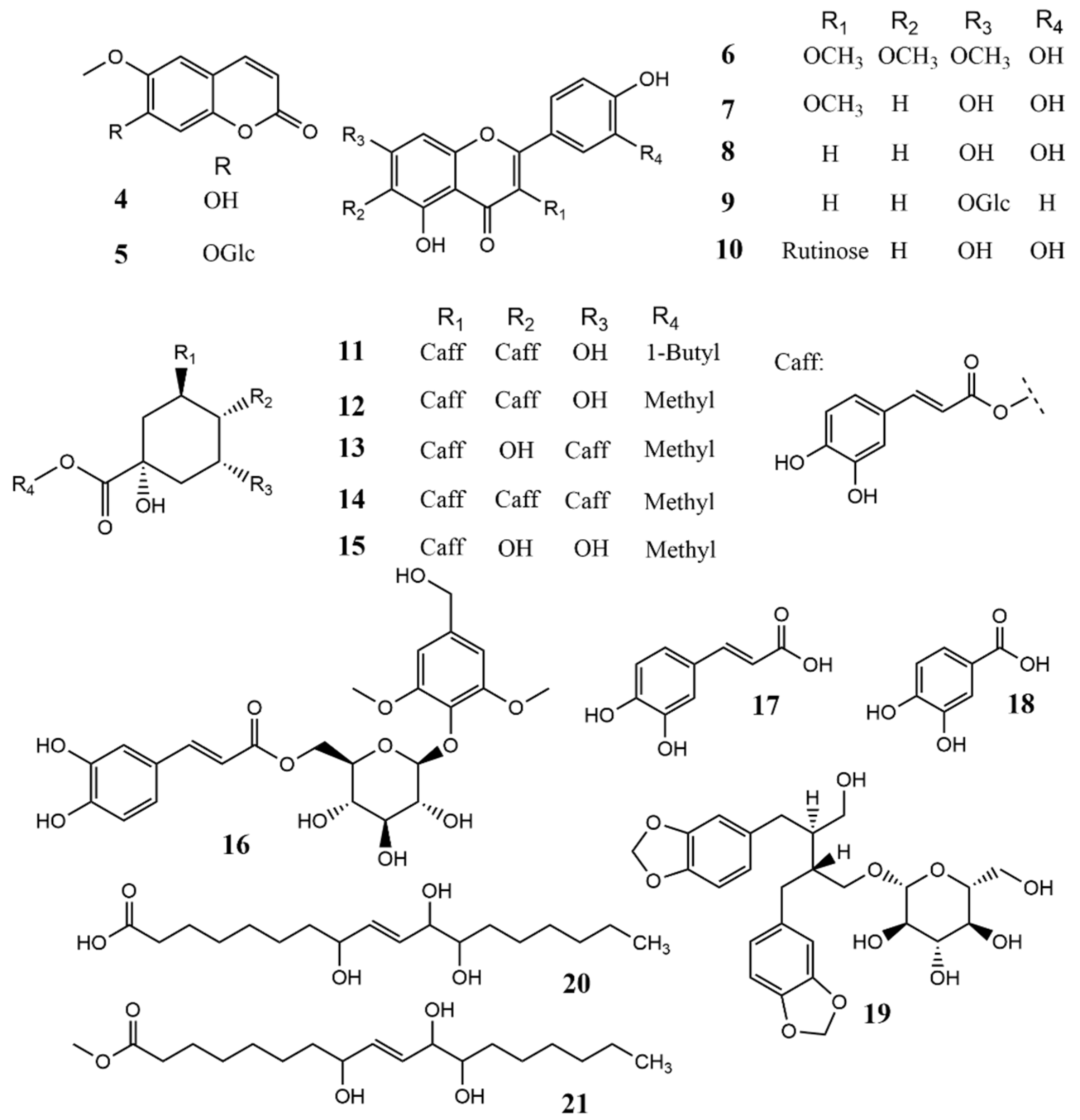
2.1.3. Structure of 6′-O-Caffeoyl Scopolin (3)

Compound 3 was isolated as white amorphous powder with negative optical rotation ( [ α ] D 26 76.5 ,   c = 0.2 ,   MeOH ). The molecular formula was determined as C25H24O12 from its HR-ESI-MS: m/z 539.1160 [M + Na]+ (calcd for C25H24O12Na: 539.1163). HPLC analysis with an optical rotation detector revealed that acid hydrolysis of 3 with 1M HCl at 80 °C released D-glucose. The results of 1H-NMR and 13C-NMR (Table 2) were similar to those published for 6′-O-sinapinoyl esculin, and indidenes F [48,49] (Table S1). The 1H-NMR spectrum showed several downfield doublets at δH 6.32 (1H, d, J = 9.5 Hz, H-3), δH 7.65 (1H, d, J = 9.5 Hz, H-4), δH 6.69 (1H, d, J = 15.9 Hz, H-8″), and δH 7.92 (1H, d, J = 15.9 Hz, H-7″), corresponding to cis- and trans-double bonds, respectively. Moreover, several characteristic low-field signals at δH 7.31 (1H, d, J = 8.3 Hz, H-5″), δH 7.570 (1H, d, J = 1.8 Hz, H-2″), and δH 7.27 (1H, dd, J = 8.3, 1.8 Hz, H-6″) were ascribed to a 1,3,4-trisubstituted benzene ring. In the 13C-NMR spectrum, two carbonyl carbons (δC 161.6 and 167.9), seven downfield quaternary carbons (δC 113.5, 127.1, 147.4, 147.8, 149.8, 150.8, and 151.5), one methoxy carbon (δC 56.5), five oxymethine carbons (δC 102.3, 78.7, 76.1, 74.9, and 71.6), and one oxymethylene carbon (δC 64.6), corresponding to D-glucose, were observed. HMBC correlations (Figure 6) from H-3 and H-4 to C-2; from H-3 to C-4a; from H-4 to C-8a and C-5; from H-5 (δH 7.01, 1H, s) to C-6 and C-8a; and from H-8 (δH 7.568, 1H, s) to C-4a, C-6, and C-7 indicated a coumarin moiety. Moreover, HMBC correlations from H-7″ and H-8″ to C-9″; from H-8″ to C-1″; and from H-7″ to C-2″ and C-6″, together with the characteristic 1,3,4-trisubstituted benzene coupling pattern of H-2″, H-5″, and H-6″, indicated the presence of a caffeoyl group. The coupling constant of the anomeric proton (δH 5.80, 1H, d, J = 7.3 Hz, H-1′) indicated the anomeric configuration of glucose should be β. Finally, the HMBC correlations from H-1′ to C-7 and from H-6′ (δH 4.91, dd, J = 11.9, 6.5 Hz; δH 5.11, dd, J = 11.9, 1.9 Hz) to C-9″ indicated the connection between coumarin, glucose, and the caffeoyl groups. Accordingly, the structure of 3 was defined as 6′-O-caffeoyl scopolin (Figure 1).

2.1.4. Plausible Biosynthetic Pathway of 1 and 2

The plausible biosynthetic pathway of crossoseamines A and B (1 and 2) is the Michael-type reaction of the nitrogen of proline to the eudesmanolide moiety (Figure 7). Yoshikawa et al. (1993) have reported a chemical synthesis of similar proline conjugates by the reaction of α-methylene γ-lactone type eudesmanolide and proline with Et3N at 90 °C [50]. The demand for a relatively strong base and high temperature for chemical synthesis indicates the involvement of the biosynthetic enzyme to form 1 and 2 in C. chinense, not as artifacts through the extraction and purification processes. The 6,7-dihydroxy coumarin moiety of 3 is thought to be the lactone form of caffeic acid, i.e., esculetin, which undergoes further methylation, glycosylation, and caffeoylation to 3. Given the other isolated compounds having caffeoyl function, C. chinense is considered a rich source of caffeic acid and its derivatives.

2.2. Cytotoxic Activities of the Isolated Compounds

All isolated compounds from the EtOAc fraction of C. chinense were evaluated for their cytotoxic activities against L. major, P. falciparum, T. brucei (gambiense and rhodesiense), and human lung cancer cell line A549 (Table 3).
The cytotoxicity against the human lung cancer cell line, A549, and the pathogen of cutaneous leishmaniasis, L. major, were evaluated by MTT method with positive control etoposide and miltefosine, respectively. The results are summarized in Table 3. Compounds 1 and 2 and some flavonoids (68) showed significant activities comparable to the positive controls.
Crossoseamine (1) and known compounds (69, 14, 18) had activities against both subspecies, T. brucei gambiense and T. brucei rhodesiense (Table 3), by ATP measurement using luciferase. Although the anti-Trypanosoma activity of compounds 69 and 18 was reported previously [32,33,34,35,36], this is the first time to show the potential of compound 14.
The anti-Plasmodium activity was evaluated by the SYBR Green I method. The activity of compounds 1, 2, 11, and 14 are reported for the first time, whereas that of compounds 69, 12, 13, 15, and 18 was previously reported [37,38,39,40]. The reported activities of the above compounds were consistent with our results, with some deviations (Table S2). The slight differences were ascribable to the differences in the assay methods and conditions and species/strains of the parasites used.

3. Experiments

3.1. General Experimental Procedure

Optical rotations were measured on a P-1030 spectropolarimeter (JASCO, Tokyo, Japan). IR and UV spectra were measured on FT-720 (HORIBA, Kyoto, Japan) and V-520 UV/Vis spectrophotometers (JASCO, Japan), respectively. 1H- and 13C-NMR spectra were measured on Avance Ⅲ HD spectrometer (Bruker, Billerica, MA, USA) at 500 and 125 MHz, respectively, with the residual solvent signal as references. Positive- and negative-ion HR-ESI-MSs were recorded on an LTQ Orbitrap XL spectrometer (Thermo Fisher Scientific, Waltham, MA, USA), and MS/MS fragments of precursor ions were detected by the CID mode with a collision energy of 35 eV.
Silica gel column chromatography (CC) and reversed-phase octadecyl silanized silica gel (ODS) CC were performed on silica gel 60 (Merck, Darmstadt, Germany) and Cosmosil 75C18-OPN (Nacalai Tesque, Kyoto, Japan). HPLC was performed on an Inertsil ODS-3 column (GL Science, Tokyo, Japan; Φ = 10 mm, L = 25 cm) or a Cosmosil πNAP column (Nacalai Tesque, Kyoto, Japan; Φ = 10 mm, L = 25 cm), and the eluate was monitored with a refractive index detector. TLC was performed on precoated silica gel 60 F254 plates (E. Merck; 0.25 mm in thickness). Sugars and proline were analyzed by HPLC on an amino and HILIC columns using a chiral detector (JASCO OR-2090 plus) (Asahipak NH2P-50 4E (Shodex, Tokyo, CH3CN-H2O (3:1), 1.0 mL/min); Cosmosil HILIC (Nacalai Tesque, Kyoto, Japan, CH3CN-H2O (4:1), 0.7 mL/min)).

3.2. Plant Material

The aerial parts of C. chinense were collected in July 2008 in Okinawa, Japan, and a voucher specimen was deposited in the Herbarium of the Department of Pharmacognosy, Graduate School of Biomedical Sciences, Hiroshima (deposition number: 08-CC-Okinawa-0708).

3.3. Extraction and Isolation

Air-dried aerial parts of C. chinense (3.5 kg) were extracted with MeOH (3 × 10 L) at room temperature. The methanol extract was concentrated to 1.5 L and then partitioned with an equal volume of n-hexane to obtain an n-hexane-soluble layer (27.7 g). The remaining layer was evaporated and resuspended in 1.5 L of water and then extracted with 1.5 L of EtOAc and 1.5 L of 1-BuOH successively to obtain EtOAC (74.3 g), 1-BuOH (30.5 g), and H2O (171.2 g) soluble fractions.
The EtOAC fraction (57.4 g of 74.3 g) was subjected to normal-phase open column CC (silica gel, Φ = 6, L = 25 cm) with increasing amounts of MeOH in CHCl3[(CHCl3, 2.0 L), CHCl3-MeOH (20:1, 2.0 L), CHCl3-MeOH (15:1, 2.0 L), CHCl3-MeOH (10:1, 2.0 L), CHCl3-MeOH (7:1, 2.0 L), CHCl3-MeOH (5:1, 2.0 L), CHCl3-MeOH (3:1, 2.0 L), CHCl3-MeOH (2:1, 2,0 L), and (MeOH, 2.0 L)] to obtain nine fractions [Fr. 1 (0.23 g), Fr. 2 (4.7 g), Fr. 3 (24.3 g), Fr. 4 (4.8 g), Fr. 5 (7.0 g), Fr. 6 (5.0 g), Fr. 7 (1.6 g), Fr. 8 (1.7 g), and Fr. 9 (4.1 g)] named CC-E 1 to CC-E 9. The fraction CC-E 3 (24.3 g) was separated by reversed-phase open column CC (ODS) with step gradient elution [(MeOH-H2O 2:3, 0.5 L), (MeOH-H2O 1:1, 0.5 L), (MeOH-H2O 3:2, 0.5 L), (MeOH-H2O 7:3, 0.5 L), (MeOH-H2O 4:1, 0.5 L), (MeOH-H2O 9:1, 0.5 L), (MeOH, 0.5 L), and (Acetone, 0.5 L)] to obtain eight subfractions [CC-E 3-1 (7.48 g), CC-E 3-2 (5.25 g), CC-E 3-3 (1.93 g), CC-E 3-4 (2.14 g), CC-E 3-5 (1.92g), CC-E 3-6, (1.28 g), CC-E 3-7, (1.05 g), and CC-E 3-8, (0.19 g)]. Of the 5.25 g of Fr. CC-E 3-2, 1.19 g was purified by ODS HPLC [Acetone-H2O (3:7, v/v)] to obtain scopoletin (4, 186.6 mg). Fr. CC-E 3-3 (1.93 g) was separated by ODS HPLC [Acetone-H2O (4.5:5.5, v/v)+ 0.1% TFA] to acquire chrysosplenol D (6, 107.4 mg). Fr. CC-E 4 (4.81 g) was fractionated by reversed-phase silica gel CC (ODS) with solvent [(MeOH-H2O 3:7, 0.5 L), (MeOH-H2O 2:3, 0.5 L), (MeOH-H2O 1:1, 0.5 L), (MeOH-H2O 3:2, 0.5 L), (MeOH-H2O 7:3, 0.5 L), (MeOH-H2O 4:1, 0.5 L), (MeOH-H2O 9:1, 0.5 L), (MeOH, 0.5 L), and (Acetone, 0.5 L)] to yield nine subfractions [CC-E 4-1 (0.78 g), CC-E 4-2 (0.38 g), CC-E 4-3 (0.61 g), CC-E 4-4 (0.51 g), CC-E 4-5 (0.36 g), and CC-E 4-6~ 4-9 (1.55 g)]. Subfraction CC-E 4-1 (0.78 g) was purified with πNAP HPLC [Acetone-H2O (3.5:6.5, v/v)] to obtain caffeic acid (17, 10.1 mg) and 3,4-dihydroxyl benzoic acid (18, 19.4 mg). CC-E 4-3 (0.61 g) [Acetone-H2O (2:3, v/v)] was separated by ODS HPLC to obtain 3-methylquercetin (7, 50.3 mg) and luteolin (8, 31.5 mg). Fr. CC-E 4-4 (0.51 g) was purified by ODS HPLC [Acetone-H2O (1:1, v/v)] to obtain 4,5-di-O-caffeoylquinic acid butyl ester (11, 32.4 mg), tianshic acid (20, 73.1 mg), and tianshic acid methyl ester (21, 8.6 mg). Fr. CC-E 5 (7.0 g) was fractionated by reversed-phase silica gel CC [(MeOH-H2O 3:7, 0.5 L), (MeOH-H2O 2:3, 0.5 L), (MeOH-H2O 1:1, 0.5 L), (MeOH-H2O 3:2, 0.5 L), (MeOH-H2O 7:3, 0.5 L), (MeOH-H2O 4:1, 0.5 L), (MeOH-H2O 9:1, 0.5 L), (MeOH, 0.5 L), and (Acetone, 0.5 L)] to afford nine subfractions [CC-E 5-1 (1.27 g), CC-E 5-2 (2.29 g), CC-E 5-3 (0.7 g), CC-E 5-4 (0.48 g), and CC-E 5-5~5-9 (1.8 g)]. Of the 2.29 g of Fr. CC-E 5-2, 1.29 g was purified by ODS HPLC [Acetone-H2O (3.5:6.5, v/v)] to acquire 6′-O-caffeoyl scopolin (3, 3.8 mg), 4,5-di-O-caffeoylquinic acid methyl ester (12, 41.3 mg), and 3,5-di-O-caffeoylquinic acid methyl ester (13, 46.7 mg). CC-E 5-4 (0.48 g) was separated by πNAP HPLC [Acetone-H2O (3.8:6.2, v/v)] to afford tetrecentronside B (19, 14.5 mg). Fraction CC-E 6 (5.03 g) was subjected to reversed-phase silica gel CC [(MeOH-H2O 1:4, 0.5 L), (MeOH-H2O 3:7, 0.5 L), (MeOH-H2O 2:3, 0.5 L), (MeOH-H2O 1:1, 0.5 L), (MeOH-H2O 3:2, 0.5 L), (MeOH-H2O 7:3, 0.5 L), (MeOH-H2O 4:1, 0.5 L), (MeOH-H2O 9:1, 0.5 L), (MeOH, 0.5 L), and (Acetone, 0.5 L)] to obtain ten subfractions [CC-E 6-1 (0.62 g), CC-E 6-2 (1.36 g), CC-E 6-3 (1.24 g), CC-E 6-4 (0.64 g), CC-E 6-5 (0.37 g), and CC-E 6-6~6-10 (1.02 g)]. Fr. CC-E 6-1 (0.62 g) was purified with πNAP HPLC [Acetone-H2O (1:4, v/v)] to obtain scopolin (5, 10.9 mg). CC-E 6-2 (1.36 g) was purified by πNAP HPLC [Acetone-H2O (1:4, v/v)] to obtain chlorogenic acid methyl ester (15, 33.6 mg) and 2,6-dimethoxyl-4-hydroxymethyl-phenol-1-O-(6-O-caffeoyl)-β-D-glucopyranoside (16, 15.1 mg). CC-E 6-3 (1.24 g) was purified with ODS HPLC [Acetone-H2O (2:3, v/v)] to obtain cosmoslin (9, 5.1 mg). CC-E 6-5 (0.37 g) was purified with πNAP HPLC [Acetone-H2O (2:3, v/v)] to afford 3,4,5-tri-O-caffeoylqunic acid methyl ester (14, 26.6 mg). Fraction CC-E 7 (1.63 g) was combined with CC-E 8 (1.68 g) and then separated by ODS CC [(MeOH-H2O 1:9, 0.5 L), (MeOH-H2O 1:4, 0.5 L), (MeOH-H2O 3:7, 0.5 L), (MeOH-H2O 2:3, 0.5 L), (MeOH-H2O 1:1, 0.5 L), (MeOH-H2O 3:2, 0.5 L), (MeOH-H2O 7:3, 0.5 L), (MeOH-H2O 4:1, 0.5 L), (MeOH-H2O 9:1, 0.5 L), (MeOH, 0.5 L), and (Acetone, 0.5 L)] to obtain eleven subfractions [CC-E 7-1 (0.15 g), CC-E 7-2 (0.12 g), CC-E 7-3 (0.26 g), CC-E 7-4 (0.98 g), and CC-E 7-5~7-11 (1.45 g)]. Fraction CC-E 7-3 (0.26 g) was purified with πNAP HPLC [Acetone-H2O (1.5:8.5, v/v)] to obtain crossoseamine B (2, 5.3 mg). CC-E 7-4 (0.98 g) was purified with πNAP HPLC [Acetone-H2O (1:1, v/v)] to obtain quercetin-3-O-(6″-O-α-rhamnosyl)-β-glucoside (10, 23.1 mg). Fraction CC-E 9 (4.13 g) was separated by reversed-phase silica gel CC [(MeOH-H2O 1:9, 0.5 L), (MeOH-H2O 1:4, 0.5 L), (MeOH-H2O 3:7, 0.5 L), (MeOH-H2O 2:3, 0.5 L), (MeOH-H2O 1:1, 0.5 L), (MeOH-H2O 3:2, 0.5 L), (MeOH-H2O 7:3, 0.5 L), (MeOH-H2O 4:1, 0.5 L), (MeOH-H2O 9:1, 0.5 L), (MeOH, 0.5 L), and (Acetone, 0.5 L)] to obtain eleven subfractions [CC-E 9-1 (1.49 g), CC-E 9-2 (0.16 g), CC-E 9-3 (0.63 g), and CC-E 9-4~9-11 (1.74 g)]. Fr. CC-E 9-3 (0.63 g) was purified with ODS HPLC [Acetone-H2O (1.8:8.2, v/v)] to obtain crossoseamine A (1, 3.1 mg).
The known compounds were identified by comparison of their physicochemical data,   [ α ] D , IR, MS, 1H, and 13C NMR) with the reported data.
Crossoseamine A (1): a colorless amorphous powder,   [ α ] D 27 97   ( c = 0.2 ,   MeOH ); IR (film) νmax: 3411, 2943, 1762, 1710, 1633, 1450, 1374, 1319, 1225, 1038. UV λmax (MeOH) nm (log ε): 205 (4.12); CD (MeOH) Δε (nm): +4.62 (218), −4.70 (295); 1H NMR (500 MHz, DMSO-d6), Table 1; 13C NMR (125 MHz, DMSO-d6), Table 1; positive-ion HR-ESI-MS: m/z 362.1962 [M + H]+ (calcd for C20H28O5N: 362.1961); ESI MS/MS: m/z 362 [M + H]+ (48), 316 [M–COOH]+ (12), 247 [M–C5H8O2N (proline)]+ (15), 128 [M–C14H17O3 (eudesmanolide part)]+ (100).
Crossoseamine B (2): colorless needles,   [ α ] D 27 47   ( c = 0.46 ,   MeOH ); mp. 210–214 °C; IR (film) νmax: 3248, 2980, 1758, 1711, 1625, 1450, 1379, 1310, 1033. UV λmax (MeOH) nm (log ε): 204 (4.13); CD (MeOH) Δε (nm): +1.84 (233), −4.61 (294); 1H NMR (500 MHz, DMSO-d6), Table 1; 13C NMR (125 MHz, DMSO-d6), Table 1; positive-ion HR-ESI-MS: m/z 378.1912 [M + H]+ (calcd for C20H28O6N: 378.1911); ESI-MS/MS: m/z 378 [M + H]+ (66), 360 [M + H-H2O]+ (34), m/z 332 [M-COOH]+ (19) and 128 [M-C14H17O4 (eudesmanolide part)]+ (100).
6′-O-caffeoyl scopolin (3): a colorless amorphous powder,   [ α ] D 27 76.5   ( c = 0.2 ,   MeOH ) ; IR (film) νmax: 3393, 2952, 2849, 1689, 1639, 1511, 1389, 1278, 1164, 1019. UV λmax (MeOH) nm (log ε) 225sh (3.92), 250sh (3.62), 292 (3.66), 331 (3.75); 1H NMR (500 MHz, pyridine-d5), Table 2; 13C NMR (125 MHz, pyridine-d5), Table 2; positive-ion HR-ESI-MS: m/z 539.1160 [M + Na]+ (calcd for C25H24O12Na: 539.1163). ESI-MS/MS: m/z 521 [M + Na–H2O]+ (4), m/z 377 [M + Na–C9H6O3 (caffeoyl)]+ (5), m/z 347 [M + Na-C10H8O4 (scopoletin)]+ (100) and 215 [M + Na–C15H16O8 (6-O-caffeoylglucose]+ (29).

3.4. Acid Hydrolysis of Compounds 1 and 2

Compounds 1 and 2 (0.2 mg each) were treated with 1% aqueous hydrochloric acid (HCl) (0.5 mL) at room temperature for 12 h. The reaction mixture was extracted with EtOAc to obtain the EtOAc and aqueous layers. The latter was subjected to HPLC analysis with an optical rotation detector (OR-2090 plus; JASCO) on a HILIC column (Cosmosil HILIC, 10 × 250 mm, CH3CN-H2O (4:1, v/v), flow rate: 0.7 mL/min). The peaks from 1 and 2 were identical with an authentic standard, L-proline (tR: 30.0 min, negative optical rotation) [51].

3.5. Sugar Analysis of Compound 3

Compound 3 (0.5 mg) was hydrolyzed with 1M HCl (0.2 mL) at 80 °C for 2 h. After cooling, EtOAc was used to extract the reaction mixture, and the aqueous layer was subjected to HPLC analysis with an optical rotation detector (OR-2090 plus; JASCO) on an amino column [Asahipak NH2P-50 4E, 4.6 × 250 mm, CH3CN-H2O (3:1, v/v), flow rate: 1 mL/min] to identify the D-glucose from 3, which was determined by comparison of its retention time and optical rotation sign with authentic sample (tR: 7.3 min, positive optical rotation).

3.6. Growth Inhibition Activity

The evaluation of cytotoxic activities against A549, L. major, P. falciparum, and T. brucei (gambiense and rhodesiense) was conducted following previous reports.
In brief, the human lung cancer cell line A549 was maintained in 10% FCS-supplemented DMEM. Various concentrations of samples in dimethylsulfoxide (DMSO) and A549 (5 × 10³ cells/well) were cultured in a CO2 incubator for 72 h. The medium was replaced with 100 µL of MTT solution and incubated for 1.5 h in the same condition. The viability was calculated from the absorbance of formed MTT formazan at 550 nm using a microplate reader [52]. Etoposide was used as a positive control.
The leishmanicidal activities of the isolated compounds were also evaluated using an MTT assay. In a 96-well plate, various concentrations of sample solutions in dimethylsulfoxide (DMSO) and L. major (2 × 10⁵ parasite/well) in 100 µL of M199 medium were incubated for 72 h at 25 °C. Then, 100 µL of MTT solution was replaced and incubated overnight. The absorbance of the formazan solution in DMSO was recorded using a microplate reader at 550 nm [11]. Miltefosine was used as a positive control.
The trypanocidal activities of the isolated compounds were performed in 96-well plates with slight modifications [53]. In brief, each well contains 100 µL of parasite culture (1 × 10⁴ parasites/well) with serial dilutions of compounds. After incubation for 72 h at 37 °C under 5% CO2, 25 µL of CellTiter-GloTM Luminescent Cell Viability Assay reagent (Promega Japan, Tokyo, Japan) was added to evaluate intracellular ATP concentration according to the instruction. Nifurtimox was used as a positive control.
Anti-Plasmodium activity of the isolated compounds was evaluated according to the previous report [54]. In brief, 100 µL of P. falciparum 3D7 parasite culture [55] was plated in a 96-well plate with various concentrations of the compounds. After incubation for 72 h at 37 °C in a humidified chamber under a gas mixture of 90% N2, 5% O2, and 5% CO2, the parasitemia was determined by SYBR Green I assay (Lonza Ltd., Basel, Switzerland) with a microplate reader at 485 and 530 nm. Artemisinin dissolved in DMSO was used as a positive control, and DMSO was used as a negative control. Human erythrocytes and plasma were obtained from the Nagasaki Red Cross Blood Center, and their usage was approved by the ethical committee of the Institute of Tropical Medicine, Nagasaki University.
The 50% inhibitory concentration (IC50) values were obtained for each compound by linear regression method.

4. Conclusions

The ethyl acetate extract of C. chinense was intensively fractionated to obtain previously undescribed sesquiterpene-amino acid conjugate crossoseamines A and B (1 and 2) and a coumarin glucoside, 6′-O-caffeoyl scopolin (3) together with 18 known compounds (421). The plausible biosynthetic pathway of crossoseamines A and B (1 and 2) is the Michael-type reaction of the nitrogen of proline to the eudesmanolide moiety (Figure 7).
Crossoseamine A (1) showed potent activity against A549 and L. major, while the introduction of the hydroxy function on position 8 (crossoseamine B (2)) decreased the activity. The polarity and/or functionality of eudesmane moiety may be necessary for the cytotoxicity against these targets. Among the evaluated compounds, chrysosplenol D (6) showed substantial toxicity for all tested organisms, probably by general toxicity, and has already been reported previously [21,25,56]. While some flavonoids (79) and caffeic acid derivatives (14) are relatively specific for both Trypanosoma and Plasmodium, compounds (1113, 15, 18) showed specificity for Plasmodium. As a result, these compounds have the potential to treat human cancer, leishmaniasis, malaria, and trypanosomiasis. However, further chemical modification and mechanism analyses are needed in the future.

Supplementary Materials

The following are available online at https://www.mdpi.com/article/10.3390/molecules28124696/s1, Figures S1–S32: 1D, 2D NMR spectra, MS and CD data of compounds 13. Table S1: Comparison of NMR data of 3 with related compounds A and B. Table S2: Reported IC50 values of the identified known compounds.

Author Contributions

Conceptualization, K.M. and Z.W.; Methodology, S.S. and S.K.; Software, Y.Y. and S.K.; Validation, K.M. and S.S.; Formal analysis, K.M. and O.K.; Investigation, Z.W., B.-Y.A.C. and K.S.; Resources, K.M. and H.O.; Data curation, Z.W., B.-Y.A.C. and K.S.; Writing—original draft preparation, Z.W.; Editing, K.M. and O.K.; Visualization, Z.W.; Supervision, K.M.; Project administration, K.M.; Funding acquisition, K.M. and O.K. All authors have read and agreed to the published version of the manuscript.

Funding

This work was supported by JSPS KAKENHI Grant Number 22K06678. This work was also partly supported by JST SPRING, Grant Number JPMJSP2132, and by the Joint Usage/Research Center on Tropical Disease, Institute of Tropical Medicine, Nagasaki University (2021-Ippan-33 and 2023-Ippan-30).

Institutional Review Board Statement

Not applicable.

Informed Consent Statement

Not applicable.

Data Availability Statement

Data is contained within the article or supplementary material.

Acknowledgments

The HR-ESI-MS, MS/MS data, and some NMR data were measured at the Natural Science Center for Basic Research and Development (N-BARD-00084), Hiroshima University.

Conflicts of Interest

The authors declare no conflict of interest.

Sample Availability

Samples of the compounds 13 are available from the authors.

References

  1. Tang, F.; Chen, F.; Chen, S.; Wang, X.-E.; Zhao, H. Molecular cytogenetic identification and relationship of the artificial intergeneric hybrid between Dendranthema indica and Crossostephium chinense by GISH. Plant Syst. Evol. 2010, 289, 91–99. [Google Scholar] [CrossRef]
  2. Wu, Q.; Zou, L.; Yang, X.-W.; Fu, D.-X. Novel sesquiterpene and coumarin constituents from the whole herbs of Crossostephium chinense. J. Asian Nat. Prod. Res. 2009, 11, 85–90. [Google Scholar] [CrossRef] [PubMed]
  3. Pan, C.-H.; YLin, P.; Wang, J.-Y.; Huang, H.-Y.; Huang, S.-C.; Lo, J.-M.; Wu, C.-H. Preventive Effect and Mechanism of Crossostephium chinense Extract on Balloon Angioplasty-Induced Neointimal Hyperplasia. Evid. Based Complement. Alternat. Med. 2021, 2021, 8466543. [Google Scholar] [CrossRef]
  4. Wang, S.-W.; Kuo, H.-C.; Hsu, H.-F.; Houng, J.-Y. Inhibitory effect of Crossostephium chinense extract on RANKL-activating osteoclastogenesis in patients with tophaceous gout. Bone Abstr. 2014, 3, PP167. [Google Scholar] [CrossRef]
  5. Huang, H.-T.; Lin, C.-C.; Kuo, T.-C.; Chen, S.-J.; Huang, R.-N. Phytochemical composition and larvicidal activity of essential oils from herbal plants. Planta 2019, 250, 59–68. [Google Scholar] [CrossRef]
  6. Uehara, A.; Kitajima, J.; Kokubugata, G.; Iwashina, T. Further characterization of foliar flavonoids in Crossostephium chinense and their geographic variation. Nat. Prod. Commun. 2014, 9, 1934578X1400900207. [Google Scholar] [CrossRef] [Green Version]
  7. Wang, Y.; Wu, Q.; Yang, X.W.; Yang, X.; Wang, K. The membrane transport of flavonoids from Crossostephium chinense across the Caco2 monolayer. Biopharm. Drug Dispos. 2011, 32, 16–24. [Google Scholar] [CrossRef]
  8. Yang, X.-W.; Zou, L.; Wu, Q.; Fu, D.-X. Studies on chemical constituents from whole plants of Crossostephium chinense. China J. Chin. Mater. Med. 2008, 33, 905–908. [Google Scholar]
  9. Sasaki, S.-I.; Aoyagi, S.; Hsii, H.-Y. The isolation of taraxerol, taraxeryl acetate, and taraxerone from Crossostephium chinense Makino (Compositae). Chem. Pharm. Bull. 1965, 13, 87–88. [Google Scholar] [CrossRef] [Green Version]
  10. Alesha, A.T.; Benjamin, J.S.; Lecia, V.S.; Justin, F.G.; Rebecca, S.H. Lung cancer. Lancet 2021, 398, 535–554. [Google Scholar]
  11. Asaumi, S.; Kawakami, S.; Sugimoto, S.; Matsunami, K.; Otsuka, H.; Shinzato, T. Alkylated Benzoquinones: Ardisiaquinones A–H from the Leaves of Ardisia quinquegona and Their Anti-Leishmania Activity. Chem. Pharm. Bull. 2018, 66, 757–763. [Google Scholar] [CrossRef] [PubMed] [Green Version]
  12. Nok, A.J. Arsenicals (melarsoprol), pentamidine and suramin in the treatment of human African trypanosomiasis. Parasitol. Res. 2003, 90, 71–79. [Google Scholar] [CrossRef] [PubMed]
  13. Kuzoe, F.A. Current situation of African trypanosomiasis. Acta Trop. 1993, 54, 153–162. [Google Scholar] [CrossRef]
  14. Balikagala, B.; Fukuda, N.; Ikeda, M.; Katuro, O.T.; Tachibana, S.-I.; Yamauchi, M.; Opio, W.; Emoto, S.; Anywar, D.A.; Kimura, E.; et al. Evidence of Artemisinin-Resistant Malaria in Africa. N. Engl. J. Med. 2021, 385, 1163–1171. [Google Scholar] [CrossRef] [PubMed]
  15. Woodrow, C.J.; White, N.J. The clinical impact of artemisinin resistance in Southeast Asia and the potential for future spread. FEMS Microbiol. Rev. 2017, 41, 34–48. [Google Scholar] [CrossRef] [Green Version]
  16. Joseph, S.; Omar, H.; Michael, B.; Srikkanth, B.; Tobias, A.O.; Ulrike, H. Improving anti-trypanosomal activity of alkamides isolated from Achillea fragrantissima. Fitoterapia 2018, 125, 191–198. [Google Scholar]
  17. Takeara, S.R.; Lopes, A.N.P.; Lopes, J.L.C. Trypanocidal activity of Lychnophora staavioides Mart. (Vernonieae, Asteraceae). Phytomedicine 2003, 10, 490–493. [Google Scholar] [CrossRef] [PubMed]
  18. Taleb-Contini, S.H.; Salvador, M.J.; Balanco, J.M.F.; Albuquerque, S.; de Oliveira, D.C.R. Antiprotozoal effect of crude extracts and flavonoids isolated from Chromolaena hirsuta (asteraceae). Phytother. Res. 2004, 18, 250–254. [Google Scholar] [CrossRef]
  19. Stipan, N.; Buyankhishig, B.; Suganuma, K.; Ishikawa, Y.; Kutsuma, M.; Abe, M.; Sasaki, K.; Davaapurev, B.O.; Batkhuu, J.; Murata, T. Phytochemical investigation of Scutellaria scordiifolia and its trypanocidal activity. Phytochemistry 2023, 209, 11361. [Google Scholar]
  20. Mamadalieva, N.Z.; Herrmann, F.; El-Readi, M.Z.; Tahrani, A.; Hamoud, R.; Egamberdieva, D.R.; Azimova, S.S.; Wink, M. Flavonoids in Scutellaria immaculata and S. ramosissima (Lamiaceae) and their biological activity. J. Pharm. Pharmacol. 2011, 63, 1346–1357. [Google Scholar] [CrossRef]
  21. Hashim, Y.; Toume, K.; Mizukami, S.; Kitami, T.; Taniguchi, M.; Teklemichael, A.A.; Tayama, Y.; Huy, N.T.; Lami, J.N.; Bodi, J.M.; et al. Phenylpropanoid-conjugated iridoid glucosides from leaves of Morinda morindoides. J. Nat. Med. 2022, 76, 281–290. [Google Scholar] [CrossRef] [PubMed]
  22. Liu, K.C.; Yang, S.L.; Roberts, M.F.; Elford, B.C.; Phillipson, J.D. Antimalarial activity of Artemisia annua flavonoids from whole plants and cell cultures. Plant Cell Rep. 1992, 11, 637–640. [Google Scholar] [CrossRef] [PubMed]
  23. Ogunlana, O.O.; Kim, H.S.; Wataya, Y.; Olagunju, J.O.; Akindahunsi, A.; Tan, N.H. Antiplasmodial flavonoid from young twigs and leaves of Caesalpinia bonduc (Linn) Roxb. J. Chem. Pharm. Res. 2015, 7, 931–937. [Google Scholar]
  24. Vitalini, S.; Beretta, G.; Iriti, M.; Orsenigo, S.; Basilico, N.; Dall’Acqua, S.; Iorizzi, M.; Fico, G. Phenolic compounds from Achillea millefolium L. and their bioactivity. Acta Biochim. Pol. 2011, 58, 203–209. [Google Scholar] [CrossRef] [PubMed]
  25. Sangsopha, W.; Lekphrom, R.; Kanokmedhakul, S.; Kanokmedhakul, K. Cytotoxic and antimalarial constituents from aerial parts of Sphaeranthus indicus. Phytochem. Lett. 2016, 17, 278–281. [Google Scholar] [CrossRef]
  26. Lagnika, L.; Weniger, B.; Attioua, B.; Jensen, O.; Anthaume, C.; Sanni, A.; Kaiser, M.; Lobstein, A.; Vonthron-Senecheau, C. Trypanocidal activity of diarylheptanoids from Schrankia leptocarpa DC. S. Afr. J. Bot. 2012, 83, 92–97. [Google Scholar] [CrossRef] [Green Version]
  27. Aldulaimi, O.; Uche, F.I.; Hameed, H.; Mbye, H.; Ullah, I.; Drijfhout, F.; Claridge, T.D.W.; Horrocks, P.; Li, W.W. A characterization of the antimalarial activity of the bark of Cylicodiscus gabunensis Harms. J. Ethnopharmacol. 2017, 198, 221–225. [Google Scholar] [CrossRef]
  28. Vasconcelos, J.M.J.; Silva, A.M.S.; Cavaleiro, J.A.S. Chromones and flavanones from artemisia campestris subsp. maritima. Phytochemistry 1998, 49, 1421–1424. [Google Scholar] [CrossRef]
  29. Tsukamoto, H.; Hisada, S.; Nishibe, S. Coumarin and Secoiridoid Glucosides from Bark of Olea africana and Olea capensis. Chem. Pharm. Bull. 1985, 33, 396–399. [Google Scholar] [CrossRef] [Green Version]
  30. Zhu, Y.; Zhang, L.-X.; Zhao, Y.; Huang, G.-D. Unusual sesquiterpene lactones with a new carbon skeleton and new acetylenes from Ajania przewalskii. Food Chem. 2010, 118, 228–238. [Google Scholar] [CrossRef]
  31. Marco, J.A.; Barberá, O.; Rodríguez, S.; Domingo, C.; Adell, J. Flavonoids and other phenolics from Artemisia hispanica. Phytochemistry 1988, 27, 3155–3159. [Google Scholar] [CrossRef]
  32. Lee, E.H.; Kim, H.J.; Song, Y.S.; Jin, C.; Lee, K.-T.; Cho, J.; Lee, Y.S. Constituents of the stems and fruits of Opuntia ficus-indica var. saboten. Arch. Pharm. Res. 2003, 26, 1018–1023. [Google Scholar] [CrossRef]
  33. Jung, M.J.; Kang, S.S.; Jung, H.A.; Kim, G.J.; Choi, J.S. Isolation of flavonoids and a cerebroside from the stem bark of Albizzia julibrissin. Arch. Pharm. Res. 2004, 27, 593–599. [Google Scholar] [CrossRef] [PubMed]
  34. Redaelli, C.; Formentini, L.; Santaniello, E. Apigenin 7-glucoside diacetates in ligulate flowers of Matricaria chamomilla. Phytochemistry 1982, 21, 1828–1830. [Google Scholar] [CrossRef]
  35. Kazuma, K.; Noda, N.; Suzuki, M. Malonylated flavonol glycosides from the petals of Clitoria ternatea. Phytochemistry 2003, 62, 229–237. [Google Scholar] [CrossRef] [PubMed]
  36. Xiao-Li, M.A.; Lin, W.B. Chemical Constituents of Osmanthus yunnanensis. Nat. Prod. Res. Dev. 2009, 21, 593–599. [Google Scholar]
  37. Nugroho, A.; Lee, K.R.; Alam, M.B.; Choi, J.S.; Park, H.J. Isolation and quantitative analysis of peroxynitrite scavengers from Artemisia princeps var. orientalis. Arch. Pharm. Res. 2010, 33, 703–708. [Google Scholar] [CrossRef] [PubMed]
  38. Kim, J.Y.; Cho, J.-Y.; Ma, Y.-K.; Park, K.Y.; Lee, S.-H.; Ham, K.-S.; Lee, H.J.; Park, K.-H.; Moon, J.-H. Dicaffeoylquinic acid derivatives and flavonoid glucosides from glasswort (Salicornia herbacea L.) and their antioxidative activity. Food Chem. 2011, 125, 55–62. [Google Scholar]
  39. Merfort, I. Caffeoylquinic acids from flowers of Arnica montana and Arnica chamissonis. Phytochemistry 1992, 31, 2111–2113. [Google Scholar] [CrossRef]
  40. Zhu, X.; Dong, X.; Wang, Y.; Ju, P.; Luo, S. Phenolic Compounds from Viburnum cylindricum. Helv. Chim. Acta 2005, 88, 339–342. [Google Scholar] [CrossRef]
  41. Yang, P.-F.; Yang, Y.-N.; Feng, Z.-M.; Jiang, J.-S.; Zhang, P.-C. Six new compounds from the flowers of Chrysanthemum morifolium and their biological activities. Bioorg. Chem. 2019, 82, 139–144. [Google Scholar] [CrossRef]
  42. Nakazawa, T.; Ohsawa, K. Metabolism of Rosmarinic Acid in Rats. J. Nat. Prod. 1998, 61, 993–996. [Google Scholar] [CrossRef] [PubMed]
  43. Zhang, H.L.; Nagatsu, A.; Okuyama, H.; Mizukami, H.; Sakakibara, J. Sesquiterpene glycosides from cotton oil cake. Phytochemistry 1998, 48, 665–668. [Google Scholar] [CrossRef]
  44. Yi, J.-H.; Zhang, G.-L.; Li, B.-G.; Chen, Y.-Z. Two glycosides from the stem bark of Tetracentron sinense. Phytochemistry 2000, 53, 1001–1003. [Google Scholar] [CrossRef] [PubMed]
  45. Yang, X.; Wong, M.; Wang, N.; Chan, A.S.C.; Yao, X. A New Eudesmane Derivative and a New Fatty Acid Ester from Sambucus williamsii. Chem. Pharm. Bull. 2006, 54, 676–678. [Google Scholar] [CrossRef] [Green Version]
  46. Matsuda, H.; Kageura, T.; Inoue, Y.; Morikawa, T.; Yoshikawa, M. Absolute Stereostructures and Syntheses of Saussureamines A, B, C, D and E, Amino Acid–Sesquiterpene Conjugates with Gastroprotective Effect, from the Roots of Saussurea lappa. Tetrahedron 2000, 56, 7763–7777. [Google Scholar] [CrossRef]
  47. Moiseeva, G.P.; Kasymov, S.Z.; Yagudaev, M.R.; Sidyakin, G.P. Determination of the absolute configuration of the asymmetric center at C11 in the eudesmanolides. Chem. Nat. Compd. 1980, 16, 254–256. [Google Scholar] [CrossRef]
  48. Zhang, D.M.; Wang, L.L.; Li, J.; Hu, L.H. Two new coumarins from Fraxinus chinensis Rexb. J. Integr. Plant Biol. 2007, 49, 218–221. [Google Scholar] [CrossRef]
  49. He, R.; Huang, X.; Zhang, Y.; Wu, L.; Nie, H.; Zhou, D.; Liu, B.; Deng, S.; Yang, R.; Huang, S.; et al. Structural Characterization and Assessment of the Cytotoxicity of 2,3-Dihydro-1H-indene Derivatives and Coumarin Glucosides from the Bark of Streblus indicus. J. Nat. Prod. 2016, 79, 2472–2478. [Google Scholar] [CrossRef]
  50. Yoshikawa, M.; Hatakeyama, S.; Inoue, Y.; Yamahara, J. Saussureamines a, b, c, d, and e, new anti-ulcer principles from chinese saussureae radix. Chem. Pharm. Bull. 1993, 41, 214–216. [Google Scholar] [CrossRef] [Green Version]
  51. Sugimoto, S.; Yamano, Y.; Desoukey, S.Y.; Katakawa, K.; Wanas, A.S.; Otsuka, H.; Matsunami, K. Isolation of Sesquiterpene–Amino Acid Conjugates, Onopornoids A–D, and a Flavonoid Glucoside from Onopordum alexandrinum. J. Nat. Prod. 2019, 82, 1471–1477. [Google Scholar] [CrossRef]
  52. Mahmoud, B.K.; Samy, M.N.; Hamed, A.N.E.; Abdelmohsen, U.R.; Hajjar, D.; Yamano, Y.; Sugimoto, S.; Matsunami, K.; Kamel, M.S. Bignanoside A “A new neolignan glucoside” and bignanoside B “A new iridoid glucoside” from Bignonia binata leaves. Phytochem. Lett. 2020, 35, 200–205. [Google Scholar] [CrossRef]
  53. Suganuma, K.; Allamanda, P.; Hakimi, H.; Zhou, M.; Angeles, J.M.; Kawazu, S.; Inoue, N. Establishment of ATP-based luciferase viability assay in 96-well plate for Trypanosoma congolense. J. Vet. Med. Sci. 2014, 76, 1437–1441. [Google Scholar] [CrossRef] [Green Version]
  54. Ishikawa, T.; Mizuta, S.; Kaneko, O.; Yahata, K. Fragment Molecular Orbital Study of the Interaction between Sarco/Endoplasmic Reticulum Ca2+-ATPase and its Inhibitor Thapsigargin toward Anti-Malarial Development. J. Phys. Chem. B 2018, 122, 7970–7977. [Google Scholar] [CrossRef]
  55. Walliker, D.; Quakyi, I.A.; Wellems, T.E.; McCutchan, T.F.; Szarfman, A.; London, W.T.; Corcoran, L.M.; Burkot, T.R.; Carter, R. Genetic Analysis of the Human Malaria Parasite Plasmodium falciparum. Science 1987, 236, 1661–1666. [Google Scholar] [CrossRef] [PubMed]
  56. Khalid, S.A.; Farouk, A.; Geary, T.G.; Jensen, J.B. Potential anti-malarial candidates from African plants: An in vitro approach using Plasmodium falciparum. J. Ethnopharmacol. 1986, 15, 201–209. [Google Scholar] [CrossRef] [PubMed]
Figure 1. Structures of novel compounds (13) from C. chinense.
Molecules 28 04696 g001
Figure 2. Structures of isolated known compounds (421) from C. chinense.
Molecules 28 04696 g002
Figure 3. 1H-1H COSY and HMBC correlations of 1 and 2.
Molecules 28 04696 g003
Figure 4. ESI-MS/MS spectrum (in-source CID fragmentation) of 1.
Molecules 28 04696 g004
Figure 5. Selected NOESY correlations of compounds 1 and 2.
Molecules 28 04696 g005
Figure 6. COSY and Key HMBC correlations of 3.
Molecules 28 04696 g006
Figure 7. Plausible biosynthetic pathway of compounds 1 and 2.
Molecules 28 04696 g007
Table 1. 13C-NMR and 1H-NMR spectroscopic data for compounds 1 and 2.
12
Position13C1H13C1H
1212.9 212.2
235.52.43 (ddd, 13.6, 7.8, 6.6) α35.22.40 (m) α
2.55 (ddd, 13.6, 7.0, 6.2) β 2.58 (m) β
332.32.35 (br dt, 17.0, 6.2) α32.42.37 (m) α
2.26 (br dt, 17.0, 6.9) β 2.27 (m) β
4125.2 126.1
5130.0 128.6
680.84.80 (dquint-like, 11.2, 1.3)77.94.87 (dquint-like, 11.4, 1.1)
748.52.02 (m)57.61.88 (m)
823.11.95 (m) α66.03.89 (td, 10.8, 4.5)
1.63 (m) β
934.61.44 (td, 14.0, 5.1) α43.11.25 (br t, 11.8) α
1.63 (m) β 1.87 (m) β
1048.4 47.4
1144.22.73 (dt-like, 12.1, 5.4)44.33.01 (m)
12177.0 175.8
1351.22.86 (dd, 13.2, 5.1)52.82.92 (m)
3.08 (dd, 13.2, 5.7) 3.15 (m)
1423.01.25 (3H, s) 24.11.24 (3H, s)
1519.41.86 (3H, br s)19.31.87 (3H, br s)
2′66.63.30 (dd, 8.7, 4.6)66.33.45 (dd, 9.0, 3.4)
3′28.61.81 (m)28.91.90 (m)
2.03 (m) 2.07 (dq-like, 12.8, 8.8)
4′23.41.68–1.79 (2H, m)23.11.74 (m)
1.80 (m)
5′53.22.58 (dd-like, 9.0, 7.8)52.82.84 (dt, 9.5, 7.5)
2.99 (ddd, 9.0, 7.5, 3.9) 3.00 (m)
6′173.9 173.6
500 MHz and 125 MHz (DMSO-d6). Chemical shifts (δ) in ppm. m: multiplet or overlapped signals.
Table 2. 13C-NMR and 1H-NMR spectroscopic data for 3.
3
Position13C1HPosition13C1H
2161.6 4′71.64.24 (br t, 8.8)
3114.56.32 (d, 9.5)5′76.14.37 (ddd, 9.5, 6.5, 1.9)
4144.07.65 (d, 9.5)6′64.64.91 (dd, 11.9, 6.5)
4a113.5 5.11 (dd, 11.9, 1.9)
5110.27.01 (s)1″127.1
6147.4 2″116.57.570 (d, 1.8)
7151.5 3″147.8
8104.67.568 (s)4″149.8
8a150.8 5″117.37.31 (d, 8.3)
6-OCH356.53.69 (3H, s)6″122.57.27 (dd, 8.3, 1.8)
1′102.35.80 (d, 7.3)7″146.67.92 (d, 15.9)
2′74.94.42 (dd-like, 8.8, 7.3)8″114.96.69 (d, 15.9)
3′78.74.44 (br t, 8.8)9″167.9
500 MHz and 125 MHz (pyridine-d5). Chemical shifts (δ) in ppm.
Table 3. IC50 of the isolated compounds against A549 and parasites (Mean ± SE, µg/mL, n = 3).
CompoundsA549L. majorT. b.
gambiense
T. b.
rhodesiense
P. falciparum
13.3 ± 0.36.9 ± 0.626.3 ± 8.527.8 ± 10.412.1 ± 1.1
212.3 ± 1.024.9 ± 2.239.5 ± 6.941.0 ± 8.915.6 ± 1.2
64.4 ± 1.55.4 ± 0.82.6 ± 0.11.7 ± 0.36.0 ± 1.1
717.7 ± 4.417.5 ± 1.24.0 ± 0.23.2 ± 0.316.5 ± 1.1
819.9 ± 5.526.9 ± 2.84.9 ± 0.33.2 ± 0.411.0 ±1.3
978.3 ± 15.658.9 ± 7.516.9 ± 0.78.6 ± 1.28.9 ± 1.0
10>10090.9 ± 5.5>100>10037.0 ± 1.1
1173.5 ± 13.0>10047.4 ± 2.734.8 ± 4.65.4 ± 1.1
12115.0 ± 19.3>10097.1 ± 0.268.9 ± 14.24.6 ± 1.1
13118.2 ± 19.7>100>10080.5 ± 18.410.4 ± 1.2
1442.6 ± 6.958.3 ± 6.611.0 ± 0.112.4 ± 0.83.6 ± 1.1
1579.4 ± 8.7>10083.8 ± 13.353.6 ± 2.210.2 ± 1.1
16>100>100>100>10035.5 ± 1.0
1742.5 ± 0.1>10053.2 ± 7.542.3 ±2.925.0 ± 1.1
18>100>10019.6 ± 2.724.8 ± 1.215.9 ± 1.2
19>100>100>10055.2 ± 0.830.6 ± 1.1
20>100>10044.1 ± 3.032.8 ± 1.826.1 ± 1.1
21>100>10058.7 ± 3.036.0 ± 0.927.1 ± 1.1
PC17.5 ± 0.37.4 ± 0.71.3 ± 0.71.3 ± 0.50.0023 ± 0.0003
PC (Positive control): A549 (Etoposide); L. major (Miltefosine); T. b. gambiense/rhodesiense (Nifurtimox). P. falciparum (Artemisinin). Compounds 35 were not active at 100 μg/mL and omitted.
Disclaimer/Publisher’s Note: The statements, opinions and data contained in all publications are solely those of the individual author(s) and contributor(s) and not of MDPI and/or the editor(s). MDPI and/or the editor(s) disclaim responsibility for any injury to people or property resulting from any ideas, methods, instructions or products referred to in the content.

