Controlling the Transverse Magneto-Optical Kerr Effect in Cr/NiFe Bilayer Thin Films by Changing the Thicknesses of the Cr Layer

 , , , , , and
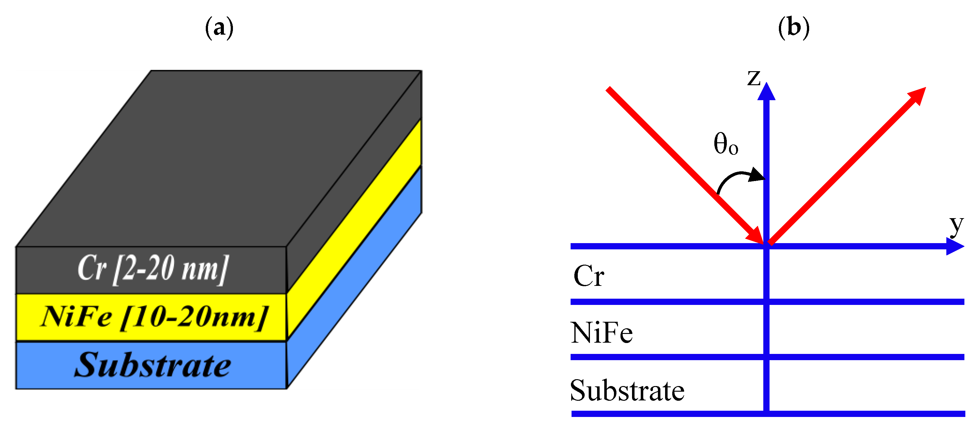
Abstract
:1. Introduction
Generalised Magneto-Optical and Ellipsometry (GMOE) Formalism
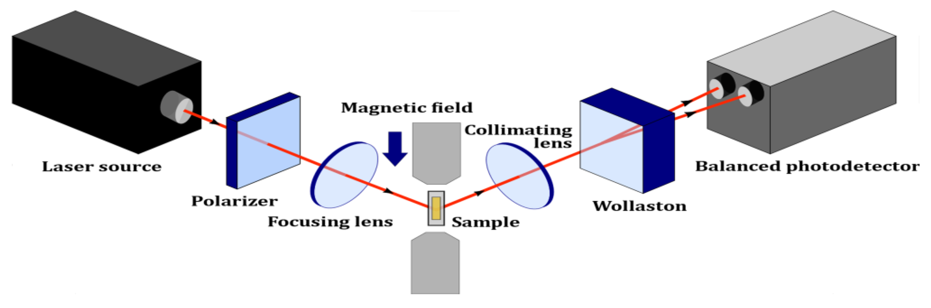
2. Materials and Methods
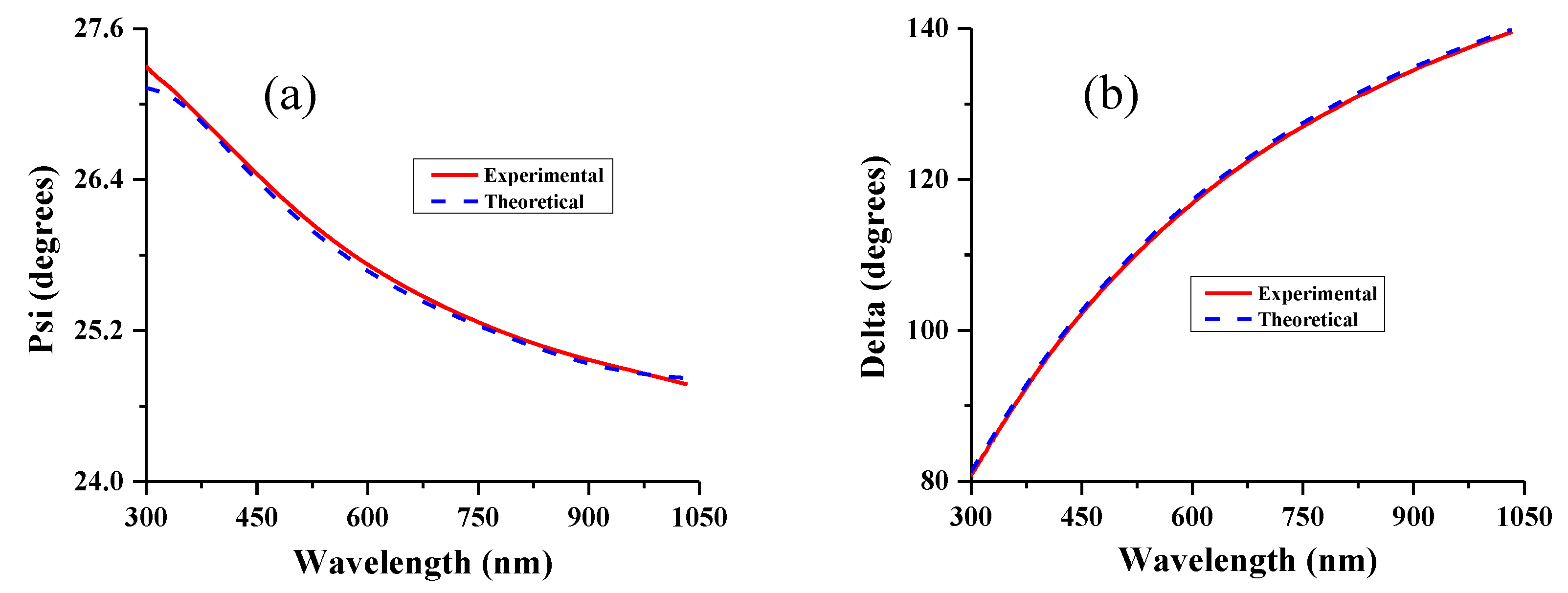
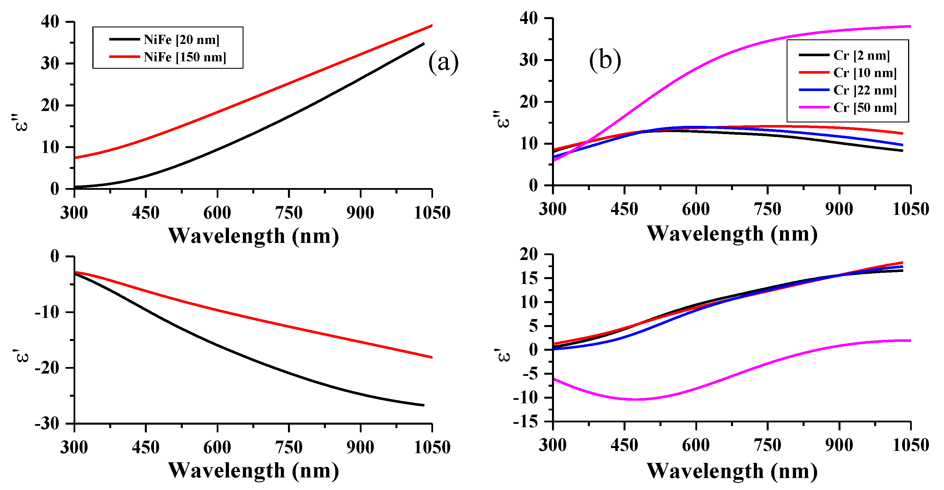
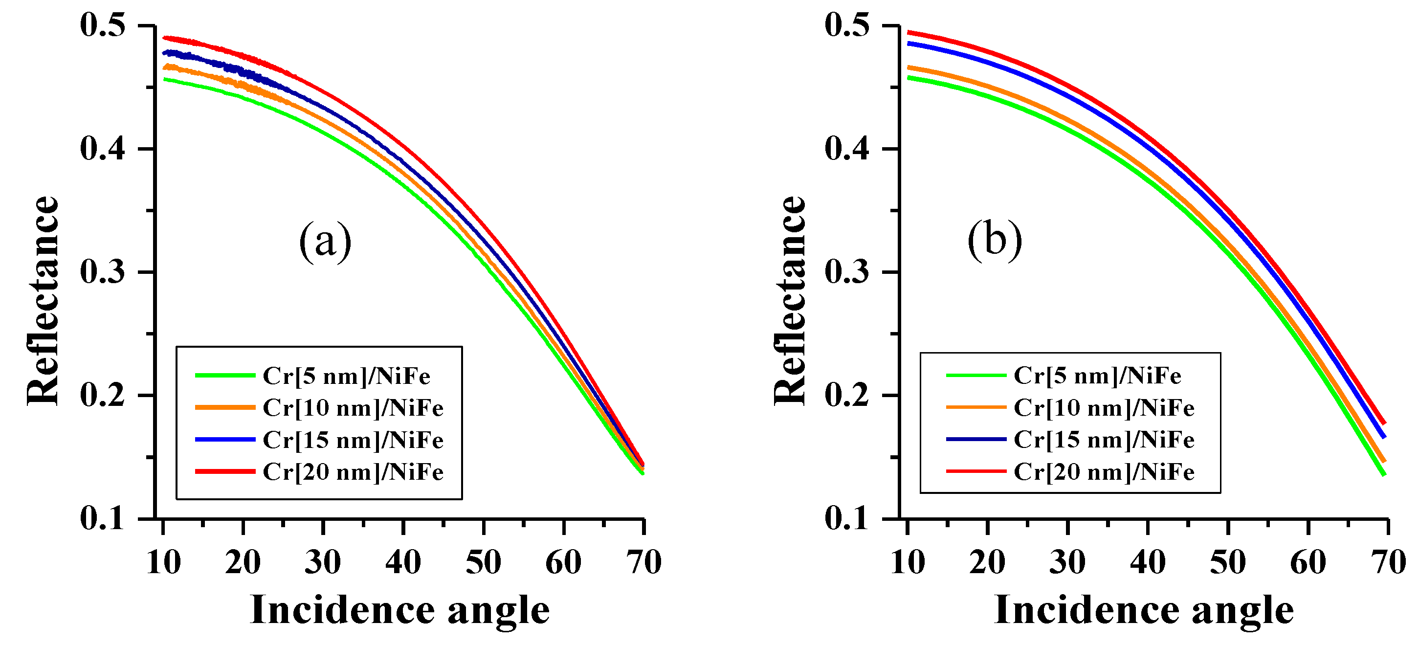
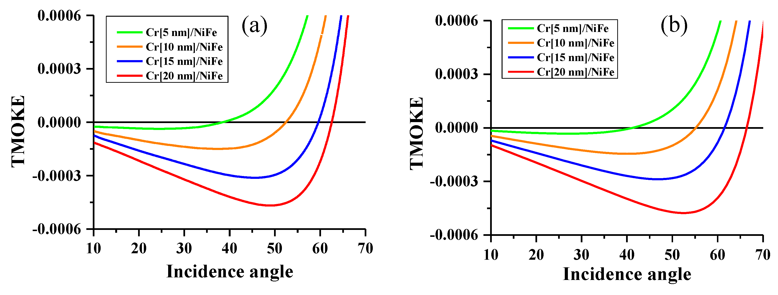
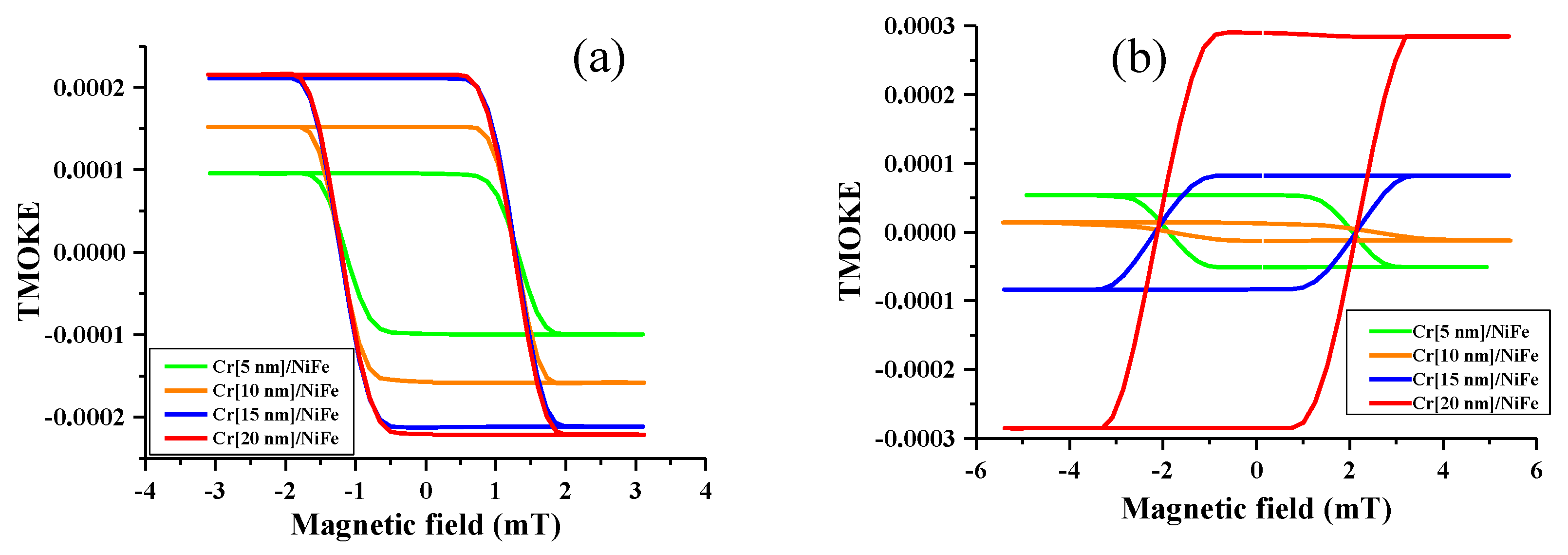
3. Results and Discussion
4. Conclusions
Author Contributions
Funding
Conflicts of Interest
References
- Hashim, H.; Singkh, S.P.; Panina, L.V.; Pudonin, F.A.; Sherstnev, I.A.; Podgornaya, S.V.; Shpetny, I.A.; Beklemisheva, A.V. Spectral ellipsometry as a method for characterization of nanosized films with ferromagnetic layers. Phys. Solid State 2017, 59, 2211. [Google Scholar] [CrossRef]
- Dyakov, S.A.; Klompmaker, L.; Spitzer, F.; Yalcin, E.; Akimov, I.; Yavsin, D.A.; Pavlov, S.I.; Pevtsov, A.B.; Verbin, S.Y.; Tikhodeev, S.G.; et al. Wide band enhancement of transverse magneto-optic Kerr effect in magnetite-based plasmonic crystals. Phys. Rev. B 2019, 100, 214411. [Google Scholar] [CrossRef] [Green Version]
- Betzig, E.; Trautman, J.K.; Wolfe, R.; Gyorgy, E.M.; Finn, P.L.; Kryder, M.H.; Chang, C.H. Near-field magneto-optics and high density data storage. Appl. Phys. Lett. 1992, 61, 142. [Google Scholar] [CrossRef]
- Mok, K.; Kovács, G.J.; McCord, J.; Li, L.; Helm, M.; Schmidt, H. Magneto-optical coupling in ferromagnetic thin films investigated by vector-magneto-optical generalized ellipsometry. Phys. Rev. B 2011, 84, 094413. [Google Scholar] [CrossRef]
- Didosyan, Y.S.; Hauser, H.; Wolfmayr, H.; Nicolics, J.; Fulmek, P. Magneto-optical rotational speed sensor. Sensor. Actuat. A Phys. 2003, 106, 168. [Google Scholar] [CrossRef]
- Sepúlveda, B.; Calle, A.; Lechuga, L.M.; Armelles, G. Highly sensitive detection of biomolecules with the magneto-optic surface-plasmon-resonance sensor. Opt. Lett. 2006, 31, 1085. [Google Scholar] [CrossRef] [PubMed]
- Regatos, D.; Sepúlveda, B.; Fariña, D.; Carrascosa, L.G.; Lechuga, L.M. Suitable combination of noble/ferromagnetic metal multilayers for enhanced magneto-plasmonic biosensing. Opt. Express 2011, 19, 8336. [Google Scholar] [CrossRef]
- Rotondaro, M.D.; Zhdanov, B.V.; Knize, R.J. Generalized treatment of magneto-optical transmission filters. Opt. Soc. Am. B 2015, 32, 2507. [Google Scholar] [CrossRef]
- Keaveney, J.; Wrathmall, S.A.; Adams, C.S.; Hughes, I.G. Optimized ultra-narrow atomic bandpass filters via magneto-optic rotation in an unconstrained geometry. Opt. Lett. 2018, 43, 4272. [Google Scholar] [CrossRef]
- Qu, D.; Huang, S.Y.; Chien, C.L. Inverse spin Hall effect in Cr: Independence of antiferromagnetic ordering. Phys. Rev. B 2015, 92, 020418. [Google Scholar] [CrossRef] [Green Version]
- Turgut, E.; Laovorakiat, C.; Shaw, J.M.; Grychtol, P.; Nembach, H.T.; Rudolf, D.; Adam, R.; Aeschlimann, M.; Schneider, C.M.; Silva, T.J.; et al. Controlling the Competition between Optically Induced Ultrafast Spin-Flip Scattering and Spin Transport in Magnetic Multilayers. Phys. Rev. Lett. 2013, 110, 197201. [Google Scholar] [CrossRef] [PubMed] [Green Version]
- Petrila, I.; Ungureanu, F.; Manta, V. Effects of laser beam modulation on all-optical switching phase diagrams in magneto-optical ultrafast storage device. J Comput. Electron. 2015, 14, 627–633. [Google Scholar] [CrossRef]
- Petrila, I.; Manta, V. Metropolis Monte Carlo analysis of all-optical switching. Comput. Phys. Commun. 2014, 185, 2874–2878. [Google Scholar] [CrossRef]
- Ghanaatshoar, M.; Moradi, M. Magneto-optical Kerr-effect enhancement in glass/Cu/SnO2/Co/SnO2 thin films. Opt. Eng. 2011, 50, 093801. [Google Scholar]
- Belotelov, V.I.; Akimov, I.A.; Pohl, M.; Kotov, V.A.; Kasture, S.; Vengurlekar, A.S.; Gopal, A.V.; Yakovlev, D.R.; Zvezdin, A.K.; Bayer, M. Enhanced magneto-optical effects in magnetoplasmonic crystals. Nat. Nanotechnol. 2011, 6, 370–376. [Google Scholar] [CrossRef]
- Barman, A.; Sinha, J. Spin Dynamics and Damping in Ferromagnetic Thin Films and Nanostructures; Springer International Publishing: Kolkata, India, 2017; Volume 3, pp. 48–50. [Google Scholar]
- Borovkova, O.V.; Hashim, H.; Kozhaev, M.A.; Dagesyan, S.A.; Chakravarty, A.; Levy, M.; Belotelov, V.I. TMOKE as efficient tool for the magneto-optic analysis of ultra-thin magnetic films. Appl. Phys. Lett. 2018, 112, 063101. [Google Scholar] [CrossRef] [Green Version]
- Pathak, S.; Sharma, M. Polar magneto-optical Kerr effect instrument for 1-dimensional magnetic nanostructures. Appl. Phys. 2014, 115, 043906. [Google Scholar] [CrossRef]
- Singh, V.; Annadi, A.; Bhoi, B.; Madugundo, R.; Muthu, M.; Bohra, M. Synthesis, Properties, and Applications of Multifunctional Magnetic Nanostructures. J. Nanomater. 2019, 2638715. [Google Scholar] [CrossRef] [Green Version]
- Hilfiker, J.N.; Singh, N.; Tiwald, T.; Convey, D.; Baker, J.H.; Tompkins, H.G. Survey of methods to characterize thin absorbing films with Spectroscopic Ellipsometry. Thin Solid Films 2008, 516, 7979. [Google Scholar] [CrossRef]
- Postava, K.; Maziewski, A.; Stupakiewicz, A.; Wawro, A.; Baczewski, L.T.; Visnovsky, S.; Yamaguchi, T. Transverse magneto-optical Kerr effect measured using phase modulation. JEOS:RP 2006, 1, 06017. [Google Scholar] [CrossRef]
- Allen, G.A.; Dionne, G.F. Accurate analysis of the magneto-optical permittivity tensor of Y3Fe5O12. Appl. Phys. 2003, 93, 6951. [Google Scholar] [CrossRef]
- Liu, J.; Singh, A.; Llandro, J.; Duffy, L.B.; Stanton, M.R.; Holmes, S.N.; Applegate, M.J.; Phillips, R.T.; Hesjedal, T.; Barnes, C.H.W. A low-temperature Kerr effect microscope for the simultaneous magneto-optic and magneto-transport study of magnetic topological insulators. Meas. Sci. Technol. 2019, 30, 125201. [Google Scholar] [CrossRef]
- Armelles, G.; Cebollada, A.; García-Martín, A.; González, M.U. Magnetoplasmonics: Combining Magnetic and Plasmonic Functionalities. Adv. Opt. Mater. 2013, 1, 10–35. [Google Scholar] [CrossRef] [Green Version]
- Piskin, H.; Akdogan, N. Interface-induced enhancement of sensitivity in NiFe/Pt/IrMn-basedplanar hall sensors with nanoTesla resolution. Sensor. Actuat. 2019, 292, 24–29. [Google Scholar] [CrossRef] [Green Version]
- Born, M.; Wolf, E. Principles of Optics, 7th ed.; Cambridge University Press: New York, NY, USA, 2002; pp. 1–70. [Google Scholar]
- Hashim, H.; Singkh, S.P.; Panina, L.V.; Pudonin, F.A.; Sherstnev, I.; Podgornaya, S.V.; Shpetny, I.A. Optical and magnetic properties of Al/NiFe and Al/Ge/NiFe nanosized films. EPJ Web Confer. 2018, 185, 04014. [Google Scholar] [CrossRef]
- Rebib, F.; Tomasella, E.; Gaston, J.P.; Eypert, C.; Cellier, J.; Jacquet, M. Determination of optical properties of a-SiOxNy thin films by ellipsometric and UV-visible spectroscopies. J. Phys. 2008, 100, 082033. [Google Scholar] [CrossRef]
- McPeak, K.M.; Jayanti, S.V.; Kress, S.; Meyer, S.; Iotti, S.; Rossinelli, A.; Norris, D.J. Plasmonic films can easily be better: Rules and recipes. ACS Photonics 2015, 2, 326. [Google Scholar] [CrossRef]
- Rakić, A.D.; Djurišić, A.B.; Elazar, J.M.; Majewski, M.L. Optical properties of metallic films for vertical-Fcavity optoelectronic devices. Appl. Opt. 1998, 37, 5271. [Google Scholar] [CrossRef]
- Neuber, G.; Rauer, R.; Kunze, J.; Backstrom, J.; Rübhausen, M. Generalized magneto-optical ellipsometry in ferromagnetic metals. Thin Solid Films 2004, 455, 39–42. [Google Scholar] [CrossRef]


© 2020 by the authors. Licensee MDPI, Basel, Switzerland. This article is an open access article distributed under the terms and conditions of the Creative Commons Attribution (CC BY) license (http://creativecommons.org/licenses/by/4.0/).
Share and Cite
Hashim, H.; Kozhaev, M.; Kapralov, P.; Panina, L.; Belotelov, V.; Víšová, I.; Chvostová, D.; Dejneka, A.; Shpetnyi, I.; Latyshev, V.; et al. Controlling the Transverse Magneto-Optical Kerr Effect in Cr/NiFe Bilayer Thin Films by Changing the Thicknesses of the Cr Layer. Nanomaterials 2020, 10, 256. https://doi.org/10.3390/nano10020256
Hashim H, Kozhaev M, Kapralov P, Panina L, Belotelov V, Víšová I, Chvostová D, Dejneka A, Shpetnyi I, Latyshev V, et al. Controlling the Transverse Magneto-Optical Kerr Effect in Cr/NiFe Bilayer Thin Films by Changing the Thicknesses of the Cr Layer. Nanomaterials. 2020; 10(2):256. https://doi.org/10.3390/nano10020256
Chicago/Turabian StyleHashim, Hisham, Mikhail Kozhaev, Pavel Kapralov, Larissa Panina, Vladimir Belotelov, Ivana Víšová, Dagmar Chvostová, Alexandr Dejneka, Ihor Shpetnyi, Vitalii Latyshev, and et al. 2020. "Controlling the Transverse Magneto-Optical Kerr Effect in Cr/NiFe Bilayer Thin Films by Changing the Thicknesses of the Cr Layer" Nanomaterials 10, no. 2: 256. https://doi.org/10.3390/nano10020256