
Smart and Multi-Functional Magnetic Nanoparticles for Cancer Treatment Applications: Clinical Challenges and Future Prospects

 and
and
Abstract
:1. Introduction
2. Synthesis of MNPs
2.1. Biological Synthesis
2.2. Physical Synthesis
2.3. Chemical Synthesis
2.3.1. Co-Precipitation
2.3.2. Thermal Decomposition
2.3.3. Hydrothermal Synthesis
2.3.4. Microemulsion
2.3.5. Polyol
2.4. One-Pot Synthesis of MNPs
3. Surface Coating
3.1. Inorganic Coating
3.2. Organic Coating
4. Stimuli-Triggered SMNPs
4.1. Thermo-Responsive MNPs
4.2. Magnetic-Responsive NPs
4.2.1. Targeted Drug Release
4.2.2. Magnetic Hyperthermia Application
4.2.3. Theranostic Application of MRI and MNPs
4.3. Electric Field-Responsive MNP
4.4. pH-Responsive MNPs
4.5. Redox-Responsive MNPs
4.6. Enzyme-Responsive MNPs
4.7. Light and Ultrasound-Responsive MNPs
4.8. Dual and Multi-Stimuli-Responsive MNPs
5. Magnetic Nanoparticle Targeting Methods
6. Interaction of MNPs with Biological System
7. Magnetic Nanoparticles in Clinical Applications
7.1. Clinical Challenges
7.2. Future Perspectives
8. Conclusions
Funding
Data Availability Statement
Acknowledgments
Conflicts of Interest
Abbreviations
| ((+)ly) | Positively |
| (NH4)2SO4 | Ammonium sulfate |
| 5-FU | 5-Fluorouracil |
| ACA | Acrylic acid |
| ACV | Aciloclovir |
| Ag | Agar |
| AIBN | Azobisisobutyronitrile |
| Aly-Imz | Allyl imidazole |
| AMF | Alternating Magnetic Field |
| APS | Ammonium persulfate |
| Apt | Aptamer |
| APTES | 3-aminopropyltriethoxysilane |
| ASGP | Ammonium sulfate gradient protocol |
| B-CD | B-cyclodextrin |
| BCT | Blood circulation time |
| BNPC | Bis(4-nitrophenyl)carbonate |
| C18TMS | Octadecyltrimethoxysilane |
| CDNSs | Cyclodextrin nanosponges |
| CDR | Controlled drug release |
| Cho | Cholesterol |
| CMCS | Carboxymethyl Chitosan |
| CMNP | Cationic magnetic nanoparticles |
| Co 6 | Coumarin 6 |
| CST | Critical Solution Temperature |
| CSC | Chitosan co-polymer |
| CTAB | Cetyltrimethylammonium bromide |
| Cu | Copper |
| CUR | Curcumin |
| DC | Dendritic cell |
| DD | Drug Delivery |
| DDS | Drug Delivery System |
| DESE | Double emulsion solvent evaporation |
| Dex | Dextran |
| DMSO | Dimethyl sulfoxide |
| DOX | Doxorubicin |
| DPPC | Dipalmitoylphosphatidylcholine |
| 1,2-DPPC | 1,2-dipalmitoyl-sn-glycero-3-phosphocholine |
| DSPC | 1,2-distearoyl-sn-glycero-3-phosphocholine |
| DSPE-PEG2000 | 1,2-distearoyl-sn-glycero-3-phosphoethanolamine-N-[ami no(polyethylene-glycol)-2000] |
| EACCs | Ehrlich ascites carcinoma cells |
| EDA | ethylene diamine |
| EFR | Electric field-responsive |
| ENO1 | Enolase 1 |
| EPI | Epirubici |
| EPR | Enhance permeability retention affect |
| ER | Enzyme Responsive |
| ETB | Erlotinib |
| FA | Folic Acid |
| FDA | Food and drug administration |
| Fe | Iron |
| FE-SEM | Field emission scanning electron microscope |
| FITC | Fluorescein isothiocyanate |
| FMT | Ferumoxytol |
| FMT | Ferumoxytol |
| FRPR | Free radical polymerization reaction |
| FTIR | Fourier transform infrared |
| FTY720 | Fingolimod |
| GaCCM | Gadolinium Consisting contrast medium |
| Ge | Gelatin |
| GM | Glioblastoma |
| GMC | Glioblastoma cancer |
| GSH | Glutathione |
| HA | Hyaluronic Acid |
| HAS | Human Serum albumin |
| HCC | Hepatocellular Carcinoma |
| HeLa | Human Cervical Carcinoma Cells |
| HEOC | Human epithelial ovarian carcinoma |
| HIFU | High Intensity focused Ultrasound |
| HPhBA | Hyperbranched phenylboronic acid |
| HRBC | Human red blood cells |
| HSO | Hydroxy silicon oil |
| HUVEC | Human Umbilical Vein Endothelial Cells |
| IONP | Iron oxide nanoparticle |
| IA | Itaconic acid |
| LCST | Lower Critical Solution Temperature |
| LF | Lactoferrin |
| LIFUS | low-intensity focused ultrasound |
| LiA | Lipoic acid |
| LMNPs | liposome magnetic nanoparticle |
| LR | Light Responsive |
| MaA | Maleic acid anhydride |
| Magnevist | Diethylenetriaminepentaacetic acid gadolinium(III) dihydrogen salt hydrate Gd(III)-DTPA |
| MB | Methylene Blue |
| MBAA | N,N-methylenebisacrylamide |
| MBBs | Microbubble |
| MDP | Muramyl dipeptide |
| MF | Magnetic field |
| MF-SPIO-NB | Multi-functional superparamagnetic iron oxide nano bubbles |
| M-HS | Magnetic-Hydrating Solution |
| MHT | Magnetic hypothermia |
| ML | Magnetic liposeme |
| MMP-2 | Matrix metalloproteinase-2 |
| MM-USI | Magnetomotive-ultrasound imaging |
| MNFs | magnetic nanofluids |
| MNP | Magnetic nano particle |
| MNS | Magnetic nano-system |
| MPNCs | Mesoporous nano-cage |
| MPDA | Mesoporous polydopamine |
| MPSNP | Mesoporous silica nanoparticles |
| M MPSNP | Magnetic Mesoporous silica nanoparticles |
| M MPS/MBBs | Magnetic Mesoporous silica microbubbles |
| MPTSA | 4-Morpholineethanesulfonic acid |
| MRI | Magnetic resonance imaging |
| Ms | Saturation Magnetization |
| MSSM | Magnetic star structured micellar |
| MTRL | Magnetic Thermo-responsive liposome |
| MTX | Methotrexate |
| MUC1 Apt | Anti mucin aptamer |
| NADPH | Nicotinamide adenine dinucleotide phosphate |
| NB | Nanobubbles |
| nCP:Fe-CA | Fe-doped nano-calcium phosphate |
| NG | Nanogel |
| NH2-BDC | 2-aminoterephthalic acid |
| NHS | N-hydroxy succinimide |
| NIPAM | N-isopropylacrylamide |
| NIR | Near infrared light |
| NP | Nano particle |
| NSCL | Non-small-cell-lung |
| O-CMCS | O-Carboxymethyl chitosan |
| OlA | Oleic Acid |
| OlAm | Oleylamine |
| OPSS-PEG-SVA | Orthopyridyl disulfide-poly(ethylene glycol)-succinimidyl valerate |
| PA | Photoacoustic |
| PhBA | Phenylboronic Acid |
| PBS | Phosphate-buffered saline |
| PCC | Pancake coil |
| PCL | Poly(ε-caprolactone) |
| PCL-diol | Polycaprolactone diol |
| PCL-g-Dex | poly(ε-caprolactone)-grafted dextran |
| PDA | Polydopamine |
| PDEAAm | Poly(N,N-diethylacrylamide) |
| PDI | Polydispersity index |
| PEG | Polyethylene Glycol |
| PEG-2000-DSPE | Poly-ethylene-glycol-2000-distearoyl-phosphatidyl-ethanolamine |
| PEG-PBA-PEG | poly (ethylene glycol)−Poly (butylene adipate)−poly (ethylene glycol) |
| PEG-PCL | Methoxy poly(ethylene glycol)-b-poly(ε-caprolactone) |
| PEI | Polyethyleneimine |
| PESM | Polyethersulfone membrane |
| PFP | Perfluoropentane |
| PG | Prodigiosin |
| pHR | pH responsive |
| PIMF | Polyethylenimine-rafted-poly (Malevich anhydride-alt-1-ocatadecene)-folic acid |
| PLH | Poly-L-histidine |
| PMX | Pemetrexed |
| PNIPAAm | Poly(N-isopropylacrylamide) |
| PNVCL | Poly (N-vinylcaprolactom) |
| PTC | Photothermal Conversion |
| PTT | Photothermal therapy |
| QDMAMEA | Quaternized ammonium alkyl halide N,N-dimethylaminoethyl methacrylate |
| R1/r2 | Relaxivity |
| RES | Reticuloendothelial System |
| RF | Radio frequency |
| RFP | Red fluorescent probes |
| RGD | Arginylglycylaspartic |
| ROS | Reactive oxygen species |
| R-P | Receptor-Positive |
| RR | Redox Responsive |
| SA | Salic acid |
| SDS | Sodium dodecyl sulfate |
| SEM | Scanning electron microscopy |
| SH | Thiol |
| SiNc | Silicon naphthalocyanine |
| SIONP | Superparamagnetic iron oxide nano particle |
| SMNP | Smart magnetic nanoparticle |
| Sn(Oct)2 | Stannous-2-ethylhexanoate Sn(Oct)2 |
| SPECT | Single photo emission computed tomography |
| T1/T2 | Transverse/Longitudinal Time |
| TDD | Targeted drug delivery |
| TDR | Targeted drug release |
| TEM | Transmission electron microscopy |
| Temp | Temperature |
| TEOS | Tetra-ethyl ortho-silicate |
| THF | Tetrahydrofuran |
| TME | Tumor micro environment |
| TMSMA | 3-(Trimethoxysilyl) propyl methacrylate |
| TMZ | Temozolomide |
| TR | Thermo-responsive |
| UCST | Upper Critical Solution Temperature |
| U87 MG | Uppsala 87 Malignant Glioma |
| US | Ultrasound |
| UST | Ultrasound thermometry |
| US-TS | Ultrasound thermometry strain imaging |
| UV | Ultraviolet |
| Vs | Visible |
| VSM | Vibrating sample magnetometry |
| XRD | X-ray diffractometry |
| ZW | Zwitteronic 99mTc |
References
- Fitzmaurice, C.; Allen, C.; Barber, R.M.; Barregard, L.; Bhutta, Z.A.; Brenner, H.; Dicker, D.J.; Chimed-Orchir, O.; Dandona, R.; Dandona, L.; et al. Global, Regional, and National Cancer Incidence, Mortality, Years of Life Lost, Years Lived with Disability, and Disability-Adjusted Life-years for 32 Cancer Groups, 1990 to 2015: A Systematic Analysis for the Global Burden of Disease Study. JAMA Oncol. 2017, 3, 524–548. [Google Scholar] [CrossRef]
- Moein, S.; Adibi, R.; da Silva Meirelles, L.; Nardi, N.B.; Gheisari, Y. Cancer regeneration: Polyploid cells are the key drivers of tumor progression. Biochim. Biophys Acta Rev. Cancer 2020, 1874, 188408. [Google Scholar] [CrossRef]
- Thitichai, N.; Thanapongpibul, C.; Theerasilp, M.; Sungkarat, W.; Nasongkla, N. Study of biodistribution and systemic toxicity of glucose functionalized SPIO/DOX micelles. Pharm. Dev. Technol. 2019, 24, 935–946. [Google Scholar] [CrossRef] [PubMed]
- Azamjah, N.; Soltan-Zadeh, Y.; Zayeri, F. Global Trend of Breast Cancer Mortality Rate: A 25-Year Study. Asian Pac. J. Cancer Prev. 2019, 20, 2015–2020. [Google Scholar] [CrossRef]
- Mattiuzzi, C.; Lippi, G. Current Cancer Epidemiology. J. Epidemiol. Glob. Health 2019, 9, 217–222. [Google Scholar] [CrossRef] [PubMed] [Green Version]
- Sung, H.; Ferlay, J.; Siegel, R.L.; Laversanne, M.; Soerjomataram, I.; Jemal, A.; Bray, F. Global Cancer Statistics 2020: GLOBOCAN Estimates of Incidence and Mortality Worldwide for 36 Cancers in 185 Countries. CA Cancer J. Clin. 2021, 71, 209–249. [Google Scholar] [CrossRef]
- Salehiniya, H.; Ghobadi Dashdebi, S.; Rafiemanesh, H.; Mohammadian-Hafshejani, A.; Enayatrad, M. Time Trend Analysis of Cancer Incidence in Caspian Sea, 2004–2009: A Population-based Cancer Registries Study (northern Iran). Casp. J. Intern. Med. 2016, 7, 25–30. [Google Scholar]
- Takeshima, H.; Ushijima, T. Accumulation of genetic and epigenetic alterations in normal cells and cancer risk. NPJ Precis. Oncol. 2019, 3, 7. [Google Scholar] [CrossRef] [PubMed] [Green Version]
- Morand, G.B.; da Silva, S.D.; Hier, M.P.; Alaoui-Jamali, M.A. Insights into Genetic and Epigenetic Determinants with Impact on Vitamin D Signaling and Cancer Association Studies: The Case of Thyroid Cancer. Front. Oncol. 2014, 4, 309. [Google Scholar] [CrossRef] [Green Version]
- Lewandowska, A.M.; Rudzki, M.; Rudzki, S.; Lewandowski, T.; Laskowska, B. Environmental risk factors for cancer—Review paper. Ann. Agric. Environ. Med. 2019, 26, 1–7. [Google Scholar] [CrossRef] [PubMed]
- Joung, M.J.; Han, M.A.; Park, J.; Ryu, S.-Y. Association between Family and Friend Smoking Status and Adolescent Smoking Behavior and E-Cigarette Use in Korea. Int. J. Environ. Res. Public Health 2016, 13, 1183. [Google Scholar] [CrossRef]
- Erenpreisa, J.; Salmina, K.; Anatskaya, O.; Cragg, M.S. Paradoxes of cancer: Survival at the brink. Semin. Cancer Biol. 2020, 81, 119–131. [Google Scholar] [CrossRef]
- Pienta, K.J.; Hammarlund, E.U.; Axelrod, R.; Amend, S.R.; Brown, J.S. Convergent Evolution, Evolving Evolvability, and the Origins of Lethal Cancer. Mol. Cancer Res. 2020, 18, 801–810. [Google Scholar] [CrossRef] [Green Version]
- Das, M.; Solanki, A.; Joshi, A.; Devkar, R.; Seshadri, S.; Thakore, S. β-cyclodextrin based dual-responsive multifunctional nanotheranostics for cancer cell targeting and dual drug delivery. Carbohydr. Polym. 2019, 206, 694–705. [Google Scholar] [CrossRef]
- Mancarella, S.; Greco, V.; Baldassarre, F.; Vergara, D.; Maffia, M.; Leporatti, S. Polymer-Coated Magnetic Nanoparticles for Curcumin Delivery to Cancer Cells. Macromol. Biosci. 2015, 15, 1365–1374. [Google Scholar] [CrossRef]
- Manatunga, D.C.; de Silva, R.M.; de Silva, K.M.N.; Malavige, G.N.; Wijeratne, D.T.; Williams, G.R.; Jayasinghe, C.D.; Udagama, P.V. Effective delivery of hydrophobic drugs to breast and liver cancer cells using a hybrid inorganic nanocarrier: A detailed investigation using cytotoxicity assays, fluorescence imaging and flow cytometry. Eur. J. Pharm. Biopharm. 2018, 128, 18–26. [Google Scholar] [CrossRef]
- Rahimi, M.; Safa, K.D.; Salehi, R. Co-delivery of doxorubicin and methotrexate by dendritic chitosan-g-mPEG as a magnetic nanocarrier for multi-drug delivery in combination chemotherapy. Polym. Chem. 2017, 8, 7333–7350. [Google Scholar] [CrossRef]
- Yetisgin, A.A.; Cetinel, S.; Zuvin, M.; Kosar, A.; Kutlu, O. Therapeutic Nanoparticles and Their Targeted Delivery Applications. Molecules 2020, 25, 2193. [Google Scholar] [CrossRef]
- Chamundeeswari, M.; Jeslin, J.; Verma, M.L. Nanocarriers for drug delivery applications. Environ. Chem. Lett. 2018, 17, 849–865. [Google Scholar] [CrossRef]
- Bai, X.R.; Wang, L.H.; Ren, J.Q.; Bai, X.W.; Zeng, L.W.; Shen, A.G.; Hu, J.M. Accurate Clinical Diagnosis of Liver Cancer Based on Simultaneous Detection of Ternary Specific Antigens by Magnetic Induced Mixing Surface-Enhanced Raman Scattering Emissions. Anal. Chem. 2019, 91, 2955–2963. [Google Scholar] [CrossRef]
- Satpathy, M.; Wang, L.; Zielinski, R.J.; Qian, W.; Wang, Y.A.; Mohs, A.M.; Kairdolf, B.A.; Ji, X.; Capala, J.; Lipowska, M.; et al. Targeted Drug Delivery and Image-Guided Therapy of Heterogeneous Ovarian Cancer Using HER2-Targeted Theranostic Nanoparticles. Theranostics 2019, 9, 778–795. [Google Scholar] [CrossRef] [PubMed]
- Wei, J.; Shuai, X.; Wang, R.; He, X.-l.; Li, Y.; Ding, M.; Li, J.; Tan, H.; Fu, Q. Clickable and imageable multiblock polymer micelles with magnetically guided and PEG-switched targeting and release property for precise tumor theranosis. Biomaterials 2017, 145, 138–153. [Google Scholar] [CrossRef] [PubMed]
- Ciofani, G. Smart Nanoparticles for Biomedicine; Elsevier: Amsterdam, The Netherlands, 2018; pp. 131–142. [Google Scholar] [CrossRef]
- Guo, T.; Lin, M.; Huang, J.; Zhou, C.; Tian, W.; Yu, H.; Jiang, X.; Ye, J.; Shi, Y.; Xiao, Y.; et al. The Recent Advances of Magnetic Nanoparticles in Medicine. J. Nanomater. 2018, 2018, 1–8. [Google Scholar] [CrossRef] [Green Version]
- Soetaert, F.; Korangath, P.; Serantes, D.; Fiering, S.N.; Ivkov, R. Cancer therapy with iron oxide nanoparticles: Agents of thermal and immune therapies. Adv. Drug Deliv. Rev. 2020, 163, 65–83. [Google Scholar] [CrossRef]
- Assa, F.; Jafarizadeh-Malmiri, H.; Ajamein, H.; Anarjan, N.; Vaghari, H.; Sayyar, Z.; Berenjian, A. A biotechnological perspective on the application of iron oxide nanoparticles. Nano Res. 2016, 9, 2203–2225. [Google Scholar] [CrossRef]
- Sasikala, A.R.; GhavamiNejad, A.; Unnithan, A.R.; Thomas, R.G.; Moon, M.; Jeong, Y.Y.; Park, C.H.; Kim, C.S. A smart magnetic nanoplatform for synergistic anticancer therapy: Manoeuvring mussel-inspired functional magnetic nanoparticles for pH responsive anticancer drug delivery and hyperthermia. Nanoscale 2015, 7, 18119–18128. [Google Scholar] [CrossRef]
- Arami, H.; Khandhar, A.; Liggitt, D.; Krishnan, K.M. In vivo delivery, pharmacokinetics, biodistribution and toxicity of iron oxide nanoparticles. Chem. Soc. Rev. 2015, 44, 8576–8607. [Google Scholar] [CrossRef] [Green Version]
- Barick, K.C.; Ekta, E.; Gawali, S.L.; Sarkar, A.; Kunwar, A.; Priyadarsini, K.I.; Hassan, P.A. Pluronic stabilized Fe3O4 magnetic nanoparticles for intracellular delivery of curcumin. RSC Adv. 2016, 6, 98674–98681. [Google Scholar] [CrossRef]
- Pourjavadi, A.; Dastanpour, L.; Tehrani, Z.M. Magnetic micellar nanocarrier based on pH-sensitive PEG-PCL-PEG triblock copolymer: A potential carrier for hydrophobic anticancer drugs. J. Nanopart. Res. 2018, 20, 282. [Google Scholar] [CrossRef]
- Sadr, S.H.; Davaran, S.; Alizadeh, E.; Salehi, R.; Ramazani, A. PLA-based magnetic nanoparticles armed with thermo/pH responsive polymers for combination cancer chemotherapy. J. Drug Deliv. Sci. Technol. 2018, 45, 240–254. [Google Scholar] [CrossRef]
- Pourjavadi, A.; Amin, S.S.; Hosseini, S.H. Delivery of Hydrophobic Anticancer Drugs by Hydrophobically Modified Alginate Based Magnetic Nanocarrier. Ind. Eng. Chem. Res. 2018, 57, 822–832. [Google Scholar] [CrossRef]
- Kang, T.; Li, F.; Baik, S.; Shao, W.; Ling, D.; Hyeon, T. Surface design of magnetic nanoparticles for stimuli-responsive cancer imaging and therapy. Biomaterials 2017, 136, 98–114. [Google Scholar] [CrossRef] [PubMed]
- Pourjavadi, A.; Kohestanian, M.; Streb, C. pH and thermal dual-responsive poly(NIPAM-co-GMA)-coated magnetic nanoparticles via surface-initiated RAFT polymerization for controlled drug delivery. Mater. Sci. Eng. C Mater. Biol. Appl. 2020, 108, 110418. [Google Scholar] [CrossRef] [PubMed]
- Garcia-Pinel, B.; Ortega-Rodriguez, A.; Porras-Alcala, C.; Cabeza, L.; Contreras-Caceres, R.; Ortiz, R.; Diaz, A.; Moscoso, A.; Sarabia, F.; Prados, J.; et al. Magnetically active pNIPAM nanosystems as temperature-sensitive biocompatible structures for controlled drug delivery. Artif. Cells Nanomed. Biotechnol. 2020, 48, 1022–1035. [Google Scholar] [CrossRef] [PubMed]
- Agrahari, V.; Agrahari, V. Facilitating the translation of nanomedicines to a clinical product: Challenges and opportunities. Drug Discov. Today 2018, 23, 974–991. [Google Scholar] [CrossRef]
- Khandel, P.; Shahi, S.K. Mycogenic nanoparticles and their bio-prospective applications: Current status and future challenges. J. Nanostruct. Chem. 2018, 8, 369–391. [Google Scholar] [CrossRef] [Green Version]
- Rahmani, R.; Gharanfoli, M.; Gholamin, M.; Darroudi, M.; Chamani, J.; Sadri, K.; Hashemzadeh, A. Plant-mediated synthesis of superparamagnetic iron oxide nanoparticles (SPIONs) using aloe vera and flaxseed extracts and evaluation of their cellular toxicities. Ceram. Int. 2020, 46, 3051–3058. [Google Scholar] [CrossRef]
- Ullah, A.; Lim, S.I. Plant extract-based synthesis of metallic nanomaterials, their applications, and safety concerns. Biotechnol. Bioeng. 2022, 119, 2273–2304. [Google Scholar] [CrossRef]
- Majeed, S.; Danish, M.; Mohamad Ibrahim, M.N.; Sekeri, S.H.; Ansari, M.T.; Nanda, A.; Ahmad, G. Bacteria Mediated Synthesis of Iron Oxide Nanoparticles and Their Antibacterial, Antioxidant, Cytocompatibility Properties. J. Clust. Sci. 2020, 32, 1083–1094. [Google Scholar] [CrossRef]
- Vainshtein, M.; Belova, N.S.; Kulakovskaya, T.; Suzina, N.E.; Sorokin, V.V. Synthesis of magneto-sensitive iron-containing nanoparticles by yeasts. J. Ind. Microbiol. Biotechnol. 2014, 41, 657–663. [Google Scholar] [CrossRef]
- Satheeshkumar, M.K.; Kumar, E.R.; Indhumathi, P.; Srinivas, C.; Deepty, M.; Sathiyaraj, S.; Suriyanarayanan, N.; Sastry, D.L. Structural, morphological and magnetic properties of algae/CoFe2O4 and algae/Ag-Fe-O nanocomposites and their biomedical applications. Inorg. Chem. Commun. 2020, 111, 107578. [Google Scholar] [CrossRef]
- Salem, D.M.S.A.; Ismail, M.M.; Aly-Eldeen, M.A.-E. Biogenic synthesis and antimicrobial potency of iron oxide (Fe3O4) nanoparticles using algae harvested from the Mediterranean Sea, Egypt. Egypt. J. Aquat. Res. 2019, 45, 197–204. [Google Scholar] [CrossRef]
- Mathur, P.; Saini, S.; Paul, E.; Sharma, C.; Mehtani, P. Endophytic fungi mediated synthesis of iron nanoparticles: Characterization and application in methylene blue decolorization. Curr. Res. Green Sustain. Chem. 2021, 4, 100053. [Google Scholar] [CrossRef]
- Mahanty, S.; Bakshi, M.; Ghosh, S.; Chatterjee, S.; Bhattacharyya, S.; Das, P.; Das, S.; Chaudhuri, P. Green Synthesis of Iron Oxide Nanoparticles Mediated by Filamentous Fungi Isolated from Sundarban Mangrove Ecosystem, India. BioNanoScience 2019, 9, 637–651. [Google Scholar] [CrossRef]
- Nie, L.; Cai, C.; Sun, M.; Zhang, F.; Zheng, L.; Peng, Q.; Shavandi, A.; Yang, S. Iron Oxide Nanoparticles Synthesized Via Green Tea Extract for Doxorubicin Delivery. Curr. Nanosci. 2021, 17, 646–657. [Google Scholar] [CrossRef]
- Bao, Y.; He, J.; Song, K.; Guo, J.; Zhou, X.; Liu, S. Plant-Extract-Mediated Synthesis of Metal Nanoparticles. J. Chem. 2021, 2021, 6562687. [Google Scholar] [CrossRef]
- Hamdy, N.M.; Boseila, A.A.; Ramadan, A.E.; Basalious, E.B. Iron Oxide Nanoparticles-Plant Insignia Synthesis with Favorable Biomedical Activities and Less Toxicity, in the “Era of the-Green”: A Systematic Review. Pharmaceutics 2022, 14, 844. [Google Scholar] [CrossRef] [PubMed]
- Salem, S.S.; Fouda, A. Green Synthesis of Metallic Nanoparticles and Their Prospective Biotechnological Applications: An Overview. Biol. Trace Elem. Res. 2020, 199, 344–370. [Google Scholar] [CrossRef] [PubMed]
- Nadeem, M.; Khan, R.; Shah, N.R.; Bangash, I.R.; Abbasi, B.H.; Hano, C.; Liu, C.; Ullah, S.; Hashmi, S.S.; Nadhman, A.; et al. A Review of Microbial Mediated Iron Nanoparticles (IONPs) and Its Biomedical Applications. Nanomaterials 2021, 12, 130. [Google Scholar] [CrossRef]
- Mathur, P.; Jha, S.; Ramteke, S.; Jain, N.K. Pharmaceutical aspects of silver nanoparticles. Artif. Cells Nanomed. Biotechnol. 2018, 46, 115–126. [Google Scholar] [CrossRef] [PubMed]
- Singh, P.; Kim, Y.-J.; Zhang, D.; Yang, D.-C. Biological Synthesis of Nanoparticles from Plants and Microorganisms. Trends Biotechnol. 2016, 34, 588–599. [Google Scholar] [CrossRef] [PubMed]
- Klis, F.; Boorsma, A.; de Groot, P.W.J. Cell wall construction in Saccharomyces cerevisiae. Yeast 2006, 23, 185–202. [Google Scholar] [CrossRef]
- Ma, G.; Zhao, Z.-C.; Liu, H. Yeast Cells Encapsulating Polymer Nanoparticles as Trojan Particles via in Situ Polymerization inside Cells. Macromolecules 2016, 49, 1545–1551. [Google Scholar] [CrossRef]
- Fawcett, D.; Verduin, J.J.; Shah, M.; Sharma, S.; Poinern, G.E.J. A Review of Current Research into the Biogenic Synthesis of Metal and Metal Oxide Nanoparticles via Marine Algae and Seagrasses. J. Nanosci. 2017, 2017, 013850. [Google Scholar] [CrossRef] [Green Version]
- Chandran, P.R.; Naseer, M.M.; Udupa, N.; Sandhyarani, N. Size controlled synthesis of biocompatible gold nanoparticles and their activity in the oxidation of NADH. Nanotechnology 2012, 23, 015602. [Google Scholar] [CrossRef] [PubMed]
- Dahoumane, S.A.; Wujcik, E.K.; Jeffryes, C.S. Noble metal, oxide and chalcogenide-based nanomaterials from scalable phototrophic culture systems. Enzym. Microb. Technol. 2016, 95, 13–27. [Google Scholar] [CrossRef]
- Parial, D.; Pal, R. Biosynthesis of monodisperse gold nanoparticles by green alga Rhizoclonium and associated biochemical changes. J. Appl. Phycol. 2014, 27, 975–984. [Google Scholar] [CrossRef]
- Sodipo, B.K.; Aziz, A.A. Recent advances in synthesis and surface modification of superparamagnetic iron oxide nanoparticles with silica. J. Magn. Magn. Mater. 2016, 416, 275–291. [Google Scholar] [CrossRef]
- Arbain, R.; Othman, M.; Palaniandy, S. Preparation of iron oxide nanoparticles by mechanical milling. Miner. Eng. 2011, 24, 1–9. [Google Scholar] [CrossRef]
- Kurapov, Y.A.; Vazhnichaya, E.M.; Litvin, S.E.; Romanenko, S.M.; Didikin, G.G.; Devyatkina, T.A.; Mokliak, Y.V.; Oranskaya, E.I. Physical synthesis of iron oxide nanoparticles and their biological activity in vivo. SN Appl. Sci. 2018, 1, 1–11. [Google Scholar] [CrossRef]
- Scharf, F.; Mikhnevich, E.A.; Safronov, A.P. Interaction of iron oxide nanoparticles synthesized by laser target evaporation with polyacrylamide in composites and ferrogels. Chim. Techno Acta 2017, 4, 128–139. [Google Scholar] [CrossRef] [Green Version]
- Sequeira, C.A.C. Electrochemical Synthesis of Iron Oxide Nanoparticles for Biomedical Application. Org. Med. Chem. Int. J. 2018, 5, 555660. [Google Scholar] [CrossRef]
- Sorvali, M.; Nikka, M.; Juuti, P.; Honkanen, M.; Salminen, T.; Hyvärinen, L.; Mäkelä, J.M. Controlling the phase of iron oxide nanoparticles fabricated from iron(III) nitrate by liquid flame spray. Int. J. Ceram. Eng. Sci. 2019, 4, 194–205. [Google Scholar] [CrossRef]
- Sathya, K.; Saravanathamizhan, R.; Baskar, G. Ultrasound assisted phytosynthesis of iron oxide nanoparticle. Ultrason. Sonochem. 2017, 39, 446–451. [Google Scholar] [CrossRef]
- Rivera-Chaverra, M.J.; Restrepo-Parra, E.; Acosta-Medina, C.D.; Mello, A.; Ospina, R. Synthesis of Oxide Iron Nanoparticles Using Laser Ablation for Possible Hyperthermia Applications. Nanomaterials 2020, 10, 2099. [Google Scholar] [CrossRef] [PubMed]
- Dadfar, S.M.; Roemhild, K.; Drude, N.I.; von Stillfried, S.; Knüchel, R.; Kiessling, F.; Lammers, T. Iron oxide nanoparticles: Diagnostic, therapeutic and theranostic applications. Adv. Drug. Deliv. Rev. 2019, 138, 302–325. [Google Scholar] [CrossRef] [PubMed]
- Ali, A.; Shah, T.; Ullah, R.; Zhou, P.; Guo, M.; Ovais, M.; Tan, Z.; Rui, Y. Review on Recent Progress in Magnetic Nanoparticles: Synthesis, Characterization, and Diverse Applications. Front. Chem. 2021, 9, 629054. [Google Scholar] [CrossRef]
- Teja, A.S.; Koh, P.-Y. Synthesis, properties, and applications of magnetic iron oxide nanoparticles. Prog. Cryst. Growth Charact. Mater. 2009, 55, 22–45. [Google Scholar] [CrossRef]
- Osial, M.; Rybicka, P.; kaBa, M.P.; Cichowicz, G.; CyrbDski, M.K.; KrysiDski, P. Easy Synthesis and Characterization of Holmium-Doped SPIONs. Nanomaterials 2018, 8, 430. [Google Scholar] [CrossRef] [PubMed] [Green Version]
- Aende, A.; Gardy, J.; Aslam, Z.; Rogers, M.; Edokali, M.; Cespedes, O.; Harbottle, D.; Hassanpour, A. A novel highly osmotic K/Fe3O4/CNF magnetic draw solution for salty water desalination. Desalination 2022, 538, 115903. [Google Scholar] [CrossRef]
- Anastasiou, A.; Strafford, S.; Thomson, C.; Gardy, J.; Edwards, T.; Malinowski, M.; Hussain, S.; Metzger, N.; Hassanpour, A.; Brown, C.J.A.B. Exogenous mineralization of hard tissues using photo-absorptive minerals and femto-second lasers; the case of dental enamel. Acta Biomater. 2018, 71, 86–95. [Google Scholar] [CrossRef] [PubMed]
- Gardy, J.; Osatiashtiani, A.; Céspedes, O.; Hassanpour, A.; Lai, X.; Lee, A.F.; Wilson, K.; Rehan, M. A magnetically separable SO4/Fe-Al-TiO2 solid acid catalyst for biodiesel production from waste cooking oil. J. Appl. Catal. B Environ. 2018, 234, 268–278. [Google Scholar] [CrossRef] [Green Version]
- Gardy, J.; Nourafkan, E.; Osatiashtiani, A.; Lee, A.F.; Wilson, K.; Hassanpour, A.; Lai, X. A core-shell SO4/Mg-Al-Fe3O4 catalyst for biodiesel production. J. Appl. Catal. B Environ. 2019, 259, 118093. [Google Scholar] [CrossRef]
- Hesas, R.H.; Baei, M.S.; Rostami, H.; Gardy, J.; Hassanpour, A. An investigation on the capability of magnetically separable Fe3O4/mordenite zeolite for refinery oily wastewater purification. J. Environ. Manag. 2019, 241, 525–534. [Google Scholar] [CrossRef] [PubMed]
- Besenhard, M.O.; LaGrow, A.P.; Hodzic, A.; Kriechbaum, M.; Panariello, L.; Bais, G.; Loizou, K.; Damilos, S.; Margarida Cruz, M.; Thanh, N.T.K.; et al. Co-precipitation synthesis of stable iron oxide nanoparticles with NaOH: New insights and continuous production via flow chemistry. Chem. Eng. J. 2020, 399, 125740. [Google Scholar] [CrossRef]
- Besenhard, M.O.; Panariello, L.; Kiefer, C.; LaGrow, A.P.; Storozhuk, L.; Perton, F.; Bégin, S.; Mertz, D.; Thanh, N.Å.T.K.; Gavriilidis, A. Small iron oxide nanoparticles as MRI T1 contrast agent: Scalable inexpensive water-based synthesis using a flow reactor. Nanoscale 2021, 13, 8795–8805. [Google Scholar] [CrossRef]
- Bhandari, R.; Gupta, P.; Dziubla, T.; Hilt, J.Z. Single step synthesis, characterization and applications of curcumin functionalized iron oxide magnetic nanoparticles. Mater. Sci. Eng. C Mater. Biol. Appl. 2016, 67, 59–64. [Google Scholar] [CrossRef] [PubMed] [Green Version]
- Fatima, H.; Kim, K.-S. Iron-based magnetic nanoparticles for magnetic resonance imaging. Adv. Powder Technol. 2018, 29, 2678–2685. [Google Scholar] [CrossRef]
- Majidi, S.; Sehrig, F.Z.; Farkhani, S.M.; Goloujeh, M.S.; Akbarzadeh, A. Current methods for synthesis of magnetic nanoparticles. Artif. Cells Nanomed. Biotechnol. 2016, 44, 722–734. [Google Scholar] [CrossRef]
- Cotin, G.; Kiefer, C.; Perton, F.; Ihiawakrim, D.; Blanco-Andujar, C.; Moldovan, S.; Lefevre, C.; Ersen, O.; Pichon, B.; Mertz, D.; et al. Unravelling the Thermal Decomposition Parameters for The Synthesis of Anisotropic Iron Oxide Nanoparticles. Nanomaterials 2018, 8, 881. [Google Scholar] [CrossRef]
- Xie, W.; Guo, Z.; Gao, F.; Gao, Q.; Wang, D.; Liaw, B.-S.; Cai, Q.; Sun, X.; Wang, X.; Zhao, L. Shape-, size- and structure-controlled synthesis and biocompatibility of iron oxide nanoparticles for magnetic theranostics. Theranostics 2018, 8, 3284–3307. [Google Scholar] [CrossRef] [PubMed]
- Liang, Y.J.; Zhang, Y.; Guo, Z.; Xie, J.; Bai, T.; Zou, J.; Gu, N. Ultrafast Preparation of Monodisperse Fe3O4 Nanoparticles by Microwave-Assisted Thermal Decomposition. Chemistry 2016, 22, 11807–11815. [Google Scholar] [CrossRef] [PubMed]
- Gyergyek, S.; Makovec, D.; Jagodj, M.; Drofenik, M.; Schenk, K.; Jordan, O.; Kovb, J.; Drb~i, G.; Hofmann, H. Hydrothermal growth of iron oxide NPs with a uniform size distribution for magnetically induced hyperthermia: Structural, colloidal and magnetic properties. J. Alloys Compd. 2017, 694, 261–271. [Google Scholar] [CrossRef]
- Salvador, M.; Gutiérrez, G.; Noriega, S.; Moyano, A.; Blanco-López, M.d.C.; Matos, M. Microemulsion Synthesis of Superparamagnetic Nanoparticles for Bioapplications. Int. J. Mol. Sci. 2021, 22, 427. [Google Scholar] [CrossRef]
- Najafi, A.; Nematipour, K. Synthesis and Magnetic Properties Evaluation of Monosized FeCo Alloy Nanoparticles Through Microemulsion Method. J. Supercond. Nov. Magn. 2017, 30, 2647–2653. [Google Scholar] [CrossRef]
- Horikoshi, S.; Serpone, N. Microwaves in Nanoparticle Synthesis: Fundamentals and Applications; John Wiley and Sons: Hoboken, NJ, USA, 2013. [Google Scholar] [CrossRef]
- Antone, A.J.; Sun, Z.; Bao, Y. Preparation and Application of Iron Oxide Nanoclusters. Magnetochemistry 2019, 5, 45. [Google Scholar] [CrossRef] [Green Version]
- Wang, P.; Sun, W.; Guo, J.; Zhang, K.; Liu, Y.; Jiang, Q.; Su, D.-K.; Sun, X. One pot synthesis of zwitteronic 99mTc doped ultrasmall iron oxide nanoparticles for SPECT/T1-weighted MR dual-modality tumor imaging. Colloids Surf. B Biointerfaces 2020, 197, 111403. [Google Scholar] [CrossRef]
- Yoo, D.; Lee, C.; Seo, B.; Piao, Y. One pot synthesis of amine-functionalized and angular-shaped superparamagnetic iron oxide nanoparticles for MR/fluorescence bimodal imaging application. RSC Adv. 2017, 7, 12876–12885. [Google Scholar] [CrossRef] [Green Version]
- Pakapongpan, S.; Poo-arporn, Y.; Tuantranont, A.; Poo-arporn, R.P. A facile one-pot synthesis of magnetic iron oxide nanoparticles embed N-doped graphene modified magnetic screen printed electrode for electrochemical sensing of chloramphenicol and diethylstilbestrol. Talanta 2022, 241, 123184. [Google Scholar] [CrossRef]
- Al-Rawi, N.N.; Anwer, B.A.; Al-Rawi, N.H.; Uthman, A.T.; Ahmed, I.S. Magnetism in drug delivery: The marvels of iron oxides and substituted ferrites nanoparticles. Saudi Pharm. J. 2020, 28, 876–887. [Google Scholar] [CrossRef]
- Mohammed, L.; Gomaa, H.G.; Ragab, D.; Zhu, J. Magnetic nanoparticles for environmental and biomedical applications: A review. Particuology 2017, 30, 1–14. [Google Scholar] [CrossRef]
- Veiseh, O.; Gunn, J.W.; Zhang, M. Design and fabrication of magnetic nanoparticles for targeted drug delivery and imaging. Adv. Drug. Deliv. Rev. 2010, 62, 284–304. [Google Scholar] [CrossRef] [Green Version]
- Zhou, H.; Yang, H.; Wang, G.; Gao, A.; Yuan, Z. Recent Advances of Plasmonic Gold Nanoparticles in Optical Sensing and Therapy. Curr. Pharm. Des. 2019, 25, 4861–4876. [Google Scholar] [CrossRef]
- Gotman, I.; Psakhie, S.G.; Lozhkomoev, A.S.; Gutmanas, E.Y. Iron oxide and gold nanoparticles in cancer therapy. AIP Conf. Proc. 2016, 1760, 020020. [Google Scholar] [CrossRef] [Green Version]
- Giménez, C.; de la Torre, C.; Gorbe, M.; Aznar, E.; Sancenón, F.; Murguía, J.R.; Martínez Máñez, R.; Marcos, M.D.; Amorós, P. Gated mesoporous silica nanoparticles for the controlled delivery of drugs in cancer cells. Langmuir ACS J. Surf. Colloids 2015, 31, 3753–3762. [Google Scholar] [CrossRef]
- Liberman, A.; Mendez, N.; Trogler, W.C.; Kummel, A.C. Synthesis and surface functionalization of silica nanoparticles for nanomedicine. Surf. Sci. Rep. 2014, 69, 132–158. [Google Scholar] [CrossRef] [Green Version]
- Murugadoss, S.; Lison, D.; Godderis, L.; van den Brûle, S.; Mast, J.; Brassinne, F.; Sebaihi, N.; Hoet, P.H.M. Toxicology of silica nanoparticles: An update. Arch. Toxicol. 2017, 91, 2967–3010. [Google Scholar] [CrossRef] [Green Version]
- Zhong, Q.; Cao, M.; Hu, H.; Yang, D.; Chen, M.; Li, P.; Wu, L.; Zhang, Q. One-Pot Synthesis of Highly Stable CsPbBr3@SiO2 Core-Shell Nanoparticles. ACS Nano 2018, 12, 8579–8587. [Google Scholar] [CrossRef]
- Hu, G.; Yang, L.; Li, Y.n.; Wang, L. Continuous and scalable fabrication of stable and biocompatible MOF@SiO2 nanoparticles for drug loading. J. Mater. Chem. B 2018, 6, 7936–7942. [Google Scholar] [CrossRef]
- Foglia, S.; Ledda, M.; Fioretti, D.; Iucci, G.; Papi, M.; Capellini, G.; Lolli, M.G.; Grimaldi, S.; Rinaldi, M.; Lisi, A. In vitro biocompatibility study of sub-U nm silica-coated magnetic iron oxide fluorescent nanoparticles for potential biomedical application. Sci. Rep. 2017, 7, 46513. [Google Scholar] [CrossRef] [Green Version]
- Jia, L.; Kitamoto, Y. Influence of silica coating process on fine structure and magnetic properties of iron oxide nanoparticles. Electrochim. Acta 2015, 183, 148–152. [Google Scholar] [CrossRef]
- Saikia, J.; Yazdimamaghani, M.; Hadipour Moghaddam, S.P.; Ghandehari, H. Differential Protein Adsorption and Cellular Uptake of Silica Nanoparticles Based on Size and Porosity. ACS Appl. Mater. Interfaces 2016, 8, 34820–34832. [Google Scholar] [CrossRef] [Green Version]
- Xue, Y.; Wu, J.; Sun, J. Four types of inorganic nanoparticles stimulate the inflammatory reaction in brain microglia and damage neurons in vitro. Toxicol. Lett. 2012, 214, 91–98. [Google Scholar] [CrossRef]
- Chen, Q.; Xue, Y.; Sun, J. Kupffer cell-mediated hepatic injury induced by silica nanoparticles in vitro and in vivo. Int. J. Nanomed. 2013, 8, 1129–1140. [Google Scholar] [CrossRef] [Green Version]
- Yu, Y.; Li, Y.; Wang, W.; Jin, M.; Du, Z.; Li, Y.; Duan, J.; Yu, Y.; Sun, Z. Acute Toxicity of Amorphous Silica Nanoparticles in Intravenously Exposed ICR Mice. PLoS ONE 2013, 8, e61346. [Google Scholar] [CrossRef] [Green Version]
- Zhuravskii, S.G.; Yukina, G.Y.; Kulikova, O.O.; Panevin, A.S.; Tomson, V.V.; Korolev, D.V.; Galagudza, M.M. Mast cell accumulation precedes tissue fibrosis induced by intravenously administered amorphous silica nanoparticles. Toxicol. Mech. Methods 2016, 26, 260–269. [Google Scholar] [CrossRef]
- Limbach, L.K.; Wick, P.; Manser, P.; Grass, R.N.; Bruinink, A.; Stark, W.J. Exposure of engineered nanoparticles to human lung epithelial cells: Influence of chemical composition and catalytic activity on oxidative stress. Environ. Sci. Technol. 2007, 41, 4158–4163. [Google Scholar] [CrossRef]
- Boraschi, D.; Fadeel, B.; Duschl, A. Nanoparticles and the immune system. Endocrinology 2010, 151, 458–465. [Google Scholar] [CrossRef]
- Jessop, F.; Hamilton, R.F.; Rhoderick, J.F.; Shaw, P.K.; Holian, A. Autophagy deficiency in macrophages enhances NLRP3 inflammasome activity and chronic lung disease following silica exposure. Toxicol. Appl. Pharmacol. 2016, 309, 101–110. [Google Scholar] [CrossRef]
- Park, J.H.; Jackman, J.A.; Ferhan, A.R.; Belling, J.N.; Mokrzecka, N.; Weiss, P.S.; Cho, N.J. Cloaking Silica Nanoparticles with Functional Protein Coatings for Reduced Complement Activation and Cellular Uptake. ACS Nano 2020, 14, 11950–11961. [Google Scholar] [CrossRef]
- Shaterabadi, Z.; Nabiyouni, G.; Soleymani, M. High impact of in situ dextran coating on biocompatibility, stability and magnetic properties of iron oxide nanoparticles. Mater. Sci. Eng. C Mater. Biol. Appl. 2017, 75, 947–956. [Google Scholar] [CrossRef] [Green Version]
- Hein, S.; Wang, K.; Stevens, W.F.; Kjems, J. Chitosan composites for biomedical applications: Status, challenges and perspectives. Mater. Sci. Technol. 2008, 24, 1053–1061. [Google Scholar] [CrossRef]
- Hammad, M.; Hardt, S.; Mues, B.; Salamon, S.; Landers, J.; Slabu, I.; Wende, H.; Schulz, C.; Wiggers, H. Gas-phase synthesis of iron oxide nanoparticles for improved magnetic hyperthermia performance. J. Alloys Compd. 2020, 824, 153814. [Google Scholar] [CrossRef]
- Tadros, T.F. An Introduction to Surfactants; De Gruyter: Berlin, Germany, 2014. [Google Scholar] [CrossRef]
- Poller, J.M.; Zaloga, J.; Schreiber, E.; Unterweger, H.; Janko, C.; Radon, P.; Eberbeck, D.; Trahms, L.; Alexiou, C.; Friedrich, R.P. Selection of potential iron oxide nanoparticles for breast cancer treatment based on in vitro cytotoxicity and cellular uptake. Int. J. Nanomed. 2017, 12, 3207–3220. [Google Scholar] [CrossRef] [PubMed] [Green Version]
- Gupta, R.; Pancholi, K.; De Sa, R.; Murray, D.; Huo, D.; Droubi, G.; White, M.L.; Njuguna, J. Effect of Oleic Acid Coating of Iron Oxide Nanoparticles on Properties of Magnetic Polyamide-6 Nanocomposite. JOM 2019, 71, 3119–3128. [Google Scholar] [CrossRef] [Green Version]
- De Oliveira, L.R.; Rodrigues, T.A.; Costa, H.L.; da Silva Jr, W.M. Scuffing resistance of polyalphaolefin (PAO)-based nanolubricants with oleic acid (OA) and iron oxide nanoparticles. Mater. Today Commun. 2022, 31, 103837. [Google Scholar] [CrossRef]
- Köçkar, H.; Karaagac, O.; Özel, F. Effects of biocompatible surfactants on structural and corresponding magnetic properties of iron oxide nanoparticles coated by hydrothermal process. J. Magn. Magn. Mater. 2019, 474, 332–336. [Google Scholar] [CrossRef]
- Lammers, T.; Subr, V.; Ulbrich, K.; Peschke, P.; Huber, P.E.; Hennink, W.E.; Storm, G. Simultaneous delivery of doxorubicin and gemcitabine to tumors in vivo using prototypic polymeric drug carriers. Biomaterials 2009, 30, 3466–3475. [Google Scholar] [CrossRef]
- González, E.; Frey, M.W. Synthesis, characterization and electrospinning of poly(vinyl caprolactam-co-hydroxymethyl acrylamide) to create stimuli-responsive nanofibers. Polymer 2017, 108, 154–162. [Google Scholar] [CrossRef] [Green Version]
- Rasool, A.; Ata, S.; Islam, A. Stimuli responsive biopolymer (chitosan) based blend hydrogels for wound healing application. Carbohydr. Polym. 2019, 203, 423–429. [Google Scholar] [CrossRef]
- Pormohammad, A.; Monych, N.K.; Ghosh, S.; Turner, D.L.; Turner, R.J. Nanomaterials in Wound Healing and Infection Control. Antibiotics 2021, 10, 473. [Google Scholar] [CrossRef]
- Municoy, S.; Álvarez Echazú, M.I.; Antezana, P.E.; Galdopórpora, J.M.; Olivetti, C.E.; Mebert, A.M.; Foglia, M.L.; Tuttolomondo, M.V.; Álvarez, G.S.; Hardy, J.G.; et al. Stimuli-Responsive Materials for Tissue Engineering and Drug Delivery. Int. J. Mol. Sci. 2020, 21, 4724. [Google Scholar] [CrossRef]
- Chatterjee, S.; Chi-Leung Hui, P. Review of Stimuli-Responsive Polymers in Drug Delivery and Textile Application. Molecules 2019, 24, 2547. [Google Scholar] [CrossRef] [Green Version]
- Wei, M.; Gao, Y.; Li, X.; Serpe, M.J. Stimuli-responsive polymers and their applications. Polym. Chem. 2017, 8, 127–143. [Google Scholar] [CrossRef] [Green Version]
- Contreras-Cáceres, R.; Cabeza, L.; Perazzoli, G.; Díaz, A.; López-Romero, J.M.; Melguizo, C.; Prados, J. Electrospun Nanofibers: Recent Applications in Drug Delivery and Cancer Therapy. Nanomaterials 2019, 9, 656. [Google Scholar] [CrossRef] [PubMed] [Green Version]
- Das, S.S.; Bharadwaj, P.; Bilal, M.; Barani, M.; Rahdar, A.; Taboada, P.; Bungau, S.; Kyzas, G.Z. Stimuli-Responsive Polymeric Nanocarriers for Drug Delivery, Imaging, and Theragnosis. Polymers 2020, 12, 1397. [Google Scholar] [CrossRef] [PubMed]
- Hajebi, S.; Rabiee, N.; Bagherzadeh, M.; Ahmadi, S.; Rabiee, M.; Roghani-Mamaqani, H.; Tahriri, M.; Tayebi, L.; Hamblin, M.R. Stimulus-responsive polymeric nanogels as smart drug delivery systems. Acta Biomater. 2019, 92, 1–18. [Google Scholar] [CrossRef]
- Liu, D.; Yang, F.; Xiong, F.; Gu, N. The Smart Drug Delivery System and Its Clinical Potential. Theranostics 2016, 6, 1306–1323. [Google Scholar] [CrossRef] [PubMed]
- Singh, J.; Kaur, H. Stimuli-Responsive Materials: Thermo- and pH-Responsive Polymers for Drug Delivery. Adv. Drug Deliv. Rev. 2006, 58, 1655–1670. [Google Scholar] [CrossRef]
- You, Y.; Kalebaila, K.K.; Brock, S.L.; Oupický, D. Temperature-controlled uptake and release in PNIPAM-modified porous silica nanoparticles. Chem. Mater. 2008, 20, 3354–3359. [Google Scholar] [CrossRef]
- Lim, E.-K.; Kim, T.; Paik, S.; Haam, S.; Huh, Y.-M.; Lee, K. Nanomaterials for theranostics: Recent advances and future challenges. Chem. Rev. 2015, 115, 327–394. [Google Scholar] [CrossRef]
- Turan, S.K.; Y1ld1zhan, H.; Barkan, N.Q.n.; Demiralp, F.D.Ö.; Uslu, B.; Ozkan, S.A. Novel diagnostic techniques: Genomic, proteomic and systems biology approaches. In Design of Nanostructures for Theranostics Applications; William Andrew Publishing: Norwich, NY, USA, 2018; pp. 1–40. [Google Scholar] [CrossRef]
- Hu, W.; Bai, X.; Wang, Y.; Lei, Z.; Luo, H.; Tong, Z. Upper critical solution temperature polymer-grafted hollow mesoporous silica nanoparticles for near-infrared-irradiated drug release. J. Mater. Chem. B 2019, 7, 5789–5796. [Google Scholar] [CrossRef] [PubMed]
- Nandwana, V.; Ryoo, S.R.; Zheng, T.; You, M.M.; Dravid, V.P. Magnetic Nanostructure-Coated Thermoresponsive Hydrogel Nanoconstruct As a Smart Multimodal Theranostic Platform. ACS Biomater. Sci. Eng. 2019, 5, 3049–3059. [Google Scholar] [CrossRef]
- Raza, A.; Rasheed, T.; Nabeel, F.; Hayat, U.; Bilal, M.; Iqbal, H.M.N. Endogenous and Exogenous Stimuli-Responsive Drug Delivery Systems for Programmed Site-Specific Release. Molecules 2019, 24, 1117. [Google Scholar] [CrossRef] [Green Version]
- Rao, N.V.; Ko, H.; Lee, J.; Park, J.H. Recent Progress and Advances in Stimuli-Responsive Polymers for Cancer Therapy. Front. Bioeng. Biotechnol. 2018, 6, 110. [Google Scholar] [CrossRef] [Green Version]
- Ferjaoui, Z.; Jamal Al Dine, E.; Kulmukhamedova, A.; Bezdetnaya, L.; Soon Chang, C.; Schneider, R.; Mutelet, F.; Mertz, D.; Bégin-Colin, S.; Quilès, F.; et al. Doxorubicin Loaded Thermo-responsive Superparamagnetic Nanocarriers for Controlled Drug Delivery and Magnetic Hyperthermia Applications. ACS Appl. Mater. Interfaces 2019, 11, 30610–30620. [Google Scholar] [CrossRef]
- Zhang, Z.; Wang, Y.; Rizk, M.M.I.; Liang, R.; Wells, C.J.R.; Gurnani, P.; Zhou, F.; Davies, G.-L.; Williams, G.R. Thermo-responsive nano-in-micro particles for MRI-guided chemotherapy. Mater. Sci. Eng. C 2022, 134, 112716. [Google Scholar] [CrossRef]
- Low, L.E.; Lim, H.-P.; Ong, Y.S.; Siva, S.P.; Sia, C.S.; Goh, B.H.; Chan, E.S.; Tey, B.T. Stimuli-controllable iron oxide nanoparticle assemblies: Design, manipulation and bio-applications. J. Control. Release: Off. J. Control. Release Soc. 2022, 345, 231–274. [Google Scholar] [CrossRef]
- Lakshmanan, S.; Gupta, G.K.; Avci, P.; Chandran, R.; Sadasivam, M.; Jorge, A.E.S.; Hamblin, M.R. Physical energy for drug delivery; poration, concentration and activation. Adv. Drug Deliv. Rev. 2014, 71, 98–114. [Google Scholar] [CrossRef] [Green Version]
- Maniotis, N.; Makridis, A.; Myrovali, E.; Theopoulos, A.; Samaras, T.; Angelakeris, M. Magneto-mechanical action of multimodal field configurations on magnetic nanoparticle environments. J. Magn. Magn. Mater. 2019, 470, 6–11. [Google Scholar] [CrossRef]
- Vangijzegem, T.; Stanicki, D.; Laurent, S. Magnetic iron oxide nanoparticles for drug delivery: Applications and characteristics. Expert. Opin. Drug. Deliv. 2019, 16, 69–78. [Google Scholar] [CrossRef] [PubMed]
- Lee, K.; David, A.E.; Zhang, J.; Shin, M.C.; Yang, V.C. Enhanced accumulation of theranostic nanoparticles in brain tumor by external magnetic field mediated in situ clustering of magnetic nanoparticles. J. Ind. Eng. Chem. 2017, 54, 389–397. [Google Scholar] [CrossRef]
- Wang, J.; Zhu, X.; Li, C.; Cai, L.; Pei, W.; Ni, M.; He, J.; Jiang, H.; Chen, J. Efficient exosome extraction through the conjugation of superparamagnetic iron oxide nanoparticles for the targeted delivery in rat brain. Mater. Today Chem. 2022, 23, 100637. [Google Scholar] [CrossRef]
- Vilas-Boas, V.; Carvalho, F.; Espina, B. Magnetic Hyperthermia for Cancer Treatment: Main Parameters Affecting the Outcome of In Vitro and In Vivo Studies. Molecules 2020, 25, 2874. [Google Scholar] [CrossRef]
- Kobayashi, T. Cancer hyperthermia using magnetic nanoparticles. Biotechnol. J. 2011, 6, 1342–1347. [Google Scholar] [CrossRef]
- Del Sol-Fernández, S.; Portilla-Tundidor, Y.; Gutiérrez, L.; Odio, O.F.; Reguera, E.; Barber, D.F.; Morales, M.P. Flower-like Mn-doped magnetic nanoparticles functionalized with αvβ3-integrin-ligand to efficiently induce intracellular heat after AMF-exposition triggering glioma cell death. ACS Appl. Mater. Interfaces 2019, 11, 26648–26663. [Google Scholar] [CrossRef] [PubMed]
- Palzer, J.; Eckstein, L.; Slabu, I.; Reisen, O.; Neumann, U.P.; Roeth, A.A. Iron Oxide Nanoparticle-Based Hyperthermia as a Treatment Option in Various Gastrointestinal Malignancies. Nanomaterials 2021, 11, 3013. [Google Scholar] [CrossRef]
- Yang, H.; Jiang, F.; Zhang, L.; Wang, L.; Luo, Y.; Li, N.; Guo, Y.; Wang, Q.; Zou, J. Multifunctional L-arginine-based magnetic nanoparticles for multiple-synergistic tumor therapy. Biomater. Sci. 2021, 9, 2230–2243. [Google Scholar] [CrossRef] [PubMed]
- Tagami, T.; Foltz, W.; Ernsting, M.J.E.; Lee, C.M.; Tannock, I.F.; May, J.P.; Li, S.-D. MRI monitoring of intratumoral drug delivery and prediction of the therapeutic effect with a multifunctional thermosensitive liposome. Biomaterials 2011, 32, 6570–6578. [Google Scholar] [CrossRef]
- Dan, M.; Bae, Y.; Pittman, T.A.; Yokel, R.A. Alternating Magnetic Field-Induced Hyperthermia Increases Iron Oxide Nanoparticle Cell Association/Uptake and Flux in Bloo Brain Barrier Models. Pharm. Res. 2014, 32, 1615–1625. [Google Scholar] [CrossRef] [Green Version]
- Carter, T.J.; Agliardi, G.; Lin, F.-Y.; Ellis, M.J.; Jones, C.; Robson, M.P.; Richard-Londt, A.; Southern, P.; Lythgoe, M.F.; Zaw Thin, M.; et al. Potential of Magnetic Hyperthermia to Stimulate Localized Immune Activation. Small 2021, 17, e2005241. [Google Scholar] [CrossRef] [PubMed]
- Espinosa, A.; Di Corato, R.; Kolosnjaj-Tabi, J.; Flaud, P.; Pellegrino, T.; Wilhelm, C. Duality of Iron Oxide Nanoparticles in Cancer Therapy: Amplification of Heating Efficiency by Magnetic Hyperthermia and Photothermal Bimodal Treatment. ACS Nano 2016, 10, 2436–2446. [Google Scholar] [CrossRef] [PubMed]
- Thorat, N.D.; Lemine, O.M.; Bohara, R.A.; Omri, K.; El Mir, L.; Tofail, S.A.M. Superparamagnetic iron oxide nanocargoes for combined cancer thermotherapy and MRI applications. Phys. Chem. Chem. Phys. PCCP 2016, 18, 21331–21339. [Google Scholar] [CrossRef] [PubMed]
- Amin, M.; Huang, W.-L.; Seynhaeve, A.L.B.; ten Hagen, T.L.M. Hyperthermia and Temperature-Sensitive Nanomaterials for Spatiotemporal Drug Delivery to Solid Tumors. Pharmaceutics 2020, 12, 1007. [Google Scholar] [CrossRef] [PubMed]
- Shi, D.; Mi, G.; Shen, Y.; Webster, T.J. Glioma-targeted dual functionalized thermosensitive Ferri-liposomes for drug delivery through an in vitro blood-brain barrier. Nanoscale 2019, 11, 15057–15071. [Google Scholar] [CrossRef]
- Habash, R.W.Y.; Bansal, R.; Krewski, D.; Alhafid, H.T. Thermal therapy, part 2: Hyperthermia techniques. Crit. Rev. Biomed. Eng. 2006, 34, 491–542. [Google Scholar] [CrossRef] [Green Version]
- Kumar, C.S.; Mohammad, F. Magnetic nanomaterials for hyperthermia-based therapy and controlled drug delivery. Adv. Drug Deliv. Rev. 2011, 63, 789–808. [Google Scholar] [CrossRef]
- Li, J.; Hu, Y.; Yang, J.; Wei, P.; Sun, W.; Shen, M.; Zhang, G.; Shi, X. Hyaluronic acid-modified Fe3O4@Au core/shell nanostars for multimodal imaging and photothermal therapy of tumors. Biomaterials 2015, 38, 10–21. [Google Scholar] [CrossRef]
- Revia, R.A.; Zhang, M. Magnetite nanoparticles for cancer diagnosis, treatment, and treatment monitoring: Recent advances. Mater. Today 2016, 19, 157–168. [Google Scholar] [CrossRef]
- Ray, S.; Cheng, C.A.; Chen, W.; Li, Z.; Zink, J.I.; Lin, Y.Y. Magnetic Heating Stimulated Cargo Release with Dose Control using Multifunctional MR and Thermosensitive Liposome. Nanotheranostics 2019, 3, 166–178. [Google Scholar] [CrossRef]
- Asl, H.M. Applications of Nanoparticles in Magnetic Resonance Imaging: A Comprehensive Review. Asian J. Pharm. 2017, 11. [Google Scholar] [CrossRef]
- Vuong, Q.L.; Gillis, P.; Roch, A.; Gossuin, Y. Magnetic resonance relaxation induced by superparamagnetic particles used as contrast agents in magnetic resonance imaging: A theoretical review. Wiley Interdiscip. Rev. Nanomed. Nanobiotechnol. 2017, 9, e1468. [Google Scholar] [CrossRef] [PubMed]
- Buxton, R.B. Introduction to Functional Magnetic Resonance Imaging: Principles and Techniques; Cambridge University Press: Cambridge, UK, 2009. [Google Scholar] [CrossRef]
- Hu, F.; Zhao, Y.S. Inorganic nanoparticle-based T1 and T1/T2 magnetic resonance contrast probes. Nanoscale 2012, 4, 6235–6243. [Google Scholar] [CrossRef] [PubMed]
- Caravan, P.; Ellison, J.J.; McMurry, T.J.; Lauffer, R.B. Gadolinium(III) Chelates as MRI Contrast Agents: Structure, Dynamics, and Applications. Chem. Rev. 1999, 99, 2293–2352. [Google Scholar] [CrossRef] [PubMed]
- Kanda, T.; Ishii, K.; Kawaguchi, H.; Kitajima, K.; Takenaka, D. High signal intensity in the dentate nucleus and globus pallidus on unenhanced T1-weighted MR images: Relationship with increasing cumulative dose of a gadolinium-based contrast material. Radiology 2014, 270, 834–841. [Google Scholar] [CrossRef] [Green Version]
- Stojanov, D.; Aracki-Trenkj, A.; Benedeto-Stojanov, D. Gadolinium deposition within the dentate nucleus and globus pallidus after repeated administrations of gadolinium-based contrast agent“ current status. Neuroradiology 2016, 58, 433–441. [Google Scholar] [CrossRef]
- Olchowy, C.; Cebulski, K.; Aasecki, M.; Chaber, R.; Olchowy, A.; KbBwak, K.; Zaleska-Dorobisz, U. The presence of the gadolinium-based contrast agent depositions in the brain and symptoms of gadolinium neurotoxicity—A systematic review. PLoS ONE 2017, 12, e0171704. [Google Scholar] [CrossRef] [Green Version]
- Peng, Y.K.; Tsang, S.C.E.; Chou, P.T. Chemical design of nanoprobes for T1-weighted magnetic resonance imaging. Mater. Today 2016, 19, 336–348. [Google Scholar] [CrossRef]
- Heine, M.; Bartelt, A.; Bruns, O.T.; Bargheer, D.; Giemsa, A.; Freund, B.; Scheja, L.; Waurisch, C.; Eychmüller, A.; Reimer, R.; et al. The cell-type specific uptake of polymer-coated or micelle-embedded QDs and SPIOs does not provoke an acute pro-inflammatory response in the liver. Beilstein J. Nanotechnol. 2014, 5, 1432–1440. [Google Scholar] [CrossRef] [Green Version]
- Sheel, R.; Kumari, P.; Panda, P.K.; Jawed Ansari, M.D.; Patel, P.; Singh, S.; Kumari, B.; Sarkar, B.; Mallick, M.A.; Verma, S.K. Molecular intrinsic proximal interaction infer oxidative stress and apoptosis modulated in vivo biocompatibility of P.niruri contrived antibacterial iron oxide nanoparticles with zebrafish. Environ. Pollut. 2020, 267, 115482. [Google Scholar] [CrossRef]
- Kwon, H.J.; Shin, K.; Soh, M.; Chang, H.; Kim, J.; Lee, J.; Ko, G.; Kim, B.H.; Kim, D.; Hyeon, T. Large-Scale Synthesis and Medical Applications of Uniform-Sized Metal Oxide Nanoparticles. Adv. Mater. 2018, 30, e1704290. [Google Scholar] [CrossRef] [PubMed]
- Bao, Y.; Sherwood, J.A.; Sun, Z. Magnetic iron oxide nanoparticles as T1 contrast agents for magnetic resonance imaging. J. Mater. Chem. C 2018, 6, 1280–1290. [Google Scholar] [CrossRef]
- Thanh, N.T.K. Clinical Applications of Magnetic Nanoparticles: From Fabrication to Clinical Applications; CRC Press: Boca Raton, FL, USA, 2018. [Google Scholar] [CrossRef]
- Corot, C.; Robert, P.; Idee, J.M.; Port, M. Recent advances in iron oxide nanocrystal technology for medical imaging. Adv. Drug. Deliv. Rev. 2006, 58, 1471–1504. [Google Scholar] [CrossRef] [PubMed]
- Shu, G.; Chen, M.; Song, J.; Xu, X.; Lu, C.; Du, Y.; Xu, M.; Zhao, Z.; Zhu, M.; Fan, K.; et al. Sialic acid-engineered mesoporous polydopamine nanoparticles loaded with SPIO and Fe. Bioact. Mater. 2021, 6, 1423–1435. [Google Scholar] [CrossRef]
- Fernández-Barahona, I.; Muñoz-Hernando, M.; Ruiz-Cabello, J.; Herranz, F.; Pellico, J. Iron Oxide Nanoparticles: An Alternative for Positive Contrast in Magnetic Resonance Imaging. Inorganics 2020, 8, 28. [Google Scholar] [CrossRef]
- Lu, J.; Sun, J.; Li, F.; Wang, J.; Liu, J.; Kim, D.; Fan, C.; Hyeon, T.; Ling, D. Highly Sensitive Diagnosis of Small Hepatocellular Carcinoma Using pH-Responsive Iron Oxide Nanocluster Assemblies. J. Am. Chem. Soc. 2018, 140, 10071–10074. [Google Scholar] [CrossRef]
- Wang, L.; Yin, H.; Bi, R.; Gao, G.; Li, K.; Liu, H.-L. ENOQ targeted superparamagnetic iron oxide nanoparticles for detecting pancreatic cancer by magnetic resonance imaging. J. Cell. Mol. Med. 2020, 24, 5751–5757. [Google Scholar] [CrossRef] [Green Version]
- Sridharan, B.; Devarajan, N.; Jobanputra, R.B.; Gowd, G.S.; Anna, I.M.; Ashokan, A.; Nair, S.V.; Koyakutty, M. nCP:Fe Nanocontrast Agent for Magnetic Resonance Imaging-Based Early Detection of Liver Cirrhosis and Hepatocellular Carcinoma. ACS Appl. Bio Mater. 2021, 4, 3398–3409. [Google Scholar] [CrossRef]
- Anna, I.M.; Sathy, B.N.; Ashokan, A.; Gowd, G.S.; Ramachandran, R.K.; Kochugovindan Unni, A.K.; Manohar, M.; Chulliyath, D.; Nair, S.V.; Bhakoo, K.K.; et al. nCP: A Biomineral Magnetic Nanocontrast Agent for Tracking Implanted Stem Cells in Brain Using MRI. ACS Appl. Bio Mater. 2019, 2, 5390–5403. [Google Scholar] [CrossRef]
- Uppalapati, D.; Boyd, B.J.; Garg, S.; Travas-sejdic, J.; Svirskis, D.M. Conducting polymers with defined micro- or nanostructures for drug delivery. Biomaterials 2016, 111, 149–162. [Google Scholar] [CrossRef]
- Casella, A.; Panitch, A.; Leach, J.K. Endogenous Electric Signaling as a Blueprint for Conductive Materials in Tissue Engineering. Bioelectricity 2021, 3, 27–41. [Google Scholar] [CrossRef]
- Kotnik, T.; Rems, L.; Tarek, M.; Miklavčič, D. Membrane Electroporation and Electropermeabilization: Mechanisms and Models. Annu. Rev. Biophys. 2019, 48, 63–91. [Google Scholar] [CrossRef] [PubMed]
- Prakash, P.; Srimathveeravalli, G. Principles and Technologies for Electromagnetic Energy Based Therapies; Academic Press: Cambridge, MA, USA, 2021. [Google Scholar] [CrossRef]
- Viratchaiboott, N.; Sakunpongpitiporn, P.; Niamlang, S.; Sirivat, A. Release of 5-FU Loaded Pectin/Fe3O4 from Porous PBSA Matrix under Magnetic and Electric Fields. J. Alloys Compd. 2022, 906, 164239. [Google Scholar] [CrossRef]
- Benson, L. Tumor Treating Fields Technology: Alternating Electric Field Therapy for the Treatment of Solid Tumors. Semin. Oncol. Nurs. 2018, 34, 137–150. [Google Scholar] [CrossRef] [PubMed]
- Raza, A.; Hayat, U.; Rasheed, T.; Bilal, M.; Iqbal, H.M.N. Redox-responsive nano-carriers as tumor-targeted drug delivery systems. Eur. J. Med. Chem. 2018, 157, 705–715. [Google Scholar] [CrossRef]
- Molaei, H.; Zaaeri, F.; Sharifi, S.; Ramazani, A.; Safaei, S.; Abdolmohammadi, J.; Khoobi, M. Polyethylenimine-graft-poly (maleic anhydride-alt-1-octadecene) coated Fe3O4 magnetic nanoparticles: Promising targeted pH-sensitive system for curcumin delivery and MR imaging. Int. J. Polym. Mater. Polym. Biomater. 2020, 70, 1–10. [Google Scholar] [CrossRef]
- Nalluri, L.P.; Popuri, S.R.; Lee, C.-H.; Terbish, N. Synthesis of biopolymer coated functionalized superparamagnetic iron oxide nanoparticles for the pH-sensitive delivery of anti-cancer drugs epirubicin and temozolomide. Int. J. Polym. Mater. Polym. Biomater. 2020, 70, 1–14. [Google Scholar] [CrossRef]
- Banstola, A.; Poudel, K.; Kim, J.O.; Jeong, J.H.; Yook, S. Recent progress in stimuli-responsive nanosystems for inducing immunogenic cell death. J. Control. Release Off. J. Control. Release Soc. 2021, 337, 505–520. [Google Scholar] [CrossRef] [PubMed]
- Saadat, M.; Mostafaei, F.; Mahdinloo, S.; Abdi, M.; Zahednezhad, F.; Zakeri-Milani, P.; Valizadeh, H. Drug delivery of pH-Sensitive nanoparticles into the liver cancer cells. J. Drug Deliv. Sci. Technol. 2021, 63, 102557. [Google Scholar] [CrossRef]
- Rosenblum, D.; Joshi, N.; Tao, W.; Karp, J.M.; Peer, D. Progress and challenges towards targeted delivery of cancer therapeutics. Nat. Commun. 2018, 9, 1410. [Google Scholar] [CrossRef] [PubMed] [Green Version]
- Guo, X.; Cheng, Y.; Zhao, X.; Luo, Y.; Chen, J.; Yuan, W.-E. Advances in redox-responsive drug delivery systems of tumor microenvironment. J. Nanobiotechnol. 2018, 16, 74. [Google Scholar] [CrossRef] [Green Version]
- Tang, Z.; Zhang, L.; Wang, Y.; Li, D.; Zhong, Z.; Zhou, S. Redox-responsive star-shaped magnetic micelles with active-targeted and magnetic-guided functions for cancer therapy. Acta Biomater. 2016, 42, 232–246. [Google Scholar] [CrossRef]
- Chen, L.; Xue, Y.; Xia, X.-M.; Song, M.; Huang, J.; Zhang, H.; Yu, B.; Long, S.; Liu, Y.; Liu, L.; et al. A redox stimuli-responsive superparamagnetic nanogel with chemically anchored DOX for enhanced anticancer efficacy and low systemic adverse effects. J. Mater. Chem. B 2015, 3, 8949–8962. [Google Scholar] [CrossRef]
- Mousavi, S.D.; Maghsoodi, F.; Panahandeh, F.; Yazdian-Robati, R.; Reisi-Vanani, A.; Tafaghodi, M. Doxorubicin delivery via magnetic nanomicelles comprising from reduction-responsive poly(ethylene glycol)bpoly(epsiloncaprolactone) (PEG-SS-PCL) and loaded with superparamagnetic iron oxide (SPIO) nanoparticles: Preparation, characterization and simulation. Mater. Sci. Eng. C Mater. Biol. Appl. 2018, 92, 631–643. [Google Scholar] [CrossRef]
- Yang, H.Y.; Jang, M.S.; Li, Y.; Lee, J.H.; Lee, D.S. Multifunctional and Redox-Responsive Self-Assembled Magnetic Nanovectors for Protein Delivery and Dual-Modal Imaging. ACS Appl. Mater. Interfaces 2017, 9, 19184–19192. [Google Scholar] [CrossRef]
- Shang, L.; Wang, Q.; Chen, K.; Qu, J.; Lin, J.; Luo, J.; Zhou, Q. Preparation of polydopamine based redox-sensitive magnetic nanoparticles for doxorubicin delivery and MRI detection. J. Bioresour. Bioprod. 2017, 2, 67–72. [Google Scholar] [CrossRef]
- Gisbert-Garzarán, M.; Valle Regí, M. Redox-Responsive Mesoporous Silica Nanoparticles for Cancer Treatment: Recent Updates. Nanomaterials 2021, 11, 2222. [Google Scholar] [CrossRef]
- Nguyen, M.M.; Carlini, A.S.; Chien, M.P.; Sonnenberg, S.; Luo, C.; Braden, R.L.; Osborn, K.G.; Li, Y.; Gianneschi, N.C.; Christman, K.L. Enzyme-Responsive Nanoparticles for Targeted Accumulation and Prolonged Retention in Heart Tissue after Myocardial Infarction. Adv. Mater. 2015, 27, 5547–5552. [Google Scholar] [CrossRef] [PubMed]
- Ramasamy, T.; Ruttala, H.B.; Gupta, B.; Poudel, B.K.; Choi, H.G.; Yong, C.S.; Kim, J.O. Smart chemistry-based nanosized drug delivery systems for systemic applications: A comprehensive review. J. Control. Release 2017, 258, 226–253. [Google Scholar] [CrossRef] [PubMed]
- Qiao, Y.; Wan, J.; Zhou, L.; Ma, W.; Yang, Y.; Luo, W.; Yu, Z.; Wang, H. Stimuli-responsive nanotherapeutics for precision drug delivery and cancer therapy. Wiley Interdiscip. Rev. Nanomed. Nanobiotechnol. 2019, 11, e1527. [Google Scholar] [CrossRef] [Green Version]
- Li, E.; Yang, Y.; Hao, G.; Yi, X.; Zhang, S.; Pan, Y.; Xing, B.; Gao, M. Multifunctional Magnetic Mesoporous Silica Nanoagents for in vivo Enzyme-Responsive Drug Delivery and MR Imaging. Nanotheranostics 2018, 2, 233–242. [Google Scholar] [CrossRef] [PubMed] [Green Version]
- Nosrati, H.; Mojtahedi, A.; Danafar, H.; Kheiri Manjili, H. Enzymatic stimuli-responsive methotrexate-conjugated magnetic nanoparticles for target delivery to breast cancer cells and release study in lysosomal condition. J. Biomed. Mater. Research. Part A 2018, 106, 1646–1654. [Google Scholar] [CrossRef] [PubMed]
- Rastegari, B.; Karbalaei-Heidari, H.R.; Zeinali, S.; Sheardown, H. The enzyme-sensitive release of prodigiosin grafted β-cyclodextrin and chitosan magnetic nanoparticles as an anticancer drug delivery system: Synthesis, characterization and cytotoxicity studies. Colloids Surf. B Biointerfaces 2017, 158, 589–601. [Google Scholar] [CrossRef]
- Hu, Q.; Katti, P.S.; Gu, Z. Enzyme-responsive nanomaterials for controlled drug delivery. Nanoscale 2014, 6, 12273–12286. [Google Scholar] [CrossRef] [PubMed]
- Khan, M.F.; Kundu, D.; Gogoi, M.; Shrestha, A.K.; Karanth, N.G.; Patra, S. Enzyme-Responsive and Enzyme Immobilized Nanoplatforms for Therapeutic Delivery: An Overview of Research Innovations and Biomedical Applications. Princ. Appl. 2020, 3, 165–200. [Google Scholar] [CrossRef]
- Navya, P.N.; Kaphle, A.; Srinivas, S.P.; Bhargava, S.K.; Rotello, V.M.; Daima, H.K. Current trends and challenges in cancer management and therapy using designer nanomaterials. Nano Converg. 2019, 6, 23. [Google Scholar] [CrossRef] [Green Version]
- Li, M.; Zhao, G.; Su, W.; Shuai, Q. Enzyme-Responsive Nanoparticles for Anti-tumor Drug Delivery. Front. Chem. 2020, 8, 647. [Google Scholar] [CrossRef]
- Meinhardt, M.; Krebs, R.; Anders, A.; Heinrich, U.; Tronnier, H.P.D. Wavelength-dependent penetration depths of ultraviolet radiation in human skin. J. Biomed. Opt. 2008, 13, 044030. [Google Scholar] [CrossRef] [Green Version]
- Zhou, J.; Gai, L.; Zhou, Z.; Yang, W.; Mack, J.; Xu, K.; Zhao, J.; Zhao, Y.; Qiu, H.; Chan, K.S.; et al. Rational Design of Emissive NIR-Absorbing Chromophores: Rh(III) Porphyrin-Aza-BODIPY Conjugates with Orthogonal Metal-Carbon Bonds. Chemistry 2016, 22, 13201–13209. [Google Scholar] [CrossRef]
- Zhang, L.; Guan, X.; Xiao, X.; Chai, Y.; Chen, Z.; Zhou, G.; Fan, Y. Near-infrared triggered injectable ferrimagnetic chitosan thermosensitive hydrogel for photo hyperthermia and precisely controlled drug release in tumor ablation. Eur. Polym. J. 2021, 162, 110879. [Google Scholar] [CrossRef]
- Feng, Q.; Zhang, Y.; Zhang, W.; Hao, Y.; Wang, Y.; Zhang, H.; Hou, L.; Zhang, Z. Programmed near-infrared light-responsive drug delivery system for combined magnetic tumor-targeting magnetic resonance imaging and chemo-phototherapy. Acta Biomater. 2017, 49, 402–413. [Google Scholar] [CrossRef] [PubMed]
- Eyvazzadeh, N.; Shakeri-Zadeh, A.; Fekrazad, R.; Amini, E.; Ghaznavi, H.; Kamran Kamrava, S. Gold-coated magnetic nanoparticle as a nanotheranostic agent for magnetic resonance imaging and photothermal therapy of cancer. Lasers Med. Sci. 2017, 32, 1469–1477. [Google Scholar] [CrossRef] [PubMed]
- Kuo, S.H.; Wu, P.T.; Huang, J.Y.; Chiu, C.P.; Yu, J.; Liao, M.Y. Fabrication of Anisotropic Cu Ferrite-Polymer Core-Shell Nanoparticles for Photodynamic Ablation of Cervical Cancer Cells. Nanomaterials 2020, 10, 2429. [Google Scholar] [CrossRef] [PubMed]
- Delalande, A.; Bastié, C.; Pigeon, L.; Manta, S.; Lebertre, M.; Mignet, N.; Midoux, P.; Pichon, C. Cationic gas-filled microbubbles for ultrasound-based nucleic acids delivery. Biosci. Rep. 2017, 37, BSR20160619. [Google Scholar] [CrossRef]
- Kooiman, K.; Roovers, S.; Langeveld, S.A.G.; Kleven, R.T.; Dewitte, H.; Reilly, M.A.O.; Escoffre, J.-M.; Bouakaz, A.; Verweij, M.D.; Hynynen, K.; et al. Ultrasound-Responsive Cavitation Nuclei for Therapy and Drug Delivery. Ultrasound Med. Biol. 2020, 46, 1296–1325. [Google Scholar] [CrossRef] [Green Version]
- Inserra, C.; Regnault, G.; Cleve, S.; Mauger, C.; Blanc-Benon, P. Induction of Microstreaming by Nonspherical Bubble Oscillations in an Acoustic Levitation System. J. Vis. Exp. JoVE 2021, 171, e62044. [Google Scholar] [CrossRef] [PubMed]
- Du, M.; Chen, Y.; Tu, J.; Liufu, C.; Yu, J.; Yuan, Z.; Gong, X.; Chen, Z. Ultrasound Responsive Magnetic Mesoporous Silica Nanoparticle-Loaded Microbubbles for Efficient Gene Delivery. ACS Biomater. Sci. Eng. 2020, 6, 2904–2912. [Google Scholar] [CrossRef]
- Tang, Y.; Wang, G. NIR light-responsive nanocarriers for controlled release. J. Photochem. Photobiol. C-Photochem. Rev. 2021, 47, 100420. [Google Scholar] [CrossRef]
- Nesbitt, H.; Sheng, Y.; Kamila, S.; Logan, K.A.; Thomas, K.G.; Callan, B.; Taylor, M.A.; Love, M.; O’Rourke, D.; Kelly, P.; et al. Gemcitabine loaded microbubbles for targeted chemo sonodynamic therapy of pancreatic cancer. J. Control. Release 2018, 279, 16. [Google Scholar] [CrossRef] [PubMed]
- Snipstad, S.; Berg, S.; Mørch, Ý.; Bjørkøy, A.; Sulheim, E.; Hansen, R.; Grimstad, I.; van Wamel, A.; Maaland, A.F.; Torp, S.H.; et al. Ultrasound Improves the Delivery and Therapeutic Effect of Nanoparticle-Stabilized Microbubbles in Breast Cancer Xenografts. Ultrasound Med. Biol. 2017, 43, 2651–2669. [Google Scholar] [CrossRef] [Green Version]
- Bhattacharya, D.; Behera, B.; Sahu, S.K.; Ananthakrishnan, R.; Maiti, T.K.; Pramanik, P. Design of dual stimuli responsive polymer modified magnetic nanoparticles for targeted anti-cancer drug delivery and enhanced MR imaging. New J. Chem. 2016, 40, 545–557. [Google Scholar] [CrossRef]
- Kaczmarek, K.; Hornowski, T.; Kubovcíková, M.; Timko, M.; Koralewski, M.; Józefczak, A. Heating Induced by Therapeutic Ultrasound in the Presence of Magnetic Nanoparticles. ACS Appl. Mater. Interfaces 2018, 10, 11554–11564. [Google Scholar] [CrossRef] [PubMed]
- Mahmoudi, K.; Bouras, A.; Bozec, D.; Ivkov, R.; Hadjipanayis, C. Magnetic hyperthermia therapy for the treatment of glioblastoma: A review of the therapy’s history, efficacy and application in humans. Int. J. Hyperth. 2018, 34, 1316–1328. [Google Scholar] [CrossRef] [PubMed] [Green Version]
- Zhu, L.; Altman, M.B.; Laszlo, A.; Straube, W.L.; Zoberi, I.; Hallahan, D.; Chen, H. Ultrasound Hyperthermia Technology for Radiosensitization. Ultrasound Med. Biol. 2019, 45, 1025–1043. [Google Scholar] [CrossRef] [PubMed]
- Sengupta, S.; Balla, V.K. A review on the use of magnetic fields and ultrasound for non-invasive cancer treatment. J. Adv. Res. 2018, 14, 97–111. [Google Scholar] [CrossRef]
- Hadadian, Y.; Uliana, J.H.; Carneiro, A.A.O.; Pavan, T.Z. A Novel Theranostic Platform: Integration of Magnetomotive and Thermal Ultrasound Imaging With Magnetic Hyperthermia. IEEE Trans. Biomed. Eng. 2021, 68, 68–77. [Google Scholar] [CrossRef] [PubMed]
- Shalaby, T.; Gawish, A.; Hamad, H. A Promising Platform of Magnetic Nanofluid and Ultrasonic Treatment for Cancer Hyperthermia Therapy: In Vitro and in Vivo Study. Ultrasound. Med. Biol. 2021, 47, 651–665. [Google Scholar] [CrossRef]
- Rühle, B.; Datz, S.; Argyo, C.; Bein, T.; Zink, J.I. A molecular nanocap activated by superparamagnetic heating for externally stimulated cargo release. Chem. Commun. 2016, 52, 1843–1846. [Google Scholar] [CrossRef] [Green Version]
- Albarqi, H.A.; Demessie, A.A.; Sabei, F.Y.; Moses, A.S.; Hansen, M.N.; Dhagat, P.; Taratula, O.R.; Taratula, O. Systemically Delivered Magnetic Hyperthermia for Prostate Cancer Treatment. Pharmaceutics 2020, 12, 1020. [Google Scholar] [CrossRef]
- Issels, R.D.; Lindner, L.H.; Verweij, J.; Wessalowski, R.; Reichardt, P.; Wust, P.; Ghadjar, P.; Hohenberger, P.; Angele, M.; Salat, C.; et al. Effect of Neoadjuvant Chemotherapy Plus Regional Hyperthermia on Long-term Outcomes Among Patients With Localized High-Risk Soft Tissue Sarcoma: The EORTC 62961-ESHO 95 Randomized Clinical Trial. JAMA Oncol. 2018, 4, 483–492. [Google Scholar] [CrossRef]
- Chen, B.; Zhang, R.; Wu, H.; Li, M.; Zhou, G.; Ji, M. Thermoresponsive magnetoliposome encapsulating doxorubicin and high performance Ferumoxytol for effective tumor synergistic therapy in vitro. J. Drug Deliv. Sci. Technol. 2020, 57, 101677. [Google Scholar] [CrossRef]
- Guo, Y.; Zhang, Y.; Ma, J.; Li, Q.; Li, Y.; Zhou, X.; Zhao, D.; Song, H.; Chen, Q.; Zhu, X. Light/magnetic hyperthermia triggered drug released from multi-functional thermo-sensitive magnetoliposomes for precise cancer synergetic theranostics. J. Control. Release 2018, 272, 145–158. [Google Scholar] [CrossRef] [PubMed]
- Pramanik, N.; Ranganathan, S.; Rao, S.; Suneet, K.; Jain, S.; Rangarajan, A.; Jhunjhunwala, S. A Composite of Hyaluronic Acid-Modified Graphene Oxide and Iron Oxide Nanoparticles for Targeted Drug Delivery and Magnetothermal Therapy. ACS Omega 2019, 4, 9284–9293. [Google Scholar] [CrossRef] [PubMed] [Green Version]
- Aseyev, V.; Tenhu, H.; Winnik, F.M. Non-Ionic Thermoresponsive Polymers in Water; Springer: Berlin/Heidelberg, Germany, 2010. [Google Scholar] [CrossRef]
- Pourjavadi, A.; Mazaheri Tehrani, Z.; Dastanpour, L. Smart magnetic self-assembled micelle: An effective nanocarrier for thermo-triggered paclitaxel delivery. Int. J. Polym. Mater. Polym. Biomater. 2018, 68, 741–749. [Google Scholar] [CrossRef]
- Afzalipour, R.; Khoei, S.; Khoee, S.; Shirvalilou, S.; Raoufi, N.J.; Motevalian, M.; Karimi, M.Y. Thermosensitive magnetic nanoparticles exposed to alternating magnetic field and heat-mediated chemotherapy for an effective dual therapy in rat glioma model. Nanomedicine 2021, 31, 102319. [Google Scholar] [CrossRef]
- Dutta, B.; Shetake, N.G.; Barick, B.K.; Barick, K.C.; Pandey, B.N.; Priyadarsini, K.I.; Hassan, P.A. pH sensitive surfactant-stabilized Fe. Colloids Surf. B Biointerfaces 2018, 162, 163–171. [Google Scholar] [CrossRef]
- Matos, R.J.R.; Chaparro, C.I.P.; Silva, J.C.; Valente, M.A.; Borges, J.P.; Soares, P.I.P. Electrospun composite cellulose acetate/iron oxide nanoparticles non-woven membranes for magnetic hyperthermia applications. Carbohydr. Polym. 2018, 198, 9–16. [Google Scholar] [CrossRef]
- Farshbaf, M.; Salehi, R.; Annabi, N.; Khalilov, R.; Akbarzadeh, A.; Davaran, S. pH- and thermo-sensitive MTX-loaded magnetic nanocomposites: Synthesis, characterization, and in vitro studies on A549 lung cancer cell and MR imaging. Drug. Dev. Ind. Pharm. 2018, 44, 452–462. [Google Scholar] [CrossRef] [Green Version]
- Alijani, H.; Noori, A.; Faridi, N.; Bathaie, S.Z.; Mousavi, M.F. Aptamer-functionalized Fe3O4@MOF nanocarrier for targeted drug delivery and fluorescence imaging of the triple-negative MDA-MB-231 breast cancer cells. J. Solid State Chem. 2020, 292, 121680. [Google Scholar] [CrossRef]
- Gholibegloo, E.; Mortezazadeh, T.; Salehian, F.; Forootanfar, H.; Firoozpour, L.; Foroumadi, A.; Ramazani, A.; Khoobi, M. Folic acid decorated magnetic nanosponge: An efficient nanosystem for targeted curcumin delivery and magnetic resonance imaging. J. Colloid. Interface Sci. 2019, 556, 128–139. [Google Scholar] [CrossRef]
- Ali, A.A.; Hsu, F.T.; Hsieh, C.L.; Shiau, C.Y.; Chiang, C.H.; Wei, Z.H.; Chen, C.Y.; Huang, H.S. Erlotinib-Conjugated Iron Oxide Nanoparticles as a Smart Cancer-Targeted Theranostic Probe for MRI. Sci. Rep. 2016, 6, 36650. [Google Scholar] [CrossRef] [PubMed] [Green Version]
- Abedi, M.; Abolmaali, S.S.; Abedanzadeh, M.; Farjadian, F.; Mohammadi Samani, S.; Tamaddon, A.M. Core-Shell Imidazoline-Functionalized Mesoporous Silica Superparamagnetic Hybrid Nanoparticles as a Potential Theranostic Agent for Controlled Delivery of Platinum(II) Compound. Int. J. Nanomed. 2020, 15, 2617–2631. [Google Scholar] [CrossRef] [PubMed] [Green Version]
- Li, D.; Zhang, M.; Xu, F.; Chen, Y.; Chen, B.-f.; Chang, Y.; Zhong, H.; Jin, H.; Huang, Y. Biomimetic albumin-modified gold nanorods for photothermo-chemotherapy and macrophage polarization modulation. Acta Pharm. Sin. B 2018, 8, 74–84. [Google Scholar] [CrossRef] [PubMed]
- Deng, X.; Liang, H.; Yang, W.; Shao, Z. Polarization and function of tumor-associated macrophages mediate graphene oxide-induced photothermal cancer therapy. J. Photochem. Photobiol. B Biol. 2020, 208, 111913. [Google Scholar] [CrossRef] [PubMed]
- Guo, X.-M.; Chen, J.-L.; Zeng, B.-H.; Lai, J.-C.; Lin, C.-Y.; Lai, M.-Y. Ultrasound-mediated delivery of RGD-conjugated nanobubbles loaded with fingolimod and superparamagnetic iron oxide nanoparticles: Targeting hepatocellular carcinoma and enhancing magnetic resonance imaging. RSC Adv. 2020, 10, 39348–39358. [Google Scholar] [CrossRef]
- Licciardi, M.; Scialabba, C.; Puleio, R.; Cassata, G.; Cicero, L.; Cavallaro, G.; Giammona, G. Smart copolymer coated SPIONs for colon cancer chemotherapy. Int. J. Pharm. 2019, 556, 57–67. [Google Scholar] [CrossRef]
- Li, D.; Deng, M.; Yu, Z.; Liu, W.; Zhou, G.; Li, W.; Wang, X.; Yang, D.P.; Zhang, W. Biocompatible and Stable GO-Coated Fe3O4 Nanocomposite: A Robust Drug Delivery Carrier for Simultaneous Tumor MR Imaging and Targeted Therapy. ACS Biomater. Sci. Eng. 2018, 4, 2143–2154. [Google Scholar] [CrossRef]
- Ghorbani, M.; Bigdeli, B.; Jalili-Baleh, L.; Baharifar, H.; Akrami, M.; Dehghani, S.; Goliaei, B.; Amani, A.; Lotfabadi, A.; Rashedi, H.; et al. Curcumin-lipoic acid conjugate as a promising anticancer agent on the surface of goldiron oxide nanocomposites: A pH-sensitive targeted drug delivery system for brain cancer theranostics. Eur. J. Pharm. Sci. 2018, 114, 175–188. [Google Scholar] [CrossRef]
- Wang, M.; Yang, Q.; Li, M.; Zou, H.; Wang, Z.; Ran, H.; Zheng, Y.; Jian, J.; Zhou, Y.; Luo, Y.; et al. Multifunctional Nanoparticles for Multimodal Imaging-Guided Low-Intensity Focused Ultrasound/Immunosynergistic Retinoblastoma Therapy. ACS Appl. Mater. Interfaces 2020, 12, 5642–5657. [Google Scholar] [CrossRef]
- Wang, X.; Huang, P.; Liu, H.; Li, C.; Shen, G.; Cui, D. Metal ion-directed solution-phase tailoring: From large-area graphene oxide into nanoscale pieces. Nanoscale Res. Lett. 2013, 8, 226. [Google Scholar] [CrossRef] [Green Version]
- Gao, F.; Wu, X.; Wu, D.; Yu, J.; Yao, J.; Qi, Q.; Cao, Z.; Cui, Q.; Mi, Y. Preparation of degradable magnetic temperature- and redox-responsive polymeric/Fe3O4 nanocomposite nanogels in inverse miniemulsions for loading and release of 5-fluorouracil. Colloids Surf. A Physicochem. Eng. Asp. 2020, 587, 124363. [Google Scholar] [CrossRef]
- Oh, Y.; Je, J.Y.; Moorthy, M.S.; Seo, H.; Cho, W.H. pH and NIR-light-responsive magnetic iron oxide nanoparticles for mitochondria-mediated apoptotic cell death induced by chemo-photothermal therapy. Int. J. Pharm. 2017, 531, 1–13. [Google Scholar] [CrossRef] [PubMed]
- Lin, X.; Song, X.; Zhang, Y.; Cao, Y.; Xue, Y.; Wu, F.; Yu, F.; Wu, M.; Zhu, X. Multifunctional theranostic nanosystems enabling photothermal-chemo combination therapy of triple-stimuli-responsive drug release with magnetic resonance imaging. Biomater. Sci. 2020, 8, 1875–1884. [Google Scholar] [CrossRef] [PubMed]
- Luo, Y.; Yang, H.; Zhou, Y.-f.; Hu, B. Dual and multi-targeted nanoparticles for site-specific brain drug delivery. J. Control. Release Off. J. Control. Release Soc. 2019, 317, 195–215. [Google Scholar] [CrossRef] [PubMed]
- Nag, O.K.; Delehanty, J.B. Active Cellular and Subcellular Targeting of Nanoparticles for Drug Delivery. Pharmaceutics 2019, 11, 543. [Google Scholar] [CrossRef] [Green Version]
- Mair, L.O.; Adam, G.; Chowdhury, S.; Davis, A.; Arifin, D.R.; Vassoler, F.M.; Engelhard, H.H.; Li, J.; Tang, X.; Weinberg, I.N.; et al. Soft Capsule Magnetic Millirobots for Region-Specific Drug Delivery in the Central Nervous System. Front. Robot. AI 2021, 8, 226. [Google Scholar] [CrossRef] [PubMed]
- Tietjen, G.T.; Bracaglia, L.G.; Saltzman, W.M.; Pober, J.S. Focus on Fundamentals: Achieving Effective Nanoparticle Targeting. Trends Mol. Med. 2018, 24, 598–606. [Google Scholar] [CrossRef]
- Lopalco, A.; Cutrignelli, A.; Denora, N.; Lopedota, A.; Franco, M.; Laquintana, V. Transferrin Functionalized Liposomes Loading Dopamine HCl: Development and Permeability Studies across an In Vitro Model of Human Blood-Brain Barrier. Nanomaterials 2018, 8, 178. [Google Scholar] [CrossRef] [Green Version]
- Banerjee, D.; Harfouche, R.; Sengupta, S. Nanotechnology-mediated targeting of tumor angiogenesis. Vasc. Cell 2011, 3, 3. [Google Scholar] [CrossRef] [Green Version]
- Attia, M.F.; Anton, N.; Wallyn, J.; Omran, Z.; Vandamme, T. An overview of active and passive targeting strategies to improve the nanocarriers efficiency to tumour sites. J. Pharm. Pharmacol. 2019, 71, 1185–1198. [Google Scholar] [CrossRef] [Green Version]
- Bazak, R.; Houri, M.T.; Achy, S.E.; Hussein, W.; Refaat, T. Passive targeting of nanoparticles to cancer: A comprehensive review of the literature. Mol. Clin. Oncol. 2014, 2, 904–908. [Google Scholar] [CrossRef] [PubMed] [Green Version]
- Cao, Y. Emerging mechanisms of tumour lymphangiogenesis and lymphatic metastasis. Nat. Rev. Cancer 2005, 5, 735–743. [Google Scholar] [CrossRef]
- Narum, S.M.; Le, T.; Le, D.-P.; Lee, J.C.; Donahue, N.D.; Yang, W.; Wilhelm, S. Passive Targeting in Nanomedicine: Fundamental Concepts, Body Interactions, and Clinical Potential; Elsevier: Amsterdam, The Netherlands, 2020. [Google Scholar]
- Albinali, K.E.; Zagho, M.M.; Deng, Y.; Elzatahry, A.A. A perspective on magnetic core-shell carriers for responsive and targeted drug delivery systems. Int. J. Nanomed. 2019, 14, 1707–1723. [Google Scholar] [CrossRef]
- El-Boubbou, K. Magnetic iron oxide nanoparticles as drug carriers: Preparation, conjugation and delivery. Nanomedicine 2018, 13, 929–952. [Google Scholar] [CrossRef]
- Jurczyk, M.; Jelonek, K.; MusibB-Kulik, M.; Beberok, A.; Wrześniok, D.; Kasperczyk, J. Single- versus Dual-Targeted Nanoparticles with Folic Acid and Biotin for Anticancer Drug Delivery. Pharmaceutics 2021, 13, 326. [Google Scholar] [CrossRef]
- Raj, S.; Khurana, S.; Choudhari, R.; Kesari, K.K.; Kamal, M.A.; Garg, N.; Ruokolainen, J.; Das, B.C.; Kumar, D. Specific targeting cancer cells with nanoparticles and drug delivery in cancer therapy. Semin. Cancer Biol. 2021, 69, 166–177. [Google Scholar] [CrossRef] [PubMed]
- Ghorbani, M.; Zarei, M.; Mahmoodzadeh, F.; Ghorbani, M. Targeted delivery of methotrexate using a new PEGylated magnetic/gold nanoplatform covered with pH-responsive shell. Int. J. Polym. Mater. Polym. Biomater. 2020, 70, 636–645. [Google Scholar] [CrossRef]
- Avedian, N.; Zaaeri, F.; Daryasari, M.P.; Akbari Javar, H.; Khoobi, M. pH-sensitive biocompatible mesoporous magnetic nanoparticles labeled with folic acid as an efficient carrier for controlled anticancer drug delivery. J. Drug Deliv. Sci. Technol. 2018, 44, 323–332. [Google Scholar] [CrossRef]
- Yoo, J.; Park, C.; Yi, G.; Lee, D.; Koo, H. Active Targeting Strategies Using Biological Ligands for Nanoparticle Drug Delivery Systems. Cancers 2019, 11, 640. [Google Scholar] [CrossRef] [PubMed] [Green Version]
- Leserman, L.D.; Barbet, J.; Kourilsky, F.M.; Weinstein, J.N. Targeting to cells of fluorescent liposomes covalently coupled with monoclonal antibody or protein A. Nature 1980, 288, 602–604. [Google Scholar] [CrossRef]
- Senapati, S.; Mahanta, A.K.; Kumar, S.; Maiti, P. Controlled drug delivery vehicles for cancer treatment and their performance. Signal Transduct. Target. Ther. 2018, 3, 7. [Google Scholar] [CrossRef] [Green Version]
- Argenziano, M.; Arpicco, S.; Brusa, P.; Cavalli, R.; Chirio, D.; Dosio, F.; Gallarate, M.; Peira, E.; Stella, B.; Ugazio, E. Developing Actively Targeted Nanoparticles to Fight Cancer: Focus on Italian Research. Pharmaceutics 2021, 13, 1538. [Google Scholar] [CrossRef]
- Fang, Z.; Li, X.; Xu, Z.; Du, F.; Wang, W.; Shi, R.; Gao, D. Hyaluronic acid-modified mesoporous silica-coated superparamagnetic Fe3O4 nanoparticles for targeted drug delivery. Int. J. Nanomed. 2019, 14, 5785–5797. [Google Scholar] [CrossRef]
- Zhong, S.; Zhang, H.; Liu, Y.; Wang, G.; Shi, C.; Li, Z.; Feng, Y.; Cui, X. Folic acid functionalized reduction-responsive magnetic chitosan nanocapsules for targeted delivery and triggered release of drugs. Carbohydr. Polym. 2017, 168, 282–289. [Google Scholar] [CrossRef]
- Birlik Demirel, G.; Aygul, E.; Dag, A.; Atasoy, S.; Cimen, Z.; Cetin, B. Folic Acid-Conjugated pH and Redox-Sensitive Ellipsoidal Hybrid Magnetic Nanoparticles for Dual-Triggered Drug Release. ACS Appl. Biol. Mater. 2020, 3, 4949–4961. [Google Scholar] [CrossRef]
- Narmani, A.; Rezvani, M.; Farhood, B.; Darkhor, P.; Mohammadnejad, J.; Amini, B.; Refahi, S.; Abdi Goushbolagh, N. Folic acid functionalized nanoparticles as pharmaceutical carriers in drug delivery systems. Drug Dev. Res. 2019, 80, 404–424. [Google Scholar] [CrossRef]
- Nabavinia, M.S.; Gholoobi, A.; Charbgoo, F.; Nabavinia, M.; Ramezani, M.; Abnous, K. Ant‰ MUC1 aptamer: A potential opportunity for cancer treatment. Med. Res. Rev. 2017, 37, 1518–1539. [Google Scholar] [CrossRef] [PubMed]
- Siminzar, P.; Omidi, Y.; Golchin, A.; Aghanejad, A.; Barar, J. Targeted delivery of doxorubicin by magnetic mesoporous silica nanoparticles armed with mucin-1 aptamer. J. Drug Target 2020, 28, 92–101. [Google Scholar] [CrossRef]
- Song, H.; Wang, C.; Zhang, H.; Yao, L.; Zhang, J.; Gao, R.; Tang, X.; Chong, T.; Liu, W.; Tang, Y. A high-loading drug delivery system based on magnetic nanomaterials modified by hyperbranched phenylboronic acid for tumor-targeting treatment with pH response. Colloids Surf. B Biointerfaces 2019, 182, 110375. [Google Scholar] [CrossRef] [PubMed]
- Song, M.M.; Xu, H.L.; Liang, J.X.; Xiang, H.H.; Liu, R.; Shen, Y.X. Lactoferrin modified graphene oxide iron oxide nanocomposite for glioma-targeted drug delivery. Mater. Sci. Eng. C Mater. Biol. Appl. 2017, 77, 904–911. [Google Scholar] [CrossRef] [PubMed]
- Ahmadi, D.; Zarei, M.; Rahimi, M.; Khazaie, M.; Asemi, Z.; Mir, S.M.; Sadeghpour, A.; Karimian, A.; Alemi, F.; Rahmati-Yamchi, M.; et al. Preparation and in-vitro evaluation of pH-responsive cationic cyclodextrin coated magnetic nanoparticles for delivery of methotrexate to the Saos-2 bone cancer cells. J. Drug Deliv. Sci. Technol. 2020, 57, 101584. [Google Scholar] [CrossRef]
- Fathi, M.; Barar, J.; Erfan-Niya, H.; Omidi, Y. Methotrexate-conjugated chitosan-grafted pH- and thermo-responsive magnetic nanoparticles for targeted therapy of ovarian cancer. Int. J. Biol. Macromol. 2020, 154, 1175–1184. [Google Scholar] [CrossRef] [PubMed]
- Ak, G.; Aksu, D.; Çapkın, E.; Sarı, Ö.; Kımız Geboloğlu, I.; Şanlıer, Ş. Delivery of pemetrexed by magnetic nanoparticles: Design, characterization,. Prep. Biochem. Biotechnology 2020, 50, 215–225. [Google Scholar] [CrossRef]
- Aghanejad, A.; Babamiri, H.; Adibkia, K.; Barar, J.; Omidi, Y. Mucin-1 aptamer-armed superparamagnetic iron oxide nanoparticles for targeted delivery of doxorubicin to breast cancer cells. Bioimpacts 2018, 8, 117–127. [Google Scholar] [CrossRef] [PubMed]
- Reichel, D.; Tripathi, M.; Perez, J.M. Biological effects of nanoparticles on macrophage polarization in the tumor microenvironment. Nanotheranostics 2019, 3, 66. [Google Scholar] [CrossRef] [Green Version]
- Karimi, M.; Ghasemi, A.; Zangabad, P.S.; Rahighi, R.; Basri, S.M.M.; Mirshekari, H.; Amiri, M.; Pishabad, Z.S.; Aslani, A.; Bozorgomid, M. Smart micro/nanoparticles in stimulus-responsive drug/gene delivery systems. Chem. Soc. Rev. 2016, 45, 1457–1501. [Google Scholar] [CrossRef] [Green Version]
- Stater, E.P.; Sonay, A.Y.; Hart, C.; Grimm, J. The ancillary effects of nanoparticles and their implications for nanomedicine. Nat. Nanotechnol. 2021, 16, 1180–1194. [Google Scholar] [CrossRef]
- Hossen, S.; Hossain, M.K.; Basher, M.; Mia, M.; Rahman, M.; Uddin, M.J. Smart nanocarrier-based drug delivery systems for cancer therapy and toxicity studies: A review. J. Adv. Res. 2019, 15, 1–18. [Google Scholar] [CrossRef]
- Tran, S.; DeGiovanni, P.-J.; Piel, B.; Rai, P. Cancer nanomedicine: A review of recent success in drug delivery. Clin. Transl. Med. 2017, 6, 44. [Google Scholar] [CrossRef] [Green Version]
- Wang, X.; Reece, S.P.; Brown, J.M. Immunotoxicological impact of engineered nanomaterial exposure: Mechanisms of immune cell modulation. Toxicol. Mech. Methods 2013, 23, 168–177. [Google Scholar] [CrossRef] [Green Version]
- Escamilla-Rivera, V.; Solorio-Rodríguez, A.; Uribe-Ramírez, M.; Lozano, O.; Lucas, S.; Chagolla-López, A.; Winkler, R.; De Vizcaya-Ruiz, A. Plasma protein adsorption on Fe3O4-PEG nanoparticles activates the complement system and induces an inflammatory response. Int. J. Nanomed. 2019, 14, 2055. [Google Scholar] [CrossRef] [Green Version]
- Behzadi, S.; Serpooshan, V.; Tao, W.; Hamaly, M.A.; Alkawareek, M.Y.; Dreaden, E.C.; Brown, D.; Alkilany, A.M.; Farokhzad, O.C.; Mahmoudi, M. Cellular uptake of nanoparticles: Journey inside the cell. Chem. Soc. Rev. 2017, 46, 4218–4244. [Google Scholar] [CrossRef] [PubMed]
- Luo, L.; Iqbal, M.Z.; Liu, C.; Xing, J.; Akakuru, O.U.; Fang, Q.; Li, Z.; Dai, Y.; Li, A.; Guan, Y.; et al. Engineered nano-immunopotentiators efficiently promote cancer immunotherapy for inhibiting and preventing lung metastasis of melanoma. Biomaterials 2019, 223, 119464. [Google Scholar] [CrossRef]
- Ferretti, A.M.; Usseglio, S.; Mondini, S.; Drago, C.; La Mattina, R.; Chini, B.; Verderio, C.; Leonzino, M.; Cagnoli, C.; Joshi, P.; et al. Towards bio-compatible magnetic nanoparticles: Immune-related effects, in-vitro internalization, and in-vivo bio-distribution of zwitterionic ferrite nanoparticles with unexpected renal clearance. J. Colloid Interface Sci. 2020, 582 Pt B, 678–700. [Google Scholar] [CrossRef]
- Kruer, M.C.; Boddaert, N. Neurodegeneration with brain iron accumulation: A diagnostic algorithm. Semin. Pediatric Neurol. 2012, 19, 67–74. [Google Scholar] [CrossRef] [PubMed] [Green Version]
- Hauser, A.K.; Mitov, M.I.; Daley, E.F.; Mcgarry, R.; Anderson, K.W.; Hilt, J.Z. Targeted iron oxide nanoparticles for the enhancement of radiation therapy. Biomaterials 2016, 105, 127–135. [Google Scholar] [CrossRef] [PubMed] [Green Version]
- Everett, J.; Collingwood, J.F.; Tjendana-Tjhin, V.; Brooks, J.; Lermyte, F.; Plascencia-Villa, G.; Hands-Portman, I.; Dobson, J.; Perry, G.; Telling, N.D. Nanoscale synchrotron X-ray speciation of iron and calcium compounds in amyloid plaque cores from Alzheimer’s disease subjects. Nanoscale 2018, 10, 11782–11796. [Google Scholar] [CrossRef] [PubMed] [Green Version]
- Liu, Q.; Guan, J.; Song, R.; Zhang, X.; Mao, S. Physicochemical properties of nanoparticles affecting their fate and the physiological function of pulmonary surfactants. Acta Biomater. 2021, 140, 76–87. [Google Scholar] [CrossRef] [PubMed]
- Ruge, C.A.; Kirch, J.; Cañadas, O.; Schneider, M.; Pérez-Gil, J.; Schaefer, U.F.; Casals, C.; Lehr, C.M. Uptake of nanoparticles by alveolar macrophages is triggered by surfactant protein A. Nanomed. Nanotechnol. Biol. Med. 2011, 7, 690–693. [Google Scholar] [CrossRef]
- Li, X.; Guo, X.; Ling, J.; Tang, Z.; Huang, G.; He, L.; Chen, T. Nanomedicine-based cancer immunotherapies developed by reprogramming tumor-associated macrophages. Nanoscale 2021, 13, 4705–4727. [Google Scholar] [CrossRef]
- Kashfi, K.; Kannikal, J.; Nath, N. Macrophage Reprogramming and Cancer Therapeutics: Role of iNOS-Derived NO. Cells 2021, 10, 3194. [Google Scholar] [CrossRef] [PubMed]
- Zhao, Y.; Muhetaerjiang, M.; An, H.-W.; Fang, X.; Zhao, Y.; Wang, H. Nanomedicine enables spatiotemporally regulating macrophage-based cancer immunotherapy. Biomaterials 2020, 268, 120552. [Google Scholar] [CrossRef] [PubMed]
- Liu, X.; Xie, X.; Jiang, J.; Lin, M.; Zheng, E.; Qiu, W.; Yeung, I.; Zhu, M.; Li, Q.; Xia, T.; et al. Use of Nanoformulation to Target Macrophages for Disease Treatment. Adv. Funct. Mater. 2021, 31, 2104487. [Google Scholar] [CrossRef]
- Nascimento, C.S.; Alves, É.A.R.; de Melo, C.P.; Corrêa-Oliveira, R.; Calzavara-Silva, C.E. Immunotherapy for cancer: Effects of iron oxide nanoparticles on polarization of tumor-associated macrophages. Nanomedicine 2021, 16, 2633–2650. [Google Scholar] [CrossRef] [PubMed]
- Kodali, V.K.; Littke, M.H.; Tilton, S.C.; Teeguarden, J.G.; Shi, L.; Frevert, C.W.; Wang, W.; Pounds, J.G.; Thrall, B.D. Dysregulation of macrophage activation profiles by engineered nanoparticles. ACS Nano 2013, 7, 6997–7010. [Google Scholar] [CrossRef] [PubMed]
- Lu, Q.; Dai, X.; Zhang, P.; Tan, X.; Zhong, Y.; Yao, C.; Song, M.; Song, G.; Zhang, Z.; Peng, G.; et al. Fe3O4@Au composite magnetic nanoparticles modified with cetuximab for targeted magneto-photothermal therapy of glioma cells. Int. J. Nanomed. 2018, 13, 2491–2505. [Google Scholar] [CrossRef] [Green Version]
- Anselmo, A.C.; Mitragotri, S. A Review of Clinical Translation of Inorganic Nanoparticles. AAPS J. 2015, 17, 1041–1054. [Google Scholar] [CrossRef] [Green Version]
- Clinical Trial. 2022. Available online: https://www.clinicaltrials.gov/ (accessed on 5 July 2022).
- Singh, D.; McMillan, J.M.; Kabanov, A.V.; Sokolsky-Papkov, M.; Gendelman, H.E. Bench-to-bedside translation of magnetic nanoparticles. Nanomedicine 2014, 9, 501–516. [Google Scholar] [CrossRef] [Green Version]
- Maier-Hauff, K.; Ulrich, F.; Nestler, D.; Niehoff, H.; Wust, P.; Thiesen, B.; Orawa, H.; Budach, V.; Jordan, A. Efficacy and safety of intratumoral thermotherapy using magnetic iron-oxide nanoparticles combined with external beam radiotherapy on patients with recurrent glioblastoma multiforme. J. Neuro-Oncol. 2010, 103, 317–324. [Google Scholar] [CrossRef] [Green Version]
- Min, Y.; Caster, J.M.; Eblan, M.J.; Wang, A.Z. Clinical Translation of Nanomedicine. Chem. Rev. 2015, 115, 11147–11190. [Google Scholar] [CrossRef] [Green Version]
- Tekade, R.K. Basic Fundamentals of Drug Delivery; Academic Press: Cambridge, MA, USA, 2018. [Google Scholar] [CrossRef]
- Guo, S.; Huang, L. Nanoparticles escaping RES and endosome: Challenges for siRNA delivery for cancer therapy. J. Nanomater. 2011, 2011, 742895. [Google Scholar] [CrossRef] [Green Version]
- Zhang, X.; Chen, X.; Guo, Y.; Jia, H.-R.; Jiang, Y.W.; Wu, F.G. Endosome/lysosome-detained supramolecular nanogels as an efflux retarder and autophagy inhibitor for repeated photodynamic therapy of multidrug-resistant cancer. Nanoscale Horiz. 2020, 5, 481–487. [Google Scholar] [CrossRef] [PubMed]
- Schleich, N.; Danhier, F.; Préat, V. Iron oxide-loaded nanotheranostics: Major obstacles to in vivo studies and clinical translation. J. Control. Release 2015, 198, 35–54. [Google Scholar] [CrossRef]
- Ross, K.A.; Brenza, T.M.; Binnebose, A.M.; Phanse, Y.; Kanthasamy, A.G.; Gendelman, H.E.; Salem, A.K.; Bartholomay, L.C.; Bellaire, B.H.; Narasimhan, B. Nano-enabled delivery of diverse payloads across complex biological barriers. J. Control. Release Off. J. Control. Release Soc. 2015, 219, 548–559. [Google Scholar] [CrossRef] [PubMed] [Green Version]
- Landry, R.S.; Jacobs, P.M.; Davis, R.; Shenouda, M.; Bolton, W.K. Pharmacokinetic Study of Ferumoxytol: A New Iron Replacement Therapy in Normal Subjects and Hemodialysis Patients. Am. J. Nephrol. 2005, 25, 400–410. [Google Scholar] [CrossRef] [PubMed]
- Feng, Q.; Liu, Y.; Huang, J.; Chen, K.; Huang, J.; Xiao, K. Uptake, distribution, clearance, and toxicity of iron oxide nanoparticles with different sizes and coatings. Sci. Rep. 2018, 8, 2082. [Google Scholar] [CrossRef] [PubMed] [Green Version]
- Gao, Y.; Lim, J.; Teoh, S.-H.; Xu, C. Emerging translational research on magnetic nanoparticles for regenerative medicine. Chem. Soc. Rev. 2015, 44, 6306–6329. [Google Scholar] [CrossRef] [Green Version]
- Barré-Sinoussi, F.o.; Montagutelli, X. Animal models are essential to biological research: Issues and perspectives. Future Sci. OA 2015, 1, FSO63. [Google Scholar] [CrossRef] [Green Version]
- Pound, P.; Ritskes-Hoitinga, M. Is it possible to overcome issues of external validity in preclinical animal research? Why most animal models are bound to fail. J. Transl. Med. 2018, 16, 304. [Google Scholar] [CrossRef]


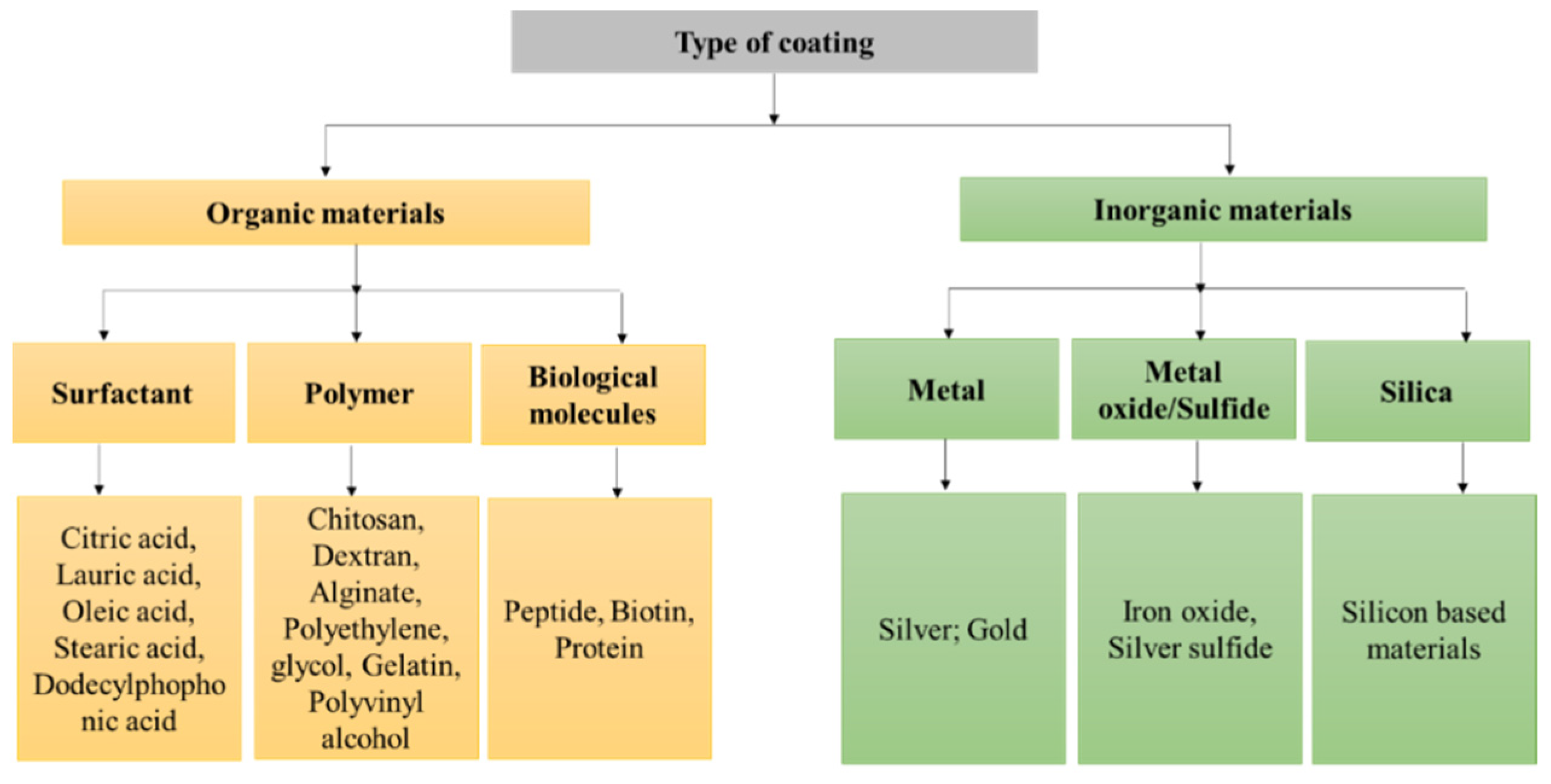
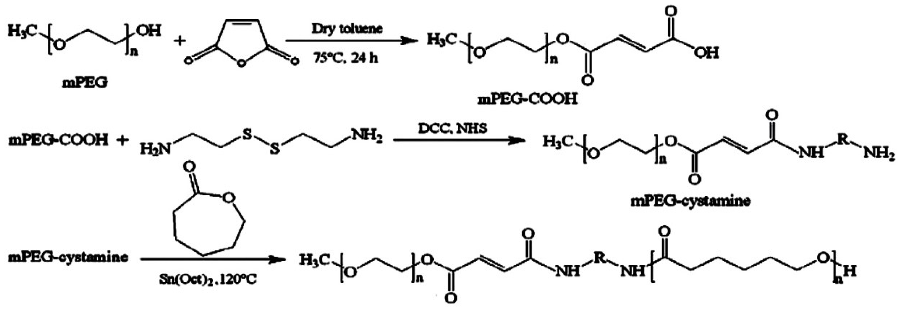
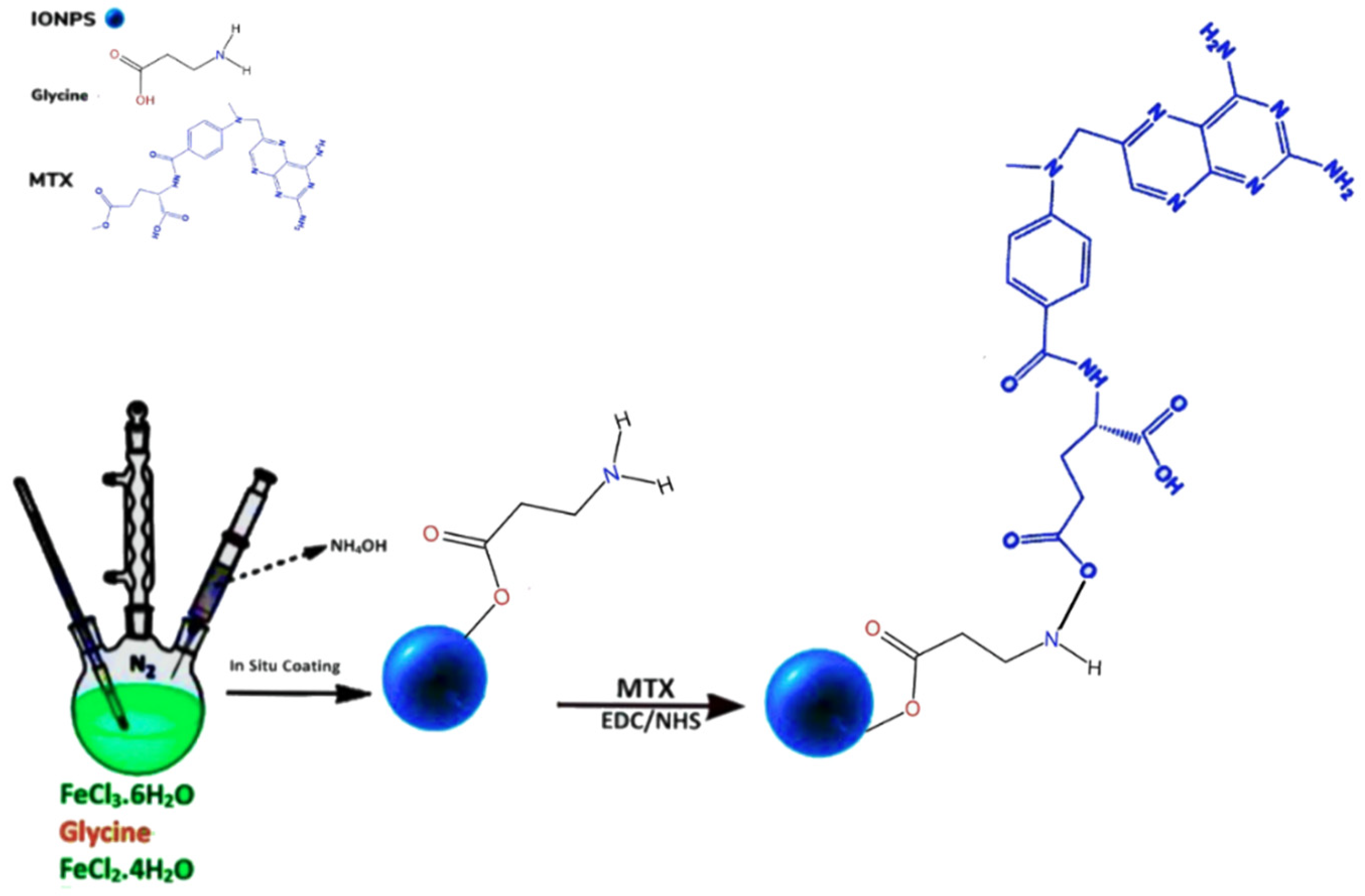
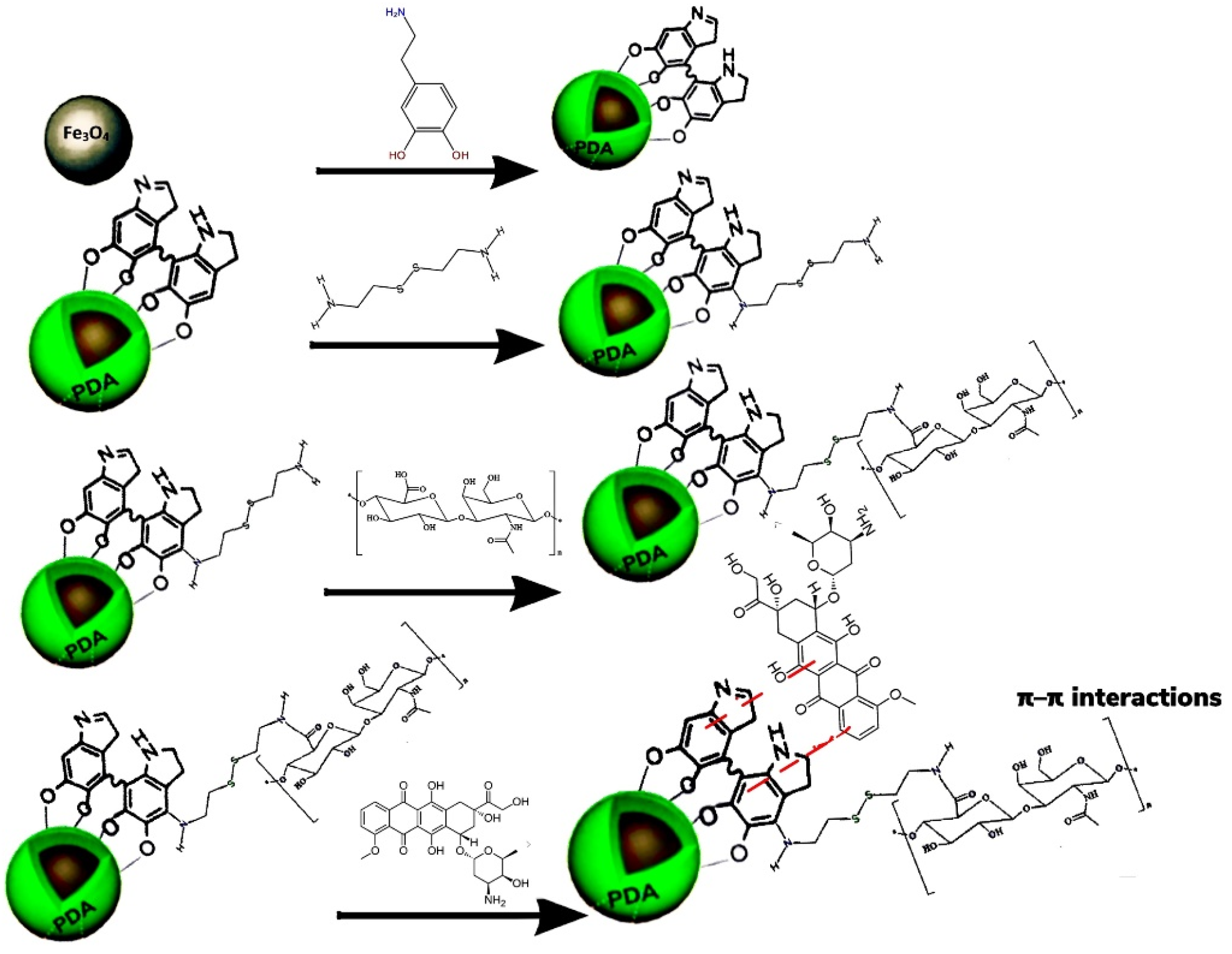
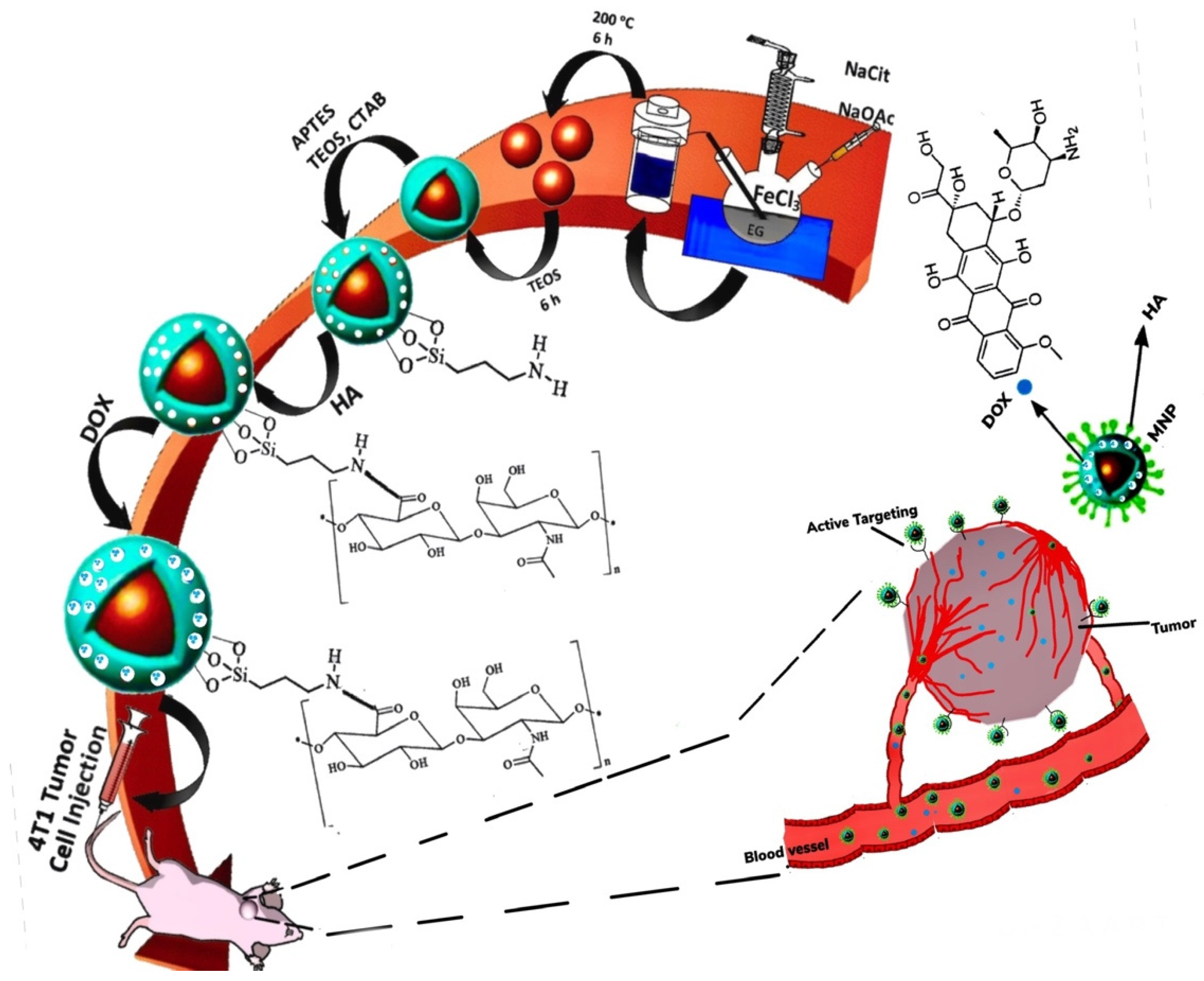
| pH | Redox | Temperature | Light | |
|---|---|---|---|---|
 Vinyl ester |  Carboxylic acid |  Disulphide |  Caprolactam |  Azobenzene |
 Amide |  Primary amine |  Diselenide |  N-isopropyl acrylamide | |
 Imine |  Tertiary amine | |||
 Oxime |  Pyridine | |||
 Hydrazone |  Orthoester | |||
 Acetal | ||||
| Fe3O4/PIMF | Fe3O4/PIMF-CUR | ||||
|---|---|---|---|---|---|
| Size (nm) (FE-SEM) | Saturation Magnetization (emu/g) | Cell Viability (%) | IC50 (mg/mL) | ||
| MCF-7 Cell Line | HeLa Cell Line | MCF-7 Cell Line | HeLa Cell Line | ||
| 20–30 | 45 | 44 | 32 | 0.23 | 0.15 |
| Fe3O4/MPSNPs | Peptide-Fe3O4/MPSNPs/DOX | ||||
|---|---|---|---|---|---|
| Size (nm) | Transverse Relaxivity (r2) (mM−1 s−1) | Loading Efficiency of Drug (%) | Drug Release (%) | ||
| TEM | DLS | Without MMP-2 Enzyme | With MMP-2 Enzyme | ||
| 114 | 600 | 135.6 | 12.2 | 15.7 | 70 |
| Samples | Size (nm) | Ms (emu/g) | EEPG (%) | PG Release over 1 h (%) | IC50 (μg/mL) | |||
|---|---|---|---|---|---|---|---|---|
| TEM | DLS | No-Enzyme | With-Enzyme | MCF-7/GFP Cell Line | HepG2 Cell Line | |||
| Fe3O4/CMCS | 9.8 | 38.1 | 65.01 | 91.78 | 6.95 | 44.61 | 0.8544 | 1.05 |
| Fe3O4/β-CD | 14.2 | 121.1 | 37.48 | 80.93 | 3.59 | 58.24 | 2.61 | 1.79 |
| Samples | Size (nm) DLS | Zeta Potential (mV) | Gene Transfection Rate (%) |
|---|---|---|---|
| M MPSNPs | 82 | −17.65 | 7 |
| M MPS/MBBs | 1120 | 31.47 | 14.87 |
| Formula | Shell | Application | Status | Procedure | Results | Ref |
|---|---|---|---|---|---|---|
| Zn0.1Fe0.9Fe2O4-Ge-Ag | Ge/Ag | -Contrast agent in MM-USI -Heat mediator in MHT | Pre-clinical MHT in phantom | -Synthesizing Zn-doped MNPs by co-precipitation (Zn0.1Fe0.9Fe2O4) -Manufacturing Ge/Ag phantom -Coating MNPs by Ge/Ag -Tissue mimicking (elasticity and acoustic) generating phantom -Applying PCC to produce powerful MF | -Cheap, non-invasive, and no ionizing effect -MM-USI provided real-time mapping of MNPs distribution, -MM-USI demonstrated successful 2D Temp monitoring prior and during MHT (for any depth within US limitation) | [233] |
| Fe3O4 | No surfactant/capping materials | -Heat mediator in MHT -Sonosensitizer in US | In vitro In vivo | -Preparing Fe3O4 MNPs by co-precipitation -Dispersing into saline to produce MNFs | -The MNFs and US-TS produced a major rise in the cytotoxicity response of EACCs in rats -Ineffective for deep-seated tumors | [234] |
| ZnMn-IONCs–PEG-PCL-SiNc | PEG-PCL | -heat mediator in MHT by AMF | In vitro In vivo | -Fabricating Zn-Mn-substituted IONPS via thermal decomposition. -Synthesizing IONCs by adding Zn-Mn-dopped IONPs and PEG-PCL in THF (Zn-MN-IONCs-PEG-PCL). -Loading SiNc onto IONCs | -Doping of Zn-Mn/Fe increased adsorption rate of IONPs and improved heat efficiency. -Encapsulation by PEG-PCL caused the MNPs to cluster inside the shell and have a higher heat-efficiency. -Over 90% apoptosis of DU145 cells by MHT. -The shrinkage and inhibition of prostate tumor following four cycles of MH treatment. -No toxicity to healthy cells. -No weight loss in DU145-bearing mice. | [236] |
| Formula | Targeting Agent | Drug | Shell | Target | Release Mechanism | Application | Status | Procedure | Result | Ref |
|---|---|---|---|---|---|---|---|---|---|---|
| NG-MNS-DOX | DOX | DOX | NIPAAm | MDA-MB-231 cells | TR | Chemotherapy and MHT by RF field | In vitro | -Preparing NG (carboxylate functionalization) by dissolving NIPAAm with ACA + MBAA (cross linker) + APS(catalyst) + SBS -Coating Fe3O4 by OlA and OlAm (MNS) -Covalently decorating NGs to MNS -Loading DOX on NG-MNS | -High stability (zerta potential = −28 mV) -High thermal stability -Passive targeting -Homogeneous heating and CDR due to the mono-dispersity in size/shape and uniform coating -Lower IC50 and enhanced therapeutic efficacy compared to free DOX | [137] |
| FMT-ML-DOX | DOX | DOX | lipid | 4T1 breast cancer | TR | chemotherapy and MHT | In vitro | -Fabricating ML to enclose FMT via lipid hydration, using DPPC + Chol+ PEG-2000-DSPE in chloroform and methanol -Preparing FMT in (NH4)2SO4, via hydrating lipid film then collected by PESM -Encapsulating DOX into ML-FMT via ASGP, then dialysis in PBS | -TR CDR profile compared to single utilization of MHT/chemotherapy. -Higher payload (DOX) release -FMT-ML-DOX uniformly distributed | [238] |
| DOX- MTX-MNPs-DPPC, Chol, SA, DSPE-MPEG2000, DSPE-PEG2000- | MTX | DOX | DSPE-PEG2000-NH2 | HeLa cells | TR | Dual imaging Dual targeting-light/MHT CDR via AMF + NIR-laser. | In vivo and In vitro | -Activating carboxylate group of MTX by NHS + DCC + DMSO -Followed by introducing DSPE-PEG2000-NH2 to it (DSPE-PEG2000-NH2-MTX) -Preparing OlA-coated-MNPs -Synthesizing MTX-MTRLs by adding DPPC, Chol, SA, DSPE-MPEG2000, DSPE-PEG2000-MTX and MNPs -Encapsulating DOX onto MTX-MTRLs | -Combination of AMF and NIR laser highly improved the DOX uptake, CDR and TDD into HeLa cells -MTX increased cytotoxicity to malignancy cells while the side effect to normal cells decreased | [239] |
| Fe3O4-Ge-HA-EDA-GO-DOX/PTX | HA | DOX/PTX | Ge | MDA-MB-231 and BT-474 | TR | -TDD and MHT | In vitro | -Preparing GO via the Hummer reaction and sonication -Aminating the HA by EDA followed by addition of NHS to activate the COOH group (HA-EDA) -Functionalizing GO by HA-EDA -Fabricating Fe3O4 MNPs via co-precipitation -Coating Fe3O4 by Ge -Adding Fe3O4—Ge to GO-HA-EDA to generate MN-composite -Loading of DOX/PTX onto Fe3O4—Ge—GO-HA-EDA | -DOX loaded MN-had higher efficacy compared to PTX. -Higher performance of HA functionalized MN-composites in destroying MDA-MB-231 with overexpressed CD44, not BT-474 cells since they lack CD44 -Incorporation of DOX and GO-HA MN-composite with MHT exhibited high killing efficacy | [240] |
| NIPAAm-PCL-NIPAA-MNP-OlA -PTX | PTX | PTX | NIPAM-PCL-NIPAM | MCF-7 | TR | -TDD, HT and chemotherapy | In vitro | -Fabricating of MNPs via co-precipitation -Adding OlA to MNPs (MNP@OlA) -Preparing PCL-diol polymer (using ε-CL + Sn(Oct)2 -Synthesizing triblock polymer by adding PCL-diol to THF + ALBN + NIPAM (PNIPAAm-PCL-PNIPAAm) (FRP) -Forming MNP micelles via solvent evaporation adding, PNIPAAm-PCL-PNIPAAm + THF + MNP@OlA -Loading PTX onto MNP-micelles | -Successful formation of PCL-diol and triblock PNIPAAm-PCL-PNIPAM -Biocompatible MNPs -PTX loaded MNPs were toxic to MCF-7 cell line. -Higher PTX release 89.3 ± 2.7% at Temp (42 °C) -Increasing Temp, decreased hydrodynamic diameter (size = 30–40 nm was thermal dependent) | [242] |
| SIONPPs-PEG-PBA-PEG-FA-TMZ | FA | TMZ | PEG-PBA-PEG | C6 GM cells | TR | -As heat mediator of AMF | In vivo and in vitro | -Fabricating of SIONPs via co-precipitation -Preparing PEG-PBA-PEG via poly-condensation and coupled with FA. -Loading TMZ onto SIONPs-PEG-PBA-PEG-FA via DESE | -Slight reduction in Ms value, due to FA and TMZ -Superparamagnetism with no magnetic hysteresis -Homo-compatible -High therapeutic efficiency, eradicating deeply placed tumors, e.g., brain GM-SIONPs-PEG-PBA-PEG-FA-TMZ remained inactive at 37°, activated when temp raised to 43° by AMF, resulting in an increase in the rate of drug release -Removal of AMF resulted in reduction in drug concentration, confirming the TR MNP facilitated the CDR -Improved thermal stability for hydrophilic phase | [243] |
| Formula | Targeting Agent | Drug | Shell | Target | Release Mechanism | Status | Procedure | Result | Ref |
|---|---|---|---|---|---|---|---|---|---|
| Dex-SIONPs-Manevist-fluorescein-1,2-DPPC, DSPC, DSPE-PEG-2000 | Fluorescein | Fluorescein and | 1,2-DPPC, DSPC, DSPE-PEG-2000 | Non-specific | TR | In vivo In vitro | -Producing M-HS via mixing commercially purchased Dex-IONPs with DTPA and fluorescein in PBS -Fabricating LMNPs using 1,2-DPPC, DSPC, DSPE-PEG-2000 -Hydrating LMNPs by M-HS -Exposing the LMNPs to AMF to release the drug model. | -High stability and dispersity, the PDI of LMNP before AMF = 0.134 and d = 231 nm, after AMF PDI = 0.1 and d = 223 nm at physiological pH = 7.4, confirming LMNPs stayed within liposome during and after the procedure. -Targeting malignancy via EPR -PEG enabled LMNPs to have prolong BCT -Applying AMF to LMNPs, fluorescein remained intact. even at 80° C -No drug release without AMF -A 100% drug release at 37 °C—37 °C by AMF -R1 showed CDR, concurrent release of DTPA and Fe -High-resolution/contrast and imaging quality. -Potential in MRI cancer theranostics | [164] |
| SA-MPDA-SPIO-DOX-Fe3+ | SA | DOX | SA-PEG-NH2 | HepG2 and Bel-7402 cells | pH and TR | In vivo In vitro | -Generating OlA-stabilized SIONPPs via thermal-decomposition. -Co-assembling MPDA and SIONPs by soft-templating (MPDA-SIONPs) -Synthesizing (SA-PEG-NH2) -Altering surface of MPDA-SIONPPs by SA-PEG-NH2 (MPDA-SIONPPs-SAPEG) -Chelating Fe3+ by MPDA-SIONPPs and MPDA-SIONPPs-SAPEG using FeCl3 -Encapsulating DOX onto MPDA-SIONPPs-SAPEG-FE3+ | -Biocompatible and dispersible in water -Successful modification of MNPs with SA in SA-PEG-MPDA-SPIO-Fe3+ -Highly precise T1/T2 MRI effect -An increased therapeutic efficacy due to active interaction of SA and E-selectin in vitro -Exceptional PTC ability and photostability. | [180] |
| Fe3O4-PDMAEMA/ PNIPAAm/MTX | MTX | MTX | MS PNIPAAm-PDMAEMA | A549 lung cancer cell | pHR and TR | In vitro | -Generating multi-modal MNPs via co-precipitation -Modifying MNPs by TMSMA to supply vinyl-link on the MNPs surface (MNPs + acetic acid + ethanol + TMSMA) -Fabricating MPSNPs via Stober method ((NH4OH + anhydrous ethanol + TEOS), precipitating with n-hexane -Producing CIL using DMAEMA and CPTMS -Preparing CIL-MPSNPs using (CIL monomer + DMSO + MPSNPs) -Conjugating MNPs-TMSMA, CIL-MPSNPs and NIPAAm to produce dual sensitive MNSs -Loading MTX (prepared in PBS) onto the MNSs (MNS-MTX) | -Increased antitumor activity of MNPs (MNS-MTX) and CDR, due to entering the cell via R-ME -Applicable as T2 MRI contrast agent in vitro -Polymerized MPNCs sustained their magnetic characteristics -MNS-MTX demonstrated potential in vivo application because of passive targeting (EPR effect) and TDD via MF -The MNPs aggregated in malignant tissue -Dose-dependent anti-neoplasm efficacy in A548 cells -Reduced adverse effects. -Biocompatible and only minor cytotoxicity due to high dose. | [246] |
| Fe3O4/MOF/CD/DOX/AS1411 Apt | AS1411 Apt | DOX | UiO-66-NH2 | MDA-MB-231 HBC cells | pHR | In vitro | -Ultra sonicating Fe3O4 in DMF and mixing with UiO-66-NH2 in NH2-BDC to generate Fe3O4@MOF core-shell MNPs -Loading DOX (DOX prepared in PBS, pH 8, 24 h) onto Fe3O4@MOF (Fe3O4@MOF-DOX) -Activating CDs by EDC and NHS (preparation in dark to activate the acid groups) for covalent conjugation with Fe3O4@MOF-DOX (Fe3O4@MOF-DOX-CDs) -Dissolving AS1411 Apt in DIW and US to conjugate with Fe3O4@MOF-DOX-CDs (Fe3O4-MOF@DOX-CDs-AS1411 Apt) | -High stability (up to 6 days in vitro) -Safe for HUVEC -Improved drug loading efficiency, tumor uptake and pHR drug release -TDD to nucleus of the triple-negative MDA-MB-231 HBC via overexpressed receptors, nucleolin -Effective anti-proliferation and promoting apoptosis in MDA-MB-231 HBC cells (77% cell apoptosis after 24 h) -Enhanced cancer cell targeting and binding affinity -Ability as FL bio-imaging | [247] |
| Fe3O4/CDNSs/FA/CUR | FA | CUR | β-CD | M109 cells | pHR | In vitro | -Polymerizing βCD and EPI as cross-linking agent to produce CDNS using anhydrous (βCD + DMSO + Et3N. EPI) -Synthesizing Fe3O4 NPs@CDNS via dispersing Fe3O4 NPs in (DMSO + βCD + Et3N + EPI) -Adding carboxyl group onto Fe3O4 NPs/CDNS using back titration method, dispersing Fe3O4 NPs-CDNS in (NaOH + HCl) -Fabricating FA-hydrazide (FA-NH-NH2) by dissolving FA in (DMSO + NHS + EDC) then hydrazine hydrate and converting into hydrochloride salt (using HCl) and precipitating by diethyl ether/acetonitrile and finally washing with ethanol -Conjugating FA-hydrazide with Fe3O4 NPs/CDNS, stirring Fe3O4/CDNS + DMSO + EDC and adding FA-hydrazide (Fe3O4/CDNS-FA NP) -Loading CUR into Fe3O4/CDNS-FA NP using (PBS + CUR in acetone) | -Fe3O4/CDNSs-FA-CUR was highly toxic to FRP M109 cells compared to FRN MCF 10A cells -Enhanced MRI (T2 negative signal) -Satisfying drug loading capacity -Acceptable agent release profile and TDD -Successful performance in theranostic platform -More in vivo studies on the potency of the nano-sponge to shrink the tumor will be required. | [248] |
| Fe3O4/Dex/ETB | ETB | ETB | Dex | CL1-5-F4 cancer cells | pHR | In vivo In vitro | -Preparing ultra-small Fe3O4 MNPs via co-precipitation -Coating Fe3O4 with Dex (Fe-D MNPs) -Crosslinking Dex coating with Epichlorohydrin/NaOH -Adding primary amino groups on the surface of IONPs (FeDN MNPs) -Treating FeDN MNPs with DMSO in buffer (pH:8.5, 0.1 M NaHCO3, dialysis with DIW for 3 days), adding MPTSA and NaCl generating FeDC MNPs -Conjugating FeDNC MNPs with ETB (FeDNC-E MNPs) | -Higher cellular uptake of ETB and intracellular drug delivery. -High re-ignition ability to kill cells with overexpressed EGFR receptors and leaving EGFR-negative cells intact -FeDNC-E MNPs suppressed EGFR–ERK–NF-κB signaling pathways -FeDNC-E MNPs inhibited, migration and metastasize of extremely intrusive CL1-5 F4 lung cancer cells in vivo xenograft. -Non-invasive real-time tracking of tumor by MRI -Decreased MRI T2 values in tumor cells compared to non-targeted cells -Promising for clinical application in targeted treatment and MRI | [249] |
| MMPSNP-Imi/Cis-Pt | Cis-Pt | Cis-Pt | TIP | Ovary cells | pHR | In vitro | -Synthesizing Fe3O4 MNPs via co-precipitation -Stabilizing MNPs by capping with CA (Fe3O4-CA) -Preparing MMPSNPs via sol-gel method, adding Fe3O4-CA+CTAB (pore constructing agent)+TEOS -Functionalizing MMPSNPs with Imi-groups using anhydrous toluene and TIP (MMPSNPs-Imi) -Replacing chloro-ligand with aqua-ligand via suspending Cis-Pt in DI water and AgNO3 to precipitate in the dark, centrifuged to remove AgCl and diluted with DIW to obtain 0.1 mg/mL -Adsorbing Cis-Pt on MMPSNPs-Imi via direct incubation (mixing at 37 °C in the dark for 24 h). | -Functionalizing MMPSNPs with Imi-groups weakened Cis-Pt interactions and resulting CDR -Replacing chloro-ligand with aqua-ligand enhanced the reactivity of Cis-Pt in aqua-solution. -Sustained and prolonged Cis-Pt release, due to the mesoporous features (e.g., pore size/opening, ratio of NPs to pore size, attraction of NPs to pore walls) prevents Cis-Pt diffusion into the pores instead adsorbed on the surface -High Cis-Pt loading capacity -With no-burst, pHR, and constant Cis-Pt release in acidic conditions resulting in growth inhibition of HEOC cancer cells -Successful apoptosis and necrosis -Enhanced T2-weighted MRI (high transverse relaxivity) -Potential in vivo application as chemo and contrast agents TDR | [250] |
| Fe3O4-FTY720-PFP-RGD | FTY720 | FTY720 | RGD | HepG2 and Huh7 cells | US-responsive | In vitro | -Synthesizing SIONPPs via organic phase decomposition -Loading drug onto SIONPs via thin film hydration pathway, dissolving 1,2-DPPC+DSPE-PEG-MAL+FTY720 in THF, adding SIONPs. Mixing with buffered saline and sonicated. Adding PFP then ultrasonicated, emulsified and dialyzed for 3 day, store at −4 °C to produce FTY720@SIONP/PFP/NBs -Mixing FTY720@SIONP/PFP/NBs + EDC + NHS then adding RGD-peptide. -Dialyzing with DIW for 3 day and keep at −4 °C to develop FTY720@SIONP/PFP/RGD-NBs | -Enhanced FTY720 release and active targeting due to LIFUS -High stability, good encapsulation, and agent loading efficiency -Low toxicity to normal fibroblast 3T3 cells -High inhibition of HepG2 and Huh7 cells -High relaxation value and T2-weighted MRI -MNBs resulted in an increase in EPR results -Induced HepG2 apoptosis via activating Caspase3, Caspase9, and p53 | [253] |
| Fe3O4-INU-LA-PEG-FA-DOX | FA | DOX | INU-LA-PEG | Colon cancer | RR | In vivo in vitro | -Synthesizing INU-LA-PEG-FA via microwave radiation, by dispersing (INU + BNPC + (±)-α-lipoic acid (LA) in DMF), adding PEG-FA -Coating SIONPs with INU-LA-PEG-FA -Loading DOX-HCl onto SIONP@INU-LA-PEG-FA | -Improved cancer inhibition due to the presence of FA -Reduced tumor volume -Enhanced MRI in vivo, promising in locoregional chemo-treatment | [254] |
| Fe3O4-PDMAEMA/ PNIPAAm/MTX | MTX | MTX | MS PNIPAAm-PDMAEMA | A549 lung cancer cell | pHR andTR | In vitro | -Generating multi-modal MNPs via co-precipitation -Modifying MNPs by TMSMA to supply vinyl-link on the MNPs surface via ultrasonicating (MNPs + acetic acid + ethanol + TMSMA) -Fabricating MPSNPs via Stober method ((NH4OH + anhydrous ethanol + TEOS), precipitating with n-hexane -Producing CIL using DMAEMA and CPTMS -Preparing CIL-MPSNPs using (CIL monomer + DMSO + MPSNPs) -Conjugating MNPs-TMSMA, CIL-MPSNPs and NIPAAm to produce dual sensitive MNSs -Loading MTX (prepared in PBS) onto the MNSs (MNS-MTX) | -Increased antitumor activity of MNPs (MNS-MTX) and CDR, due to entering the cell via R-ME -High relaxivities in vitro, indicating the ability of MNPs to sustain their magnetic characteristics. -MNS-MTX demonstrated potential in vivo application because of passive targeting (EPR effect) and TDD via MF -The MNPs aggregated in malignant tissue -Dose-dependent anti-neoplasm efficacy in A548 cells -Reduced adverse effects. -Biocompatible and non-cytotoxic effect on A549 cell line in vitro, -Minor cytotoxicity due to high dose. | [246] |
| Fe3O4-GO-DOX | FA | DOX | GO | MGC-803 cells | pHR | In vitro In vivo | -Fabricating NGO by cutting GO sheet and adding AgNO3 (redox reaction) -Preparing Fe3O4@NGO NPs via one-pot hydrothermal pathway (FeCl3·6H2O + ethylene glycol + NaOH + NGO) -Conjugating FA with Fe3O4@NGO MNPs via coupling reaction (EDC and NHS), followed by adding Fe3O4@NGO Loading DOX onto FA-Fe3O4@nGO via precipitating with a permanent magnet | -Good targeting capability for solid tumors in vivo -Biocompatible, safe and no cytotoxicity to normal cell -Higher uptake by tumor cells and lower uptake by residual organs, e.g., liver, kidneys -Increased cancer cell targeting -Due to the nano-size and conjugation with FA, the sharpness of GO NPs reduced (cell membrane damage decreased) and had lower oxidative stress in vitro and in vivo -MRI performed after 8–24 h of administration of NPs to tumor bearing mice; -showed decrease in MRI signal in FA-Fe3O4@nGO-DOX -and decrease in MRI signal in Fe3O4@nGO-DOX, due to the EPR effect. | [255] |
| Fe3O4-Au-LA-CUR | GSH | LA-CUR | Au LA-CUR-GSH | U87MG cells | pHR | In vivo in vitro | -Synthesizing Fe3O4 via co-precipitation -Functionalizing of Fe3O4 by APTES (Fe3O4-NH2) -Fabricating Fe3O4—Au NCs via wet-chemical, using (HAuCl4.2H2O and tri-sodium citrate+NaBH4) -Conjugating LA onto CUR (LA-CUR), using CUR + LA + DMAP in DCM, adding EDC -Bonding GSH with Fe3O4—Au NCs via swapping its thiol group with citrate group of AU-NPs -Attaching LA-CUR onto the Fe3O4-Au NCs via ligand swap of S-S bond in LA with citrate of Au NPs (Fe3O4-Au-LiA-CUR) -Adding LA-CUR and GSH simultaneously to Fe3O4-Au NCs forming Fe3O4-Au-LA-CUR-GSH | -Enhanced cytotoxicity -Great LA-CUR loading efficiency -Efficient and CDR in vitro -Highly biocompatible due to minimum protein adsorption and good hemocompatibility -Capable of crossing BBB -Successful TDD due to GSH targeting agent -High cellular uptake by GSH receptor positive astrocyte cell -Highly promising for MRI negative contrast media for brain tumor imaging/therapy | [256] |
| AuNCs−Fe3O4/MDP-FITC/PFP | DCs | MDP | PEG2000-NH2 | Y79 cells | TR | In vivo in vitro | -Activating carboxyl group of citrated Au-NCs using EDC and NHS. -Conjugating PEG2000@Fe3O4 (−NH2) to Au-NCs by amide bond. -Adding MDP and FITC in PBS to Au-NCs–Fe3O4 at room temp and absence of light. -Loading PFP into Au-NCs/Fe3O4/MDP-FITC | -MNPs exhibited homogeneous size, good dispersity and superparamagnetism -Potential for multi-modal application, T2-MRI, MHT, TDD and CDR. -Facile transportation of MNPs in blood stream and assembly in RB tumor via EPR effect. -Biocompatible and non-toxic to healthy cells at high Temp (43.2 °C), leading to apoptosis/necrosis of Y79 cells. -Irradiation by LIFU resulted liquid PFP changes to gas that improve LIFU efficacy and US imaging and release MDPs which bound to NOD2 receptor resulting activation of DCs to identify and eliminate RB tumor cells. | [257] |
| Size (nm) DLS | Saturation Magnetization (emu/g) | 5-FU Loading Capacity (mg/g) | The Viability of SW620 Cells (%) |
|---|---|---|---|
| 423.5 | 7.7 | 400 | 90 |
| Size (nm) DLS | Saturation Magnetization (emu/g) | DOX Loading Capacity (%) | DOX Release within 24 h (%) | Transverse Relaxation Rate (mM−1s−1) | ||
|---|---|---|---|---|---|---|
| In the Absence of Any Stimulus | In the Presence of pH = 5.5, GSH | In the Presence of pH = 5.5, GSH and Laser | ||||
| 120 | 28.5 | 7.13 | 3.6 | 9.96 | 32.7 | 171.76 |
| Formula | Targeting Agent | Drug | Shell | Target | Release Mechanism | Procedure | Result | Ref |
|---|---|---|---|---|---|---|---|---|
| Fe3O4/Au/OPSS-PEG-SVA-/PDMAEMA/MTX | MTX | MTX | OPSS-PEG-SVA/PDMAEMA | MCF7 and MDA-MB-231 BC cells | pHR | -Preparing polymer solution via NH2-terminating DMAEMA ((+)ly charged, pHR polymer) (NH2-PDMAEMA) -Fabricating Fe3O4 via co-precipitation -Developing Au-Fe3O4 MNPs via standard citrate reduction -PEGylating MNPs using OPSS-PEG-SVA (MNPs@OPSS-PEG-SVA) -Adding PDMAEMA onto MNPs@OPSS-PEG-SVA under Ar gas (MNPs@polymer) -Loading MTX onto MNPs@polymer | -Improved EPR effect -Prolonged blood circulation -Safe and CDR at physiological pH -Improved cytotoxic activity MCF7 and MDAMB231 cell lines -Potential candidate for passive and active TDD | [276] |
| Fe3O4/MSN/PEI/ETB | FA | ETB | MS and PEI | HeLa cells | pHR | -Synthesizing of Fe3O4 via co-precipitation -Coating Fe3O4 NPs with silica to produce (Fe3O4@MSN) using TEOS and CTAB -Conjugating FA with PEI to produce PEI-FA using FA+DCC+NHS+PEI in coupling reaction -Preparing Fe3O4@ MSN/PEI-FA, using Fe3O4@MSN+modified PEI-FA+acetic acid -Loading ETB onto Fe3O4@ MSN/PEI-FA via dissolving ETB in DMSO | -Fe3O4@MSN/PEI-FA-ETB demonstrated higher cytotoxicity effect on HeLa cells compared to Fe3O4@MSN-ETB/PEI (due to lacking TA) -Non-toxic effect -TDD and CDR with minimum side effects -Promising TDD tool for in vivo applications | [277] |
| Fe3O4/CS/SH/FA/Co 6 | FA | Co 6 | CS | HeLa cells | RR | -Thiolating Fe3O4 and CS separately. (Fe3O4-SH) and (CS-SH) -Coupling CS-SH with (1) FA (FA-CS-SH) (2) RFP (RFP-CS-SH) -Followed by mixing (1) and (2) in HSO, dispersing Co 6 and ultrasonicating to generate Fe3O4/CS/SH/FA/Co 6 | -Higher internalization into HeLa cells compared to the non-targeted MNPs | [283] |
| Fe3O4/PLH−PEG −LiA/FA-PEG/DOX | FA | DOX | Si | MCF-7, MDA-MB-231, MCF-10A cells | pHR and RR | -Preparing MNPs via hydrothermal reaction -Reacting MNPs with TEOS to produce Fe3O4@SiO2 -Preparing MPS coating (using C18TMS), to produce Fe3O4@SiO2@MSiO2 -Loading Dox onto Fe3O4@SiO2@MSiO2 -Preparing polymer (PLH−PEG− NH2) using coupling reaction (EDC and NHS reagents) -Connecting LiA to PLH−PEG−NH2 using DCC coupling reagent to produce PLH−PEG−LiA polymer -Synthesizing FA−PEG−NH2 via coupling reaction | -Elipsoidal-shaped MNPsenhanced uptake and cell morbidity compared to the non-targeted NPs against BC cell -Substantial increase in DOX release in the presence of 10 mM GSH at pH 5.5 (97.1%) as compared to pH 7.4 (31.7%) within 24 h | [284] |
| Fe3O4/SiO2/MUC1 Apt/DOX | MUC1 Apt | DOX | SiO2 | MCF-7 and MDA-MB-231 BC cells | pHR | -Preparing SIONP using thermal decomposition -Developing SIONPs@SiO2-NH2 using TEOS and APTEs in the presence of CTAB -Fabricating SIONPs@SiO2-COOH via reacting SIONPs@SiO2-NH2 with succinic anhydrate in DMF -Anchoring DOX onto SIONP-SiO2-COOH -Decorating SIONP@SiO2-COOH/DOX by MUC1 Apt -Labelling SIONP@SiO2/DOX/MUC1 Apt with fluorophore using PBS and FITC | -Remarkably high drug release in acidic TME -Potential multi-modal candidate used for diagnosis and treatment of MUC1 overexpressed malignant cells. -Higher toxicity and internalization by MUC1 expressing MCF-7 cells | [287] |
| Fe3O4/PEI/HPhBA/DOX | PhBA | DOX | PEI | U-87 MG | pHR | -Functionalizing Fe3O4 by NH2 groups (Fe3O4-NH2) -Preparing NH2 groups of Fe3O4-NH2 by PEI -Reacting with PhBA to fabricate HPhBA-MNPs | -Increased therapeutic effect on U-87 MG malignant glioma cells. -Improved cellular uptake and CDR | [288] |
| Fe3O4/GO multi/Lf DOX | LF | DOX | -GO | C6 glioma cells | pHR | -Encapsulating Fe3O4 by GO (GO/Fe3O4) -Functionalizing by LF via click chemistry for TDD -Loading DOX onto MNPs | -Intracellular delivery efficiency -Increased cytotoxicity against C6 glioma cells | [289] |
| Fe3O4/TMSMA/QDMAMEA/Aly-Imz/CD/MTX | MTX | MTX | β-CD | Saos-2 bone cancer cells | pHR | -Grafting β-CD onto Aly-Imz (via reflux-coprecipitation) -Preparing CMNPs via FRPR (using β-CD/Aly-Imz/QDMAME/TMSMA in DMSO under N2), followed by co-precipitation -Loading with MTX for TDD to Saos-2 cells | -Successful pH-responsive characteristics -Increased cytotoxicity, -Enhanced cellular uptake in Saos-2 cells -No major cytotoxicity effects on HRBCs. | [290] |
| Fe3O4/CS/PNIPAAm-Co-IA/MTX/ETB | MTX | ETB | CS-NIPAAm-IA | OVCAR-3 cells | pHR and TR | -Modifying CS with SDS and MaA, (generating polymerizable organo-soluble precursor) -Grafting NIPAAm and IA (TR and pHR monomers) onto CS via FRPR (co-polymerization) -synthesizing MNPs via co-precipitation -Developing MNPs@CSC -Activating MTX via coupling reaction (using EDC and NHS reagents) -Conjugating MTX with MNPs@CSC (producing MNPs@CSC-MTX) -Loading ETB onto MNPs@CSC-MTX (producing MNPs@CSC-MTX-ETB) | -spherical-shaped MNPs -High loading efficiency for ETB. -Increased cytotoxicity and -Higher cellular uptake of MTX, by FR-positive cells -TDD and improved drug release -Potential theranostic nano-system for the targeted imaging/treatment of solid tumors, e.g., ovarian | [291] |
| O-CMCS-Fe3O4-PEM | PEM | PEM | O-CMCS | A549-luc-C8 and CRL5807 cell | pHR | -Improving CS solubility by O-CM (O-CMCS) -Encapsulating O-CMCS by Fe3O4 to produce MNPs (O-CMCS MNPs) -Loading PEM onto O-CMCS MNPs | -Highly promising therapy for NSCL carcinoma | [292] |
| Fe3O4/MUC-1/PEG//DOX | MUC-1 Apt | DOX | PEG | MDA-MB-231 and MCF-7 cells | pHR | -PEGlayting SIONPs -Decorating by MUC1-Apt | -Higher uptake as compared to non-specific targeted NPs, -Increased death rate in MCF-7 cells | [293] |
| Name | Coating | Application | Status | Ref |
|---|---|---|---|---|
| Ferumoxytol (Feraheme®) | Carboxymethyl dextran | Head and Neck imaging | Clinical trial (Early Phase I) | [317] |
| Magnablate I | Not available | MHT on prostate cancer | Clinical trial (Early Phase I) | |
| Ferumoxytol/Gadobutrol | carbohydrate | Urinary bladder imaging | Clinical trial (Early Phase I) | |
| Ferumoxytol (FerahemeTM) | Carboxymethyl dextran | Imaging for lymph node staging in esophageal cancer | Clinical trial (Phase I)/discontinued | |
| Ferumoxytol (Feraheme®) | Carboxymethyl dextran | Imaging of lymph node involvement in prostate cancer | Clinical trial (Phase I) | |
| Ferumoxytol (Feraheme®) | Carboxymethyl dextran | Imaging of lymph nodes in patients with primary prostate or breast cancer | Clinical trials (observational study) | |
| Ferumoxytol (Feraheme®) | Carboxymethyl dextran | Imaging of lymph node metastases in prostate, bladder, and kidney cancers | Clinical trial (phase II) | |
| Ferumoxytol (Feraheme®) | Carboxymethyl dextran | Brain tumor imaging | Clinical trial (phase II) | |
| Ferumoxytol/MM-398 | Carboxymethyl dextran | Imaging of solid tumors | Clinical trial (phase I) | |
| Ferumoxytol (Feraheme®, Ferumoxytol non-stoichiometric magnetite)/Gadolinium | Carboxymethyl dextran | Imaging of primary or metastatic brain tumors | Clinical trial (phase II) | |
| Ferumoxytol (Feraheme®, Ferumoxytol non-stoichiometric magnetite) | Carboxymethyl dextran | Imaging of lymph nodes in patients with advanced rectal cancer | Clinical trial (Early phase I) | |
| Ferumoxytol (Feraheme®, Ferumoxytol non-stoichiometric magnetite) | Carboxymethyl dextran | Imaging of inflammatory (macrophage) responses in patients with malignant brain tumors | Clinical trial (Early phase I) | |
| Ferumoxytol (Feraheme™, Ferumoxytol non-stoichiometric magnetite) | Carboxymethyl dextran | Imaging of lymph nodes in patients with stage IIB-IIIC esophageal cancer | Clinical trial (Early phase I) | [317] |
| Ferumoxytol(Feraheme™) | Carboxymethyl dextran | Radiotherapy with SIONP on MR-Linac for hepatic cancers | Clinical trial (Observational) | |
| Ferumoxytol (Feraheme™, Ferumoxytol non-stoichiometric magnetite) | Carboxymethyl dextran | Imaging of lung carcinoma metastatic in the brain | Clinical trial (Phase II) | |
| Ferumoxytol (Feraheme®) | Carboxymethyl dextran | Imaging of prostate tumor | Clinical trial (Early Phase I) | |
| Ferumoxytol (AMAG Pharmaceuticals, Inc. (Waltham, MA, USA), Code 7228) | Carboxymethyl dextran | Imaging of brain tumors in Patients Receiving Chemotherapy | Clinical trial (Phase I) | |
| Ferumoxytol (Feraheme®) | Carboxymethyl dextran | Bone imaging | Clinical trial (Phase II) | |
| Ferumoxytol (Feraheme™, Ferumoxytol non-stoichiometric magnetite) | Carboxymethyl dextran | Imaging of glioblastoma tumors after treatment with pembrolizumab | Clinical trial (Phase II) | |
| Magnetic nanoparticles | anti-EpCAM or anti-CD52 antibodies | Removing of blood tumor cells in patients suffering from prostate, colon, lung, or pancreatic cancer | Clinical trial (Observational) | |
| Ferumoxytol (Feraheme®) | Carboxymethyl dextran | Imaging of abnormal lymph nodes in patients with thyroid cancer | Clinical trial (unknown status) | |
| Ferumoxytol (Feraheme®) | Carboxymethyl dextran | Imaging of lymph node metastases in pancreatic cancer | Clinical trial(phase IV) | |
| Ferumoxsil (Lumirem®, GastroMARK®, AMI-121) | Poly [N-(2-aminoethyl)-3- aminopropyl] siloxane | GI tract, abdominal tissue and bowel imaging | Used and discontinued | [273,318] |
| Ferumoxide (Feridex®, EndoremTM, AMI-25) | Dextran | Liver imaging | Used and discontinued | [67,318] |
| Ferucarbotran (Resovist®, CliavistTM, SHU 555A) | Carboxydextran | Liver/spleen imaging | Approved | [67,273] |
| Ferristene (Abdoscan®, OMP) | Polystyrene (sulfonated styrene-divinylbenzene copolymer) | Gastrointestinal, abdominal imaging | Approved and discontinued | [319] |
| Ferumoxtran-10 (Ferrotran®) | Dextran | Prostate imaging | Clinical trial (Phase III) | [317] |
| Ferumoxtran (Combidex®, Sinerem®) | Dextran | Lymph node imaging | Clinical trial | [67] |
| SIONP-epirubicin | Anhydroglucose | Magnetic targeted delivery of 4-epidoxorubicin into solid tumors | Clinical trials (terminated) | [320] |
| Nanotherm® (MagForce) | Aminosilane | Magnetic hyperthermia on brain tumors | Approved/in use in Europe | [318,319] |
| MTC-Dox | Metallic iron and activated carbon | Magnetic drug targeting | Clinical trials (terminated) | [273,320] |
Publisher’s Note: MDPI stays neutral with regard to jurisdictional claims in published maps and institutional affiliations. |
© 2022 by the authors. Licensee MDPI, Basel, Switzerland. This article is an open access article distributed under the terms and conditions of the Creative Commons Attribution (CC BY) license (https://creativecommons.org/licenses/by/4.0/).
Share and Cite
Aram, E.; Moeni, M.; Abedizadeh, R.; Sabour, D.; Sadeghi-Abandansari, H.; Gardy, J.; Hassanpour, A. Smart and Multi-Functional Magnetic Nanoparticles for Cancer Treatment Applications: Clinical Challenges and Future Prospects. Nanomaterials 2022, 12, 3567. https://doi.org/10.3390/nano12203567
Aram E, Moeni M, Abedizadeh R, Sabour D, Sadeghi-Abandansari H, Gardy J, Hassanpour A. Smart and Multi-Functional Magnetic Nanoparticles for Cancer Treatment Applications: Clinical Challenges and Future Prospects. Nanomaterials. 2022; 12(20):3567. https://doi.org/10.3390/nano12203567
Chicago/Turabian StyleAram, Elham, Masome Moeni, Roya Abedizadeh, Davood Sabour, Hamid Sadeghi-Abandansari, Jabbar Gardy, and Ali Hassanpour. 2022. "Smart and Multi-Functional Magnetic Nanoparticles for Cancer Treatment Applications: Clinical Challenges and Future Prospects" Nanomaterials 12, no. 20: 3567. https://doi.org/10.3390/nano12203567