
Bacterial Pathogens in the Food Industry: Antibiotic Resistance and Virulence Factors of Salmonella enterica Strains Isolated from Food Chain Links
 , , , ,
  , , and
Abstract
:1. Introduction
2. Materials and Methods
2.1. Taxonomic Identification of the Salmonella Strains
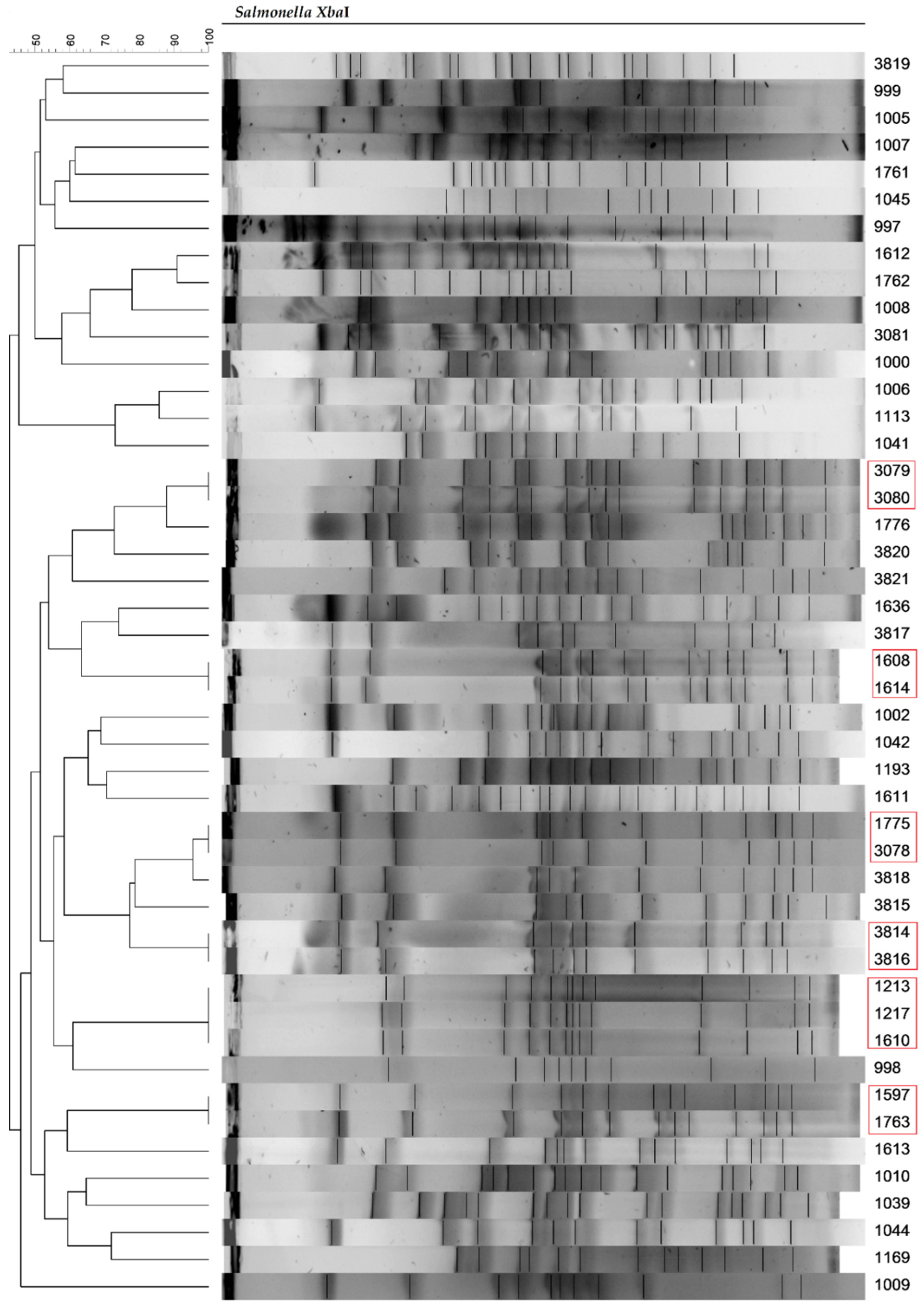
2.2. Subtyping Salmonella Strains Using Pulsed-Field Gel Electrophoresis (PFGE)
2.3. Detection of Virulence Genes in Salmonella Strains
2.4. Antimicrobial Sensitivity Testing
2.5. Determination of Antibiotics Resistance Profile of Salmonella Strains
2.6. Screening for Phenotypic and Genotypic Detection of β-lactamases-Producing Salmonella Strains
3. Results and Discussion
3.1. Source of Isolation and Taxonomic Identification of the Salmonella Strains
3.2. Subtyping Salmonella Strains Using Pulsed-Field Gel Electrophoresis (PFGE)
3.3. Detection of Virulence Genes in Salmonella Strains
3.4. Antibiotic Resistance Profiles in Salmonella Strains
3.5. Genotypic Resistance Profiles in Salmonella Strains
3.6. Screening for Phenotypic and Genotypic Detection of β-lactamases-Producing Salmonella Strains
4. Conclusions
Supplementary Materials
Author Contributions
Funding
Institutional Review Board Statement
Informed Consent Statement
Data Availability Statement
Conflicts of Interest
References
- Heredia, N.; García, S. Animals as sources of food–borne pathogens: A review. Anim. Nutr. 2018, 4, 250–255. [Google Scholar] [CrossRef] [PubMed]
- Ferrari, R.G.; Rosario, D.K.A.; Cunha–Neto, A.; Mano, S.B.; Figueiredo, E.E.S.; Conte–Junior, C.A. Worldwide Epidemiology of Salmonella Serovars in Animal–Based Foods: A Meta–analysis. Appl. Environ. Microb. 2019, 85, e00591-19. [Google Scholar] [CrossRef] [PubMed] [Green Version]
- Wang, X.; Biswas, S.; Paudyal, N.; Pan, H.; Li, X.; Fang, W.; Yue, M. Antibiotic Resistance in Salmonella Typhimurium Isolates Recovered From the Food Chain Through National Antimicrobial Resistance Monitoring System Between 1996 and 2016. Front. Microbiol. 2019, 10, 985. [Google Scholar] [CrossRef] [Green Version]
- Shelobolina, E.S.; Sullivan, S.A.; O’Neill, K.R.; Nevin, K.P.; Lovley, D.R. Isolation, characterization, and U (VI)–reducing potential of a facultatively anaerobic, acid–resistant bacterium from low–pH, nitrate– and U (VI)–contaminated subsurface sediment and description of Salmonella subterranea sp. nov. Appl. Environ. Microb. 2004, 70, 2959–2965. [Google Scholar] [CrossRef] [PubMed] [Green Version]
- Hata, H.; Natori, T.; Mizuno, T.; Kanazawa, I.; Eldesouky, I.; Hayashi, M.; Miyata, M.; Fukunaga, H.; Ohji, S.; Hosoyama, A.; et al. Phylogenetics of family Enterobacteriaceae and proposal to reclassify Escherichia hermannii and Salmonella subterranea as Atlantibacter hermannii and Atlantibacter subterranea gen. nov., comb. nov. Microbiol. Immunol. 2016, 60, 303–311. [Google Scholar] [CrossRef]
- Hurley, D.; McCusker, M.P.; Fanning, S.; Martins, M. Salmonella—Host interactions—Modulation of the host innate immune system. Front. Immunol. 2014, 5, 481. [Google Scholar] [CrossRef] [Green Version]
- Pławińska–Czarnak, J.; Wódz, K.; Kizerwetter–Świda, M.; Bogdan, J.; Kwieciński, P.; Nowak, T.; Strzałkowska, Z.; Anusz, K. Multi–Drug Resistance to Salmonella spp. When Isolated from Raw Meat Products. Antibiotics 2022, 11, 876. [Google Scholar] [CrossRef] [PubMed]
- Qin, X.; Yang, M.; Cai, H.; Liu, Y.; Gorris, L.; Aslam, M.Z.; Jia, K.; Sun, T.; Wang, X.; Dong, Q. Antibiotic Resistance of Salmonella Typhimurium Monophasic Variant 1,4,[5],12:i:– in China: A Systematic Review and Meta–Analysis. Antibiotics 2022, 11, 532. [Google Scholar] [CrossRef]
- Shaheen, A.; Tariq, A.; Shehzad, A.; Iqbal, M.; Mirza, O.; Maslov, D.A.; Rahman, M. Transcriptional regulation of drug resistance mechanisms in Salmonella: Where we stand and what we need to know. World J. Microbiol. Biotechnol. 2020, 36, 85. [Google Scholar] [CrossRef]
- Dos Santos, A.M.P.; Ferrari, R.G.; Conte–Junior, C.A. Virulence Factors in Salmonella Typhimurium: The Sagacity of a Bacterium. Curr. Microbiol. 2018, 76, 762–773. [Google Scholar] [CrossRef]
- Luo, Y.; Yi, W.; Yao, Y.; Zhu, N.; Qin, P. Characteristic diversity and antimicrobial resistance of Salmonella from gastroenteritis. J. Infect. Chemother. 2018, 24, 251–255. [Google Scholar] [CrossRef] [PubMed]
- Kurtz, J.R.; Goggins, J.A.; McLachlan, J.B. Salmonella infection: Interplay between the bacteria and host immune system. Immunol. Lett. 2017, 190, 42–50. [Google Scholar] [CrossRef] [PubMed]
- Eng, S.K.; Pusparajah, P.; Mutalib, N.S.A.; Ser, H.L.; Chan, K.G.; Lee, L.H. Salmonella: A review on pathogenesis epidemiology and antibiotic resistance. Front. Life Sci. 2015, 8, 284–293. [Google Scholar] [CrossRef] [Green Version]
- Khan, M.A.S.; Rahman, S.R. Use of Phages to Treat Antimicrobial-Resistant Salmonella Infections in Poultry. Vet. Sci. 2022, 9, 438. [Google Scholar] [CrossRef] [PubMed]
- European Food Safety Authority; European Centre for Disease Prevention and Control. The European Union One Health 2020 Zoonoses Report. EFSA J. 2021, 19, e06971. [Google Scholar]
- Chief Sanitary Inspectorate. Sanitary Condition of the Country in 2020. Available online: https://www.gov.pl/web/gis/raport---stan-sanitarny-kraju (accessed on 30 September 2022).
- Chief Sanitary Inspectorate. Sanitary Condition of the Country in 2021. Available online: https://www.gov.pl/web/gis/raport---stan-sanitarny-kraju (accessed on 30 September 2022).
- Połaska, M.; Sokołowska, B. Bacteriophages—A new hope or huge problem in the food industry. AIMS Microbiol. 2019, 5, 324–346. [Google Scholar] [CrossRef]
- Wójcicki, M.; Świder, O.; Daniluk, K.J.; Średnicka, P.; Akimowicz, M.; Roszko, M.Ł.; Sokołowska, B.; Juszczuk–Kubiak, E. Transcriptional Regulation of the Multiple Resistance Mechanisms in Salmonella—A Review. Pathogens 2021, 10, 801. [Google Scholar] [CrossRef]
- Wójcicki, M.; Błażejak, S.; Gientka, I.; Brzezicka, K. The concept of using bacteriophages to improve the microbiological quality of minimally–processed foods. Acta Sci. Pol. Technol. Aliment. 2019, 18, 373–383. [Google Scholar]
- WHO. Salmonella (Non–Typhoidal). Available online: https://www.who.int/news-room/fact-sheets/detail/salmonella-(non-typhoidal) (accessed on 3 October 2022).
- European Food Safety Authority; European Centre for Disease Prevention and Control. The European Union Summary Report on Antimicrobial Resistance in zoonotic and indicator bacteria from humans, animals and food in 2019–2020. EFSA J. 2022, 20, 7209. [Google Scholar]
- European Food Safety Authority; European Centre for Disease Prevention and Control. The European Union Summary Report on Antimicrobial Resistance in zoonotic and indicator bacteria from humans, animals and food in 2018–2019. EFSA J. 2021, 19, 6490. [Google Scholar]
- Tang, H.; Wang, M.J.; Gan, X.F.; Li, Y.Q. Funneling lignin–derived compounds into polyhydroxyalkanoate by Halomonas sp. Y3. Bioresour. Technol. 2022, 362, 127837. [Google Scholar] [CrossRef] [PubMed]
- PulseNet. Standard Operating Procedure for PulseNet PFGE of Escherichia coli O157:H7, Escherichia coli Non–O157 (STEC), Salmonella Serotypes, Shigella sonnei and Shigella flexneri. Available online: https://pulsenetinternational.org/protocols/pfge/ (accessed on 15 May 2022).
- Nadi, Z.R.; Salehi, T.Z.; Tamai, I.A.; Foroushani, A.R.; Sillanpaa, M.; Dallal, M.M.S. Evaluation of antibiotic resistance and prevalence of common Salmonella enterica serovars isolated from foodborne outbreaks. Microchem. J. 2020, 155, 104660. [Google Scholar] [CrossRef]
- Moussa, I.M.; Aleslamboly, Y.S.; Al–Arfaj, A.A.; Hessain, A.M.; Gouda, A.S.; Kamal, R.M. Molecular characterization of Salmonella virulence genes isolated from different sources relevant to human health. J. Food Agric. Environ. 2013, 11, 197–201. [Google Scholar]
- Kang, X.; Wang, M.; Meng, C.; Li, A.; Jiao, X.; Pan, Z. Prevalence and whole–genome sequencing analysis of Salmonella reveal its spread along the duck production chain. Poult. Sci. 2022, 101, 101993. [Google Scholar] [CrossRef]
- Fardsanei, F.; Dallal, M.M.S.; Douraghi, M.; Salehi, T.Z.; Mahmoodi, M.; Memariani, H.; Nikkhahi, F. Genetic diversity and virulence genes of Salmonella enterica subspecies enterica serotype Enteritidis isolated from meats and eggs. Microb. Pathog. 2017, 107, 451–456. [Google Scholar] [CrossRef]
- Pasmans, F.; Van Immerseel, F.; Heyndrickx, M.; Godard, C.; Wildemauwe, C.; Ducatelle, R.; Haesebrouck, F. Host adaptation of pigeon isolates of Salmonella serovar Typhimurium var. Copenhagen PT99 is associated with macrophage cytotoxicity. Infect. Immunol. 2003, 71, 6068–6074. [Google Scholar] [CrossRef] [Green Version]
- Haneda, T.; Okada, N.; Nakazawa, N.; Kawakami, T.; Danbara, H. Complete DNA sequence and comparative analysis of the 50–kilobase virulence plasmid of Salmonella enterica serovar Choleraesuis. Infect. Immunol. 2001, 69, 2612–2620. [Google Scholar] [CrossRef] [Green Version]
- The European Committee on Antimicrobial Susceptibility Testing. Breakpoint Tables for Interpretation of MICs and Zone Diameters.Version 12.0, 2022. Available online: http://www.eucast.org (accessed on 22 September 2022).
- CLSI. Performance Standards for Antimicrobial Disk Susceptibility Tests; CLSI Standard Mo2; Clinical and Laboratory Standards Institute: Wayne, PA, USA, Version 13.0; 2018; Available online: https://clsi.org/standards/products/microbiology/documents/m02/ (accessed on 22 September 2022).
- Akinola, S.A.; Mwanza, M.; Ateba, C.N. Occurrence, genetic diversities and antibiotic resistance profiles of Salmonella serovars isolated from chickens. Infect. Drug Resist. 2019, 12, 3327–3342. [Google Scholar] [CrossRef] [Green Version]
- Khan, S.B.; Khan, M.A.; Ahmad, I.; ur Rehman, T.; Ullah, S.; Dad, R.; Sultan, A.; Memon, A.M. Phentotypic, gentotypic antimicrobial resistance and pathogenicity of Salmonella enterica serovars Typimurium and Enteriditis in poultry and poultry products. Microb. Pathog. 2019, 129, 118–124. [Google Scholar] [CrossRef]
- Vuthy, Y.; Lay, K.S.; Seiha, H.; Kerleguer, A.; Aidara–Kane, A. Antibiotic susceptibility and molecular characterization of resistance genes among Escherichia coli and among Salmonella subsp. in chicken food chains. Asian Pac. J. Trop. Biomed. 2017, 7, 670–674. [Google Scholar] [CrossRef]
- Abdeen, E.; Elmonir, W.; Suelam, I.I.A.; Mousa, W.S. Antibiogram and genetic diversity of Salmonella enterica with zoonotic potential isolated from morbid native chickens and pigeons in Egypt. J. Appl. Microbiol. 2018, 124, 1265–1273. [Google Scholar] [CrossRef] [PubMed]
- Ćwiek, K.; Korzekwa, K.; Tabiś, A.; Bania, J.; Bugla–Płoskońska, G.; Wieliczko, A. Antimicrobial Resistance and Biofilm Formation Capacity of Salmonella enterica Serovar Enteritidis Strains Isolated from Poultry and Humans in Poland. Pathogens 2020, 9, 643. [Google Scholar] [CrossRef] [PubMed]
- Dang, S.T.T.; Truong, D.T.Q.; Olsen, J.E.; Tran, N.T.; Truong, G.T.H.; Vu, H.T.K.; Dalsgaard, A. Research note: Occurrence of mcr–encoded colistin resistance in Escherichia coli from pigs and pig farm workers in Vietnam. FEMS Microbes 2020, 1, xtaa003. [Google Scholar] [CrossRef]
- Wajid, M.; Awan, A.B.; Saleemi, M.K.; Weinreich, J.; Schierack, P.; Sarwar, Y.; Ali, A. Multiple drug resistance and virulence profiling of Salmonella enterica serovars Typhimurium and Enteritidis from poultry farms of Faisalabad, Pakistan. Microb. Drug Resist. 2019, 25, 133–142. [Google Scholar] [CrossRef]
- Herrera–Sánchez, M.P.; Rodríguez–Hernández, R.; Rondón–Barragán, I.S. Molecular characterization of antimicrobial resistance and enterobacterial repetitive intergenic consensus–PCR as a molecular typing tool for Salmonella spp. isolated from poultry and humans. Vet. World 2020, 13, 1771. [Google Scholar] [CrossRef]
- Ziech, R.E.; Lampugnani, C.; Perin, A.P.; Sereno, M.J.; Sfaciotte, R.A.P.; Viana, C.; Soares, V.M.; Pinto, J.P.D.A.N.; Bersot, L.D.S. Multidrug resistance and ESBL–producing Salmonella spp. isolated from broiler processing plants. Braz. J. Microbiol. 2016, 47, 191–195. [Google Scholar] [CrossRef] [Green Version]
- Waghamare, R.N.; Paturkar, A.M.; Vaidya, V.M.; Zende, R.J.; Dubal, Z.N.; Dwivedi, A.; Gaikwad, R.V. Phenotypic and genotypic drug resistance profile of Salmonella serovars isolated from poultry farm and processing units located in and around Mumbai city, India. Vet. World 2018, 11, 1682. [Google Scholar] [CrossRef] [PubMed] [Green Version]
- Ahmed, A.M.; Nakano, H.; Shimamoto, T. The first characterization of extended–spectrum β–lactamase–producing Salmonella in Japan. J. Antimicrob. Chemoth. 2004, 54, 283–284. [Google Scholar] [CrossRef] [Green Version]
- Mazurek, J.; Bok, E.; Pusz, P.; Stosik, M.; Baldy–Chudzik, K. Phenotypic and genotypic characteristics of antibiotic resistance of commensal Escherichia coli isolates from healthy pigs. Bull Vet. Inst. Pulawy 2014, 58, 211–218. [Google Scholar] [CrossRef] [Green Version]
- Dawangpa, A.; Lertwatcharasarakul, P.; Ramasoota, P.; Boonsoongnern, A.; Ratanavanichrojn, N.; Sanguankiat, A.; Phatthanakunanan, S.; Tulayakul, P. Genotypic and phenotypic situation of antimicrobial drug resistance of Escherichia coli in water and manure between biogas and non–biogas swine farms in central Thailand. J. Environ. Manag. 2021, 279, 111659. [Google Scholar] [CrossRef]
- Nikiema, M.E.M.; Kakou–Ngazoa, S.; Ky/By, A.; Sylla, A.; Bako, E.; Addablah, A.Y.A.; Ouoba, J.B.; Sampo, E.; Gnada, K.; Zongo, O.; et al. Characterization of virulence factors of Salmonella isolated from human stools and street food in urban areas of Burkina Faso. BMC Microbiol. 2021, 21, 1–12. [Google Scholar] [CrossRef] [PubMed]
- Li, Q.; Cheng, W.; Zhang, D.; Yu, T.; Yin, Y.; Ju, H.; Ding, S. Rapid and sensitive strategy for Salmonella detection using an InvA gene–based electrochemical DNA sensor. Int. J. Electrochem. Sci. 2012, 7, 844–856. [Google Scholar]
- El–Sebay, N.A.; Abu Shady, H.M.; El–Rashed El–Zeedy, S.A.; Samy, A.A. InvA gene sequencing of Salmonella Typhimurium isolated from Egyptian poultry. Asian J. Sci. Res. 2017, 10, 194–202. [Google Scholar] [CrossRef] [Green Version]
- Song, X.; Zhang, H.; Ma, S.; Song, Y.; Lv, R.; Liu, X.; Yang, B.; Huang, D.; Jiang, L. Transcriptome analysis of virulence gene regulation by the ATP–dependent Lon protease in Salmonella Typhimurium. Future Microbiol. 2019, 14, 1109–1122. [Google Scholar] [CrossRef]
- Culler, H.F.; Couto, S.C.F.; Higa, J.S.; Ruiz, R.M.; Yang, M.J.; Bueris, V.; Franzolin, M.R.; Sircili, M.P. Role of SdiA on Biofilm Formation by Atypical Enteropathogenic Escherichia coli. Genes 2018, 9, 253. [Google Scholar] [CrossRef] [Green Version]
- Li, J.; Li, N.; Ning, C.; Guo, Y.; Ji, C.; Zhu, X.; Zhang, X.; Meng, Q.; Shang, Y.; Xiao, C.; et al. sRNA STnc150 is involved in virulence regulation of Salmonella Typhimurium by targeting fimA mRNA. FEMS Microbiol. Lett. 2021, 368, fnab124. [Google Scholar] [CrossRef]
- Prager, R.; Fruth, A.; Tschäpe, H. Salmonella enterotoxin (stn) gene is prevalent among strains of Salmonella enterica, but not among Salmonella bongori and other Enterobacteriaceae. FEMS Immunol. Med. Mic. 1995, 12, 47–50. [Google Scholar] [CrossRef]
- Khalefa, H.S.; Ahmed, Z.S.; Abdel–Kader, F.; Ismail, E.M.; Elshafiee, E.A. Sequencing and phylogenetic analysis of the stn gene of Salmonella species isolated from different environmental sources at Lake Qarun protectorate: The role of migratory birds and public health importance. Vet. World 2021, 14, 2764. [Google Scholar] [CrossRef]
- Krzyzanowski, F.; Zappelini, L.; Martone–Rocha, S.; Dropa, M.; Matté, M.H.; Nacache, F.; Razzolini, M.T.P. Quantification and characterization of Salmonella spp. isolates in sewage sludge with potential usage in agriculture. BMC Microbiol. 2014, 14, 1–9. [Google Scholar] [CrossRef]
- Bolton, D.J.; Ivory, C.; McDowell, D. A study of Salmonella in pigs from birth to carcass: Serotypes, genotypes, antibiotic resistance and virulence profiles. Int. J. Food Microbiol. 2013, 160, 298–303. [Google Scholar] [CrossRef]
- Koczerka, M.; Douarre, P.E.; Kempf, F.; Holbert, S.; Mistou, M.Y.; Grépinet, O.; Virlogeux–Payant, I. The invasin and complement–resistance protein Rck of Salmonella is more widely distributed than previously expected. Microbiol. Spectr. 2021, 9, e01457-21. [Google Scholar] [CrossRef] [PubMed]
- Barilleau, E.; Védrine, M.; Koczerka, M.; Burlaud–Gaillard, J.; Kempf, F.; Grépinet, O.; Virlogeux–Payant, I.; Velge, P.; Wiedemann, A. Investigation of the invasion mechanism mediated by the outer membrane protein PagN of Salmonella Typhimurium. BMC Microbiol. 2021, 21, 1–18. [Google Scholar] [CrossRef] [PubMed]
- Chaudhary, J.; Nayak, J.; Brahmbhatt, M.; Makwana, P. Virulence genes detection of Salmonella serovars isolated from pork and slaughterhouse environment in Ahmedabad, Gujarat. Vet. World 2015, 8, 121–124. [Google Scholar] [CrossRef] [PubMed]
- Somda, N.S.; Bonkoungou, I.J.O.; Sambe–Ba, B.; Drabo, M.S.; Wane, A.A.; Sawadogo–Lingani, H.; Savadogo, A. Diversity and antimicrobial drug resistance of non–typhoid Salmonella serotypes isolated in lettuce, irrigation water and clinical samples in Burkina Faso. J. Agric. Food Res. 2021, 5, 100167. [Google Scholar] [CrossRef]
- Deguenon, E.; Dougnon, V.; Lozes, E.; Maman, N.; Agbankpe, J.; Abdel–Massih, R.M.; Djegui, F.; Baba–Moussa, J.; Dougnon, J. Resistance and virulence determinants of faecal Salmonella spp. isolated from slaughter animals in Benin. BMC Res. Notes 2019, 12, 1–7. [Google Scholar] [CrossRef]
- Qiao, J.; Alali, W.Q.; Liu, J.; Wang, Y.; Chen, S.; Cui, S.; Yang, B. Prevalence of Virulence Genes in Extended–Spectrum β–lactamases (ESBLs)–Producing Salmonella in Retail Raw Chicken in China. J. Food Sci. 2018, 83, 1048–1052. [Google Scholar] [CrossRef]
- Larsson, D.G.; Flach, C.F. Antibiotic resistance in the environment. Nat. Rev. Microbiol. 2022, 20, 257–269. [Google Scholar] [CrossRef]
- Zhang, R.; Yang, S.; An, Y.; Wang, Y.; Lei, Y.; Song, L. Antibiotics and antibiotic resistance genes in landfills: A review. Sci. Total Environ. 2022, 806, 150647. [Google Scholar] [CrossRef]
- Aslam, B.; Wang, W.; Arshad, M.I.; Khurshid, M.; Muzammil, S.; Rasool, M.H.; Nisar, M.A.; Alvi, R.F.; Aslam, M.A.; Qamar, M.U.; et al. Antibiotic resistance: A rundown of a global crisis. Infect. Drug Resist. 2018, 11, 1645. [Google Scholar] [CrossRef] [Green Version]
- Zhang, Z.; Zhang, Q.; Wang, T.; Xu, N.; Lu, T.; Hong, W.; Penuelas, J.; Gillings, M.; Wang, M.; Gao, W.; et al. Assessment of global health risk of antibiotic resistance genes. Nat. Commun. 2022, 13, 1–11. [Google Scholar] [CrossRef]
- Mora–Ochomogo, M.; Lohans, C.T. β–Lactam antibiotic targets and resistance mechanisms: From covalent inhibitors to substrates. RSC Med. Chem. 2021, 12, 1623–1639. [Google Scholar] [CrossRef] [PubMed]
- Krishnamoorthy, R.; Athinarayanan, J.; Periyasamy, V.S.; Alshuniaber, M.A.; Alshammari, G.; Hakeem, M.J.; Ahmed, M.A.; Alshatwi, A.A. Antibacterial Mechanisms of Zinc Oxide Nanoparticle against Bacterial Food Pathogens Resistant to Beta–Lactam Antibiotics. Molecules 2022, 27, 2489. [Google Scholar] [CrossRef] [PubMed]
- De Rosa, M.; Verdino, A.; Soriente, A.; Marabotti, A. The Odd Couple(s): An Overview of Beta–Lactam Antibiotics Bearing More Than One Pharmacophoric Group. Int. J. Mol. Sci. 2021, 22, 617. [Google Scholar] [CrossRef] [PubMed]
- Zango, U.U.; Ibrahim, M.; Shawai, S.A.A.; Shamsuddin, I.M. A review on β–lactam antibiotic drug resistance. MOJ Drug Des. Develop. Ther. 2019, 3, 52–58. [Google Scholar]
- Behzadi, P.; García–Perdomo, H.A.; Karpiński, T.M.; Issakhanian, L. Metallo–β–lactamases: A review. Mol. Biol. Rep. 2020, 47, 6281–6294. [Google Scholar] [CrossRef]
- Nazek, A.G.; Ridha, B.A. Fluoroquinolones Resistance Salmonella: State of Knowledge. Adv. Biotechnol. Microbiol. 2017, 7, 555717. [Google Scholar]
- Zhang, C.Z.; Ren, S.Q.; Chang, M.X.; Chen, P.X.; Ding, H.Z.; Jiang, H.X. Resistance mechanisms and fitness of Salmonella Typhimurium and Salmonella Enteritidis mutants evolved under selection with ciprofloxacin in vitro. Sci. Rep. 2017, 7, 9113. [Google Scholar] [CrossRef] [Green Version]
- Velhner, M. Mechanisms of Resistance to Quinolones and Epidemiological Significance of Salmonella spp. Acta Vet. 2016, 66, 147–159. [Google Scholar] [CrossRef]
- Zhang, C.Z.; Zhang, Y.; Ding, X.M.; Lin, X.L.; Lian, X.L.; Trampari, E.; Thomson, N.M.; Ding, H.Z.; Webber, M.A.; Jiang, H.X. Emergence of ciprofloxacin heteroresistance in foodborne Salmonella enterica serovar Agona. J. Antimicrob. Chemother. 2020, 75, 2773–2779. [Google Scholar] [CrossRef]
- Cuypers, W.L.; Jacobs, J.; Wong, V.; Klemm, E.J.; Deborggraeve, S.; Puyvelde, S.V. Fluoroquinolone resistance in Salmonella: Insights by whole–genome sequencing. Microb. Genom. 2018, 4, e000195. [Google Scholar] [CrossRef]
- Fu, X.; Wan, P.; Li, P.; Wang, J.; Guo, S.; Zhang, Y.; An, Y.; Ye, C.; Liu, Z.; Gao, J.; et al. Mechanism and prevention of ototoxicity induced by aminoglycosides. Front. Cell. Neurosci. 2021, 15, 692762. [Google Scholar] [CrossRef] [PubMed]
- Van Duijkeren, E.; Schwarz, C.; Bouchard, D.; Catry, B.; Pomba, C.; Baptiste, K.E.; Moreno, M.A.; Rantala, M.; Ružauskas, M.; Sandres, P.; et al. The use of aminoglycosides in animals within the EU: Development of resistance in animals and possible impact on human and animal health: A review. J. Antimicrob. Chemother. 2019, 74, 2480–2496. [Google Scholar] [CrossRef] [PubMed]
- Basant, B.N.; Kumar, S.; Gorachiya, P.R.; Panwar, R. Antimicrobial resistance profile of Escherichia coli isolates from mastitic milk samples. Pharma Innov. J. 2022, 11, 1328–1331. [Google Scholar]
- Urban–Chmiel, R.; Marek, A.; Stępień–Pyśniak, D.; Wieczorek, K.; Dec, M.; Nowaczek, A.; Osek, J. Antibiotic Resistance in Bacteria–A Review. Antibiotics 2022, 11, 1079. [Google Scholar] [CrossRef]
- Meşeli, T.; Doğan, Ş.D.; Gündüz, M.G.; Kökbudak, Z.; Bogojevic, S.S.; Noonan, T.; Vojnovic, S.; Wolber, G.; Nikodinovic–Runic, J. Design, synthesis, antibacterial activity evaluation and molecular modeling studies of new sulfonamides containing a sulfathiazole moiety. New J. Chem. 2021, 45, 8166–8177. [Google Scholar] [CrossRef]
- Fernández–Villa, D.; Aguilar, M.R.; Rojo, L. Folic Acid Antagonists: Antimicrobial and Immunomodulating Mechanisms and Applications. Int. J. Mol. Sci. 2019, 20, 4996. [Google Scholar] [CrossRef] [Green Version]
- Shahzad, S.; Qadir, M.A.; Ahmed, M.; Ahmad, S.; Khan, M.J.; Gulzar, A.; Muddassar, M. Folic acid–sulfonamide conjugates as antibacterial agents: Design, synthesis and molecular docking studies. RSC Adv. 2020, 10, 42983–42992. [Google Scholar] [CrossRef]
- Alam, S.B.; Mahmud, M.; Akter, R.; Hasan, M.; Sobur, A.; Nazir, K.N.H.; Noreddin, A.; Rahman, T.; El Zowalaty, M.E.; Rahman, M. Molecular Detection of Multidrug Resistant Salmonella Species Isolated from Broiler Farm in Bangladesh. Pathogens 2020, 9, 201. [Google Scholar] [CrossRef] [Green Version]
- Hughes, D.L. Patent review of manufacturing routes to fifth–generation cephalosporin drugs. Part 2, ceftaroline fosamil and ceftobiprole medocaril. Org. Process. Res. Dev. 2017, 21, 800–815. [Google Scholar] [CrossRef]
- Mehta, D.; Sharma, A.K. Cephalosporins: A review on imperative class of antibiotics. Inventi Rapid Mol. Pharmacol. 2016, 1, 1–6. [Google Scholar]
- Marin, C.; Lorenzo–Rebenaque, L.; Laso, O.; Villora–Gonzalez, J.; Vega, S. Pet reptiles: A potential source of transmission of multidrug–resistant Salmonella. Front. Vet. Sci. 2021, 7, 613718. [Google Scholar] [CrossRef] [PubMed]
- Chen, T.; Jiang, J.; Ye, C.; Xie, J.; Chen, X.; Xu, D.; Zeng, Z.; Peng, Y.; Hu, D.–L.; Fang, R. Genotypic characterization and antimicrobial resistance profile of Salmonella isolated from chicken, pork and the environment at abattoirs and supermarkets in Chongqing, China. BMC Vet. Res. 2019, 15, 1–8. [Google Scholar] [CrossRef] [PubMed] [Green Version]
- Maka, L.; Popowska, M. Antimicrobial resistance of Salmonella spp. isolated from food. Rocz. Państwowego Zakładu Hig. 2016, 67, 343–358. [Google Scholar]
- Iredell, J.; Brown, J.; Tagg, K. Antibiotic resistance in Enterobacteriaceae: Mechanisms and clinical implications. BMJ 2016, 352, h6420. [Google Scholar] [CrossRef]
- Meshref, A.–M.E.; Eldesoukey, I.E.; Alouffi, A.S.; Alrashedi, S.A.; Osman, S.A.; Ahmed, A.M. Molecular Analysis of Antimicrobial Resistance among Enterobacteriaceae Isolated from Diarrhoeic Calves in Egypt. Animals 2021, 11, 1712. [Google Scholar] [CrossRef]
- Philippon, A.; Slama, P.; Dény, P.; Labia, R. A structure–based classification of class A β–lactamases, a broadly diverse family of enzymes. Clin. Microbiol. Rev. 2016, 29, 29–57. [Google Scholar] [CrossRef] [Green Version]
- Sabry, M.A.; Abdel–Moein, K.A.; Abdel–Kader, F.; Hamza, E. Extended–spectrum β–lactamase–producing Salmonella serovars among healthy and diseased chickens and their public health implication. J. Glob. Antimicrob. Resist. 2020, 22, 742–748. [Google Scholar] [CrossRef]
- Ali, T.; Ali, I.; Khan, N.A.; Han, B.; Gao, J. The growing genetic and functional diversity of extended spectrum beta–lactamases. BioMed Res. Int. 2018, 2018, 9519718. [Google Scholar]
- Meini, S.; Tascini, C.; Cei, M.; Sozio, E.; Rossolini, G.M. AmpC β–lactamase–producing Enterobacterales: What a clinician should know. Infection 2019, 47, 363–375. [Google Scholar] [CrossRef]
- Rensing, K.L.; Abdallah, H.M.; Koek, A.; Elmowalid, G.A.; Vandenbroucke–Grauls, C.M.; Al Naiemi, N.; van Dijk, K. Prevalence of plasmid–mediated AmpC in Enterobacteriaceae isolated from humans and from retail meat in Zagazig, Egypt. Antimicrob. Resist. Infect. Control. 2019, 8, 1–8. [Google Scholar] [CrossRef] [Green Version]
- Jeon, H.Y.; Kim, Y.B.; Lim, S.K.; Lee, Y.J.; Seo, K.W. Characteristics of cephalosporin–resistant Salmonella isolates from poultry in Korea, 2010–2017. Poult. Sci. 2019, 98, 957–965. [Google Scholar] [CrossRef] [PubMed]


| Target Gene | Primer Sequences 5′–3′ | Annealing Temperature | Product Size | Reference |
|---|---|---|---|---|
| invA | F–GTGAAATTATCGCCACGTTCGGGCAA R–TCATCGCACCGTCAAAGGAACC | 63 °C | 284 bp | [26] |
| fimA | F–CCTTTCTCCATCGTCCTGAA R–TGGTGTTATCTGCCTGACCA | 56 °C | 85 bp | [27] |
| stn | F–CTTTGGTCGTAAAATAAGGCG R–TGCCCAAAGCAGAGAGATTC | 56 °C | 260 bp | [28] |
| spvC | F–ACTCCTTGCACAACCAAATGCGGA R–TGTCTTCTGCATTTCGCCACCATCA | 63 °C | 571 bp | [29] |
| spvR | F–CAGGTTCCTTCAGTATCGCA R–TTTGGCCGGAAATGGTCAGT | 56 °C | 310 bp | [30] |
| rck | F–CTGACCACCCATTCCGTGT R–GTAACCGACACCAACGTT | 56 °C | 479 bp | [31] |
| Target Gene/ Antibiotic | Resistance Mechanism | Primer Sequences 5′-3′ | Annealing Temperature | Product Size | Reference |
|---|---|---|---|---|---|
| strA/strB streptomycin | Aminoglicoside phosphotransferase | F–ATGGTGGACCCTAAAACTCT R–CGTCTAGGATCGAGACAAAG | 63 °C | 891 bp | [35] |
| aadA streptomycin | Streptomycin adenyltransferase | F–GTGGATGGCGGCCTGAAGCC R–AATGCCCAGTCGGCAGCG | 63 °C | 525 bp | [36] |
| aadB gentamicin | Aminoglycoside transferase | F–GAGGAGTTGGACTATGGATT R–CTTCATCGGCATAGTAAAAG | 60 °C | 208 bp | [35] |
| aacC gentamicin | Aminoglycoside acetyltransferase | F–GGCGCGATCAACGAATTTATCCGA R–CCATTCGATGCCGAAGGAAACGAT | 58 °C | 448 bp | [37] |
| floF florfenicol | Efflux | F–CACGTTGAGCCTCTATATGG R–ATGCAGAAGTAGAACGCGAC | 61 °C | 888 bp | [7] |
| floR chloramphenicol | Efflux | F–AACCCGCCCTCTGGATCAAGTCAA R–CAAATCACGGGCCACGCTGTATC | 60 °C | 548 bp | [38] |
| cat1 chloramphenicol | Chloramphenicol acetyltransferase | F–CCTATAACCAGACCGTTCAG R–TCACAGACGGCATGATGAAC | 56 °C | 491 bp | [38] |
| cat2 chloramphenicol | Chloramphenicol acetyltransferase | F–CCGGATTGACCTGAATACCT R–TCACATACTGCATGATGAAC | 56 °C | 456 bp | [38] |
| mcr1 colistin | Phosphoetanolamine transferase | F–AGTCCGTTTGTTCTTGTGGC R–AGATCCTTGGTCTCGGCTTG | 58 °C | 320 bp | [39] |
| mcr2 colistin | Phosphoetanolamine transferase | F–CAAGTGTGTTGGTCGCAGTT R–TCTAGCCCGACAAGCATACC | 58 °C | 715 bp | [39] |
| mcr3 colistin | Phosphoetanolamine transferase | F–AAATAAAAATTGTTCCGCTTATG R–AATGGAGATCCCCGTTTTT | 58 °C | 929 bp | [39] |
| mcr4 colistin | Phosphoetanolamine transferase | F–TCACTTTCATCACTGCGTTG R–TTGGTCCATGACTACCAATG | 58 °C | 1116 bp | [39] |
| mcr5 colistin | Phosphoetanolamine transferase | F–ATGCGGTTGTCTGCATTTATC R–TCATTGTGGTTGTCCTTTTCTG | 58 °C | 1644 bp | [39] |
| aphAI-IAB kanamycin | Aminoglycoside phosphoryltranferase | F–AAACGTCTTGCTCGAGGC R–CAAACCGTTATTCATTCGTGA | 55 °C | 461 bp | [40] |
| aphA1 neomycin | Aminoglicoside phosphotransferase | F–ATGGGCTCGCGATAATGTC R–CTCACCGAGGCAGTTCCAT | 60 °C | 634 bp | [7] |
| aphA2 neomycin | Aminoglicoside phosphotransferase | F–GATTGAACAAGATGGATTGC R–CCATGATGGATACTTTCTCG | 60 °C | 347 bp | [7] |
| tetA tetracycline | Efflux | F–GCTACATCCTGCTTGCCTTC R–CATAGATCGCCGTGAAGAGG | 56 °C | 210 bp | [38] |
| tetB tetracycline | Efflux | F–TTGGTTAGGGGCAAGTTTTG R–GTAATGGGCCAATAACACCG | 53 °C | 659 bp | [38] |
| tetC tetracycline | Efflux | F–CTTGAGAGCCTTCAACCCAG R–ATGGTCGTCATCTACCTGCC | 56 °C | 417 bp | [38] |
| sul1 sulfamethoxazole | Dihydropteroate synthase inhibitor | F–CGGCGTGGGCTACCTGAACG R–GCCGATCGCGTGAAGTTCCG | 66 °C | 433 bp | [35] |
| sul2 sulfamethoxazole | Dihydropteroate synthase inhibitor | F–CGGCATCGTCAACATAACCT R–TGTGCGGATGAAGTCAGCTC | 66 °C | 721 bp | [35] |
| sul3 sulfamethoxazole | Dihydropteroate synthase inhibitor | F–GGGAGCCGCTTCCAGTAAT R–TCCGTGACACTGCAATCATTA | 57 °C | 500 bp | [7] |
| dfrA1 trimethoprim | Dihydrofolate reductase | F–CAATGGCTGTTGGTTGGAC R–CCGGCTCGATGTCTATTGT | 62 °C | 253 bp | [41] |
| dfrA10 trimethoprim | Dihydrofolate reductase | F–TCAAGGCAAATTACCTTGGC R–ATCTATTGGATCACCTACCC | 59 °C | 433 bp | [41] |
| dfrA12 trimethoprim | Dihydrofolate reductase | F–TTCGCAGACTCACTGAGGG R–CGGTTGAGACAAGCTCGAAT | 63 °C | 330 bp | [41] |
| Target Gene | Resistance Mechanism | Primer Sequences 5′-3′ | Annealing Temperature | Product Size | Reference |
|---|---|---|---|---|---|
| blaTEM | TEM-type ESBL | F–ATGAGTATTCAACATTTCCG R–CTGACAGTTACCAATGCTTA | 55 °C | 867 bp | [43] |
| blaCTX-M | CTX-type ESBL | F–CGCTTTGCGATGTGCAG R–ACCGCGATATCGTTGGT | 60 °C | 585 bp | [44] |
| blaSHV | SHV-type ESBL | F–AGGATTGACTGCCTTTTTG R–ATTTGCTGATTTCGCTCG | 55 °C | 393 bp | [45] |
| blaCMY-2 | AmpC | F–GACAGCCTCTTTCTCCACA R–TGGACACGAAGGCTACGTA | 55 °C | 1000 bp | [45] |
| blaPSE-1 | Efflux | F–GCAAGTAGGGCAGGCAATCA R–GAGCTAGATAGATGCTCACAA | 60 °C | 422 bp | [46] |
| Bacterial Strain Number | Year of Isolation | Source of Isolation | Bacteria Identification Acc. to MALDI–TOF MS | Bacteria Identification Acc. to 16S rRNA Sequencing | GenBank Accession Number |
|---|---|---|---|---|---|
| KKP 996 | 1981 | HP/fecal sample | Salmonella enterica subsp. enterica | Salmonella enterica subsp. enterica | ON627842 |
| KKP 997 | 1981 | FP | Salmonella enterica subsp. enterica | Salmonella enterica subsp. enterica | MW046052 |
| KKP 998 | 1991 | FP | Salmonella enterica subsp. enterica | Salmonella enterica subsp. enterica | ON764274 |
| KKP 999 | 1991 | FP | Salmonella enterica subsp. enterica | Salmonella enterica subsp. enterica | ON627845 |
| KKP 1000 | 2005 | FP | Salmonella enterica subsp. enterica | Salmonella enterica subsp. enterica | ON312999 |
| KKP 1001 | 2005 | FP | Salmonella enterica subsp. enterica | Salmonella enterica subsp. enterica | MW332255 |
| KKP 1002 | 2005 | FP | Salmonella sp. | Salmonella enterica subsp. enterica | ON340716 |
| KKP 1003 | 2005 | FP | Salmonella enterica subsp. enterica | Salmonella enterica subsp. enterica | ON756138 |
| KKP 1004 | 2005 | FP | Salmonella enterica subsp. enterica | Salmonella enterica subsp. enterica | ON627844 |
| KKP 1005 | 2005 | FP | Salmonella enterica subsp. enterica | Salmonella enterica subsp. enterica | ON627847 |
| KKP 1006 | 2005 | FP | Salmonella sp. | Salmonella enterica subsp. enterica | ON764251 |
| KKP 1007 | 2005 | FP | Salmonella enterica subsp. enterica | Salmonella enterica subsp. enterica | ON627846 |
| KKP 1008 | 2005 | FP | Salmonella enterica subsp. enterica | Salmonella enterica subsp. enterica | ON340717 |
| KKP 1009 | 2005 | FP | Salmonella enterica subsp. enterica | Salmonella enterica subsp. enterica | ON764277 |
| KKP 1010 | 2005 | FP | Salmonella enterica subsp. enterica | Salmonella enterica subsp. enterica | ON764279 |
| KKP 1039 | 2005 | FP | Salmonella enterica subsp. enterica | Salmonella enterica subsp. enterica | ON764252 |
| KKP 1040 | 2005 | FP | Salmonella enterica subsp. enterica | Salmonella enterica subsp. enterica | ON764280 |
| KKP 1041 | 2005 | FP | Salmonella sp. | Salmonella enterica subsp. enterica | ON764253 |
| KKP 1042 | 2005 | FP | Salmonella enterica subsp. enterica | Salmonella enterica subsp. enterica | ON798424 |
| KKP 1043 | 2005 | FP | Salmonella enterica subsp. enterica | Salmonella enterica subsp. enterica | ON764281 |
| KKP 1044 | 2005 | FP | Salmonella enterica subsp. enterica | Salmonella enterica subsp. enterica | ON764287 |
| KKP 1045 | 2005 | FP | Salmonella enterica subsp. enterica | Salmonella enterica subsp. enterica | ON764254 |
| KKP 1113 | 2005 | FP/halvah | Salmonella enterica subsp. enterica | Salmonella enterica subsp. enterica | ON775567 |
| KKP 1169 | 2006 | FP/sesame seeds | Salmonella enterica subsp. enterica | Salmonella enterica subsp. enterica | ON764259 |
| KKP 1193 | 1987 | HP/fecal sample | Salmonella enterica subsp. enterica | Salmonella enterica subsp. enterica | ON764258 |
| KKP 1213 | 2009 | FP/caraway seeds | Salmonella enterica subsp. enterica | Salmonella enterica subsp. enterica | ON764805 |
| KKP 1217 | 2009 | FP/coriander | Salmonella enterica subsp. enterica | Salmonella enterica subsp. enterica | ON764807 |
| KKP 1514 | 2009 | FPL/pump filter | Salmonella enterica subsp. enterica | Salmonella enterica subsp. enterica | ON756136 |
| KKP 1597 | 2009 | FP | Salmonella enterica subsp. enterica | Salmonella enterica subsp. enterica | ON461374 |
| KKP 1608 | 2009 | FP | Salmonella enterica subsp. enterica | Salmonella enterica subsp. enterica | ON312943 |
| KKP 1610 | 2006 | FP | Salmonella enterica subsp. enterica | Salmonella enterica subsp. enterica | ON313000 |
| KKP 1611 | 2009 | FP | Salmonella enterica subsp. enterica | Salmonella enterica subsp. enterica | ON764857 |
| KKP 1612 | 2009 | FP | Salmonella enterica subsp. enterica | Salmonella enterica subsp. enterica | ON764858 |
| KKP 1613 | 2009 | FP | Salmonella enterica subsp. enterica | Salmonella enterica subsp. enterica | ON766359 |
| KKP 1614 | 2009 | FP | Salmonella enterica subsp. enterica | Salmonella enterica subsp. enterica | ON312941 |
| KKP 1636 | 2010 | FP | Salmonella enterica subsp. enterica | Salmonella enterica subsp. enterica | ON773156 |
| KKP 1761 | 2010 | FP | Salmonella enterica subsp. enterica | Salmonella enterica subsp. enterica | ON798425 |
| KKP 1762 | 2010 | FP | Salmonella enterica subsp. enterica | Salmonella enterica subsp. enterica | ON340720 |
| KKP 1763 | 2010 | FP | Salmonella enterica subsp. enterica | Salmonella enterica subsp. enterica | ON773159 |
| KKP 1775 | 1997 | HP/fecal sample | Salmonella enterica subsp. enterica | Salmonella enterica subsp. enterica | ON832663 |
| KKP 1776 | 1995 | ABR/poultry | Salmonella enterica subsp. enterica | Salmonella enterica subsp. enterica | ON461376 |
| KKP 3078 | 2019 | FP/confectionery industry | Salmonella enterica subsp. enterica | Salmonella enterica subsp. enterica | MW034593 |
| KKP 3079 | 2019 | FPL/conveyor belt | Salmonella enterica subsp. enterica | Salmonella enterica subsp. enterica | MW033548 |
| KKP 3080 | 2019 | FP/confectionery industry | Salmonella enterica subsp. enterica | Salmonella enterica subsp. enterica | MW033536 |
| KKP 3081 | 2019 | FPL/production tank | Salmonella enterica subsp. enterica | Salmonella enterica subsp. enterica | MW033602 |
| KKP 3814 | 2016 | ABR/henhouse | Salmonella enterica subsp. enterica | Salmonella enterica subsp. enterica | ON732733 |
| KKP 3815 | 2016 | ABR/henhouse | Salmonella enterica subsp. enterica | Salmonella enterica subsp. enterica | ON732742 |
| KKP 3816 | 2016 | ABR/henhouse | Salmonella enterica subsp. enterica | Salmonella enterica subsp. enterica | ON756119 |
| KKP 3817 | 2016 | ABR/henhouse | Salmonella enterica subsp. enterica | Salmonella enterica subsp. enterica | ON756120 |
| KKP 3818 | 2016 | ABR/henhouse | Salmonella enterica subsp. enterica | Salmonella enterica subsp. enterica | ON756135 |
| KKP 3819 | 2018 | ABR/poultry | Salmonella enterica subsp. enterica | Salmonella enterica subsp. enterica | ON732745 |
| KKP 3820 | 2018 | ABR/poultry | Salmonella enterica subsp. enterica | Salmonella enterica subsp. enterica | ON732744 |
| KKP 3821 | 2018 | ABR/poultry | Salmonella enterica subsp. enterica | Salmonella enterica subsp. enterica | ON732827 |
| Salmonella Strain Number | Virulence Genes | |||||
|---|---|---|---|---|---|---|
| invA | fimA | stn | spvC | spvR | rck | |
| KKP 996 | + | + | + | + | + | + |
| KKP 997 | + | + | + | - | − | − |
| KKP 998 | + | + | + | − | − | + |
| KKP 999 | + | + | + | - | - | + |
| KKP 1000 | + | + | + | + | + | + |
| KKP 1001 | + | + | + | − | − | + |
| KKP 1002 | + | + | + | − | − | − |
| KKP 1003 | + | + | + | − | − | − |
| KKP 1004 | + | + | + | − | − | − |
| KKP 1005 | + | + | + | − | − | + |
| KKP 1006 | + | + | + | − | − | − |
| KKP 1007 | + | + | + | − | − | − |
| KKP 1008 | + | + | + | − | − | − |
| KKP 1009 | + | + | + | − | − | − |
| KKP 1010 | + | + | + | − | − | − |
| KKP 1039 | + | + | + | − | − | − |
| KKP 1040 | + | + | + | − | − | − |
| KKP 1041 | + | + | + | − | − | − |
| KKP 1042 | + | + | + | − | − | − |
| KKP 1043 | + | + | + | − | − | − |
| KKP 1044 | + | + | + | − | − | − |
| KKP 1045 | + | + | + | − | − | − |
| KKP 1113 | + | + | + | − | − | − |
| KKP 1169 | + | + | + | − | − | − |
| KKP 1193 | + | + | + | − | − | − |
| KKP 1213 | + | + | + | − | − | + |
| KKP 1217 | + | + | + | − | − | − |
| KKP 1514 | + | + | + | − | − | + |
| KKP 1597 | + | + | + | − | − | − |
| KKP 1608 | + | + | + | − | − | − |
| KKP 1610 | + | + | + | − | − | − |
| KKP 1611 | + | + | + | − | − | − |
| KKP 1612 | + | + | + | − | − | − |
| KKP 1613 | + | + | + | − | − | − |
| KKP 1614 | + | + | + | − | − | − |
| KKP 1636 | + | + | + | + | + | + |
| KKP 1761 | + | + | + | − | − | + |
| KKP 1762 | + | + | + | − | − | − |
| KKP 1763 | + | + | + | − | − | − |
| KKP 1775 | + | + | + | + | + | + |
| KKP 1776 | + | + | + | + | + | + |
| KKP 3078 | + | + | + | + | + | + |
| KKP 3079 | + | + | + | − | − | − |
| KKP 3080 | + | + | + | − | − | − |
| KKP 3081 | + | + | + | − | − | − |
| KKP 3814 | + | + | + | + | + | + |
| KKP 3815 | + | + | + | + | + | + |
| KKP 3816 | + | + | + | + | + | + |
| KKP 3817 | + | + | + | + | + | + |
| KKP 3818 | + | + | + | + | + | + |
| KKP 3819 | + | + | + | + | + | + |
| KKP 3820 | + | + | + | + | + | + |
| KKP 3821 | + | + | + | − | − | − |
| Salmonella Strain Number | Antibiotic Resistance Pattern | MAR Index | MDR |
|---|---|---|---|
| KKP 996 | no resistance * | - | |
| KKP 997 | no resistance * | - | |
| KKP 998 | AMC-TTC-FEP-CTX-CPT-CAZ-CT-CRO-ETP-IMP-ATM-PEF-MXF-OFX-AK-CN-TOB | 0.61 | + |
| KKP 999 | PRL-CPT-CAZ-CRO-PEF-MXF-C | 0.25 | + |
| KKP 1000 | AMP-SAM-AMC-PRL-TTC-CPT-CN-TOB-C | 0.32 | + |
| KKP 1001 | CPT-CT-AK-CN-TOB | 0.18 | |
| KKP 1002 | PRL-CPT-ATM-CIP-PEF-MXF-NOR-CN | 0.29 | + |
| KKP 1003 | CN-TOB | 0.07 | |
| KKP 1004 | AMC-TZP-TTC-FEP-CTX-CPT-CT-MXF-OFX-AK-TOB | 0.39 | + |
| KKP 1005 | CPT-AK | 0.07 | |
| KKP 1006 | CPT-AK | 0.07 | |
| KKP 1007 | AMC-TTC-CPT-CT-CRO-MXF-AK-CN-TOB-SXT | 0.36 | + |
| KKP 1008 | no resistance * | - | |
| KKP 1009 | CPT-CIP-MXF-CN | 0.14 | + |
| KKP 1010 | CPT-PEF-OFX-AK-SXT | 0.18 | + |
| KKP 1039 | MXF-AK-TOB | 0.11 | |
| KKP 1040 | no resistance * | - | |
| KKP 1041 | CPT-AK | 0.07 | |
| KKP 1042 | CPT | - | |
| KKP 1043 | CPT-ETP-CN-TOB | 0.14 | + |
| KKP 1044 | AMC-PRL-TZP-TTC-CPT-CRO-IMP-MXF-AK-CN-TOB | 0.39 | + |
| KKP 1045 | CPT-MXF | 0.07 | |
| KKP 1113 | AK | - | |
| KKP 1169 | no resistance * | - | |
| KKP 1193 | CT-CN-TOB | 0.11 | |
| KKP 1213 | PRL-TZP-CPT-CT-CRO-ETP-OFX-AK-CN-TOB | 0.36 | + |
| KKP 1217 | CPF-CN-TOB | 0.11 | |
| KKP 1514 | CPT-CT-CRO-ETP-ATM-CIP-MXF-AK-CN-TOB | 0.36 | + |
| KKP 1597 | CPT-ETP-CIP-MXF-AK-CN-TOB | 0.25 | + |
| KKP 1608 | no resistance * | - | |
| KKP 1610 | FEP-AK | 0.07 | |
| KKP 1611 | CPT-AK-TOB | 0.11 | |
| KKP 1612 | AMC-TTC-CPT-CRO-IMP-PEF-MXF-AK-CN-TOB | 0.36 | + |
| KKP 1613 | AK | - | |
| KKP 1614 | no resistance * | - | |
| KKP 1636 | PRL-CRO-PEF-MXF-AK-CN-TOB | 0.25 | + |
| KKP 1761 | CT-CRO-PEF-MXF-NOR-AK-CN-TOB | 0.29 | + |
| KKP 1762 | AMC-CPT-CT-CRO-MXF-AK-CN-TOB | 0.29 | + |
| KKP 1763 | CN | - | |
| KKP 1775 | PRL-TZP-FEP-CPT-CT-MXF-CN | 0.25 | + |
| KKP 1776 | TTC-FEP-AK-TOB | 0.14 | + |
| KKP 3078 | CPT-MXF-CN | 0.11 | + |
| KKP 3079 | PRL-CPT-CT-CRO-AK-CN-TOB | 0.25 | + |
| KKP 3080 | AMC-FEP-CTX-CPT-CT-CRO-MXF-OFX-AK-CN-TOB | 0.39 | + |
| KKP 3081 | TZP-TTC-FEP-PEF-MXF-AK-CN-TOB | 0.29 | + |
| KKP 3814 | AK | - | |
| KKP 3815 | AMC-CPT-CT-CRO-CIP-PEF-MXF-AK-CN | 0.32 | + |
| KKP 3816 | CPT-AK | 0.07 | |
| KKP 3817 | CPT-ETP-ATM-CIP-MXF-CN | 0.21 | + |
| KKP 3818 | AK | - | |
| KKP 3819 | PEF | - | |
| KKP 3820 | AMP-SAM-PRL-TTC-CPT-AK-C | 0.25 | + |
| KKP 3821 | FEP-CTX-CPT-CAZ-CZA-CT-CRO-ETP-ATM-PEF-AK-TOB | 0.43 | + |
| Antimicrobial Class (n = 7) | Antimicrobial Agent (n = 28) | Number of Resistant Strains (n = 53) | Percentage of Resistant Strains (%) | |
|---|---|---|---|---|
| β–lactam Antibiotics | Penicillins | ampicillin | 2 | 3.8 |
| sulbactam/ampicillin | 2 | 3.8 | ||
| amoxicillin/clavulanic acid | 9 | 17.0 | ||
| piperacillin | 9 | 17.0 | ||
| piperacillin/tazobactam | 5 | 9.4 | ||
| ticarcillin/clavulanic acid | 9 | 17.0 | ||
| Cephalosporins | cefepime | 8 | 15.1 | |
| cefotaxime | 4 | 7.6 | ||
| ceftaroline | 32 | 60.4 | ||
| ceftazidime | 3 | 5.7 | ||
| ceftazidime/avibactam | 1 | 1.9 | ||
| ceftolozane/tazobactam | 14 | 26.4 | ||
| ceftriaxone | 14 | 26.4 | ||
| Carbapenems | ertapenem | 7 | 13.2 | |
| imipenem | 3 | 5.7 | ||
| meropenem | 0 | 0.0 | ||
| Monobactams | aztreonam | 5 | 9.4 | |
| Fluoroquinolones | ciprofloxacin | 6 | 11.3 | |
| pefloxacin | 11 | 20.8 | ||
| levofloxacin | 0 | 0.0 | ||
| moxifloxacin | 20 | 37.7 | ||
| ofloxacin | 6 | 11.3 | ||
| norfloxacin | 2 | 3.8 | ||
| Aminoglycosides | amikacin | 31 | 58.5 | |
| gentamycin | 26 | 49.1 | ||
| tobramycin | 24 | 45.3 | ||
| Phenicols | chloramphenicol | 3 | 5.7 | |
| Sulfonamides | sulphamethoxazole/trimethoprim | 2 | 3.8 | |
| Salmonella Strain Number | Phenotypic Antibiotic Resistance Pattern | Genotypic Antibiotic Resistance Profile |
|---|---|---|
| KKP 996 | no resistance * | floF, tetC |
| KKP 997 | no resistance * | tetC |
| KKP 998 | AMC-TTC-FEP-CTX-CPT-CAZ-CT-CRO-ETP-IMP-ATM- PEF-MXF-OFX-AK-CN-TOB | strA/strB, floF, aphA1, tetC, sul1 |
| KKP 999 | PRL-CPT-CAZ-CRO-PEF-MXF-C | aadA, floR, sul1 |
| KKP 1000 | AMP-SAM-AMC-PRL-TTC-CPT-CN-TOB-C | aadA, floF, floR, tetA, tetC, sul1 |
| KKP 1001 | CPT-CT-AK-CN-TOB | floF, tetA |
| KKP 1002 | PRL-CPT-ATM-CIP-PEF-MXF-NOR-CN | tetA, tetC |
| KKP 1003 | CN-TOB | ND** |
| KKP 1004 | AMC-TZP-TTC-FEP-CTX-CPT-CT-MXF-OFX-AK-TOB | aadA, floR, tetA, tetB, tetC, sul1 |
| KKP 1005 | CPT-AK | floR, tetB, tetC, sul1 |
| KKP 1006 | CPT-AK | tetC, sul1 |
| KKP 1007 | AMC-TTC-CPT-CT-CRO-MXF-AK-CN-TOB-SXT | aadA, floR, tetB, sul1 |
| KKP 1008 | no resistance * | tetB, tetC |
| KKP 1009 | CPT-CIP-MXF-CN | tetB, tetC, sul1 |
| KKP 1010 | CPT-PEF-OFX-AK-SXT | strA/strB, aadA, tetA, tetC, sul1 |
| KKP 1039 | MXF-AK-TOB | tetB, tetC |
| KKP 1040 | no resistance * | ND** |
| KKP 1041 | CPT-AK | aadA, tetB, tetC, sul1 |
| KKP 1042 | CPT | strA/strB, tetB, tetC, sul1 |
| KKP 1043 | CPT-ETP-CN-TOB | tetC, sul1 |
| KKP 1044 | AMC-PRL-TZP-TTC-CPT-CRO-IMP-MXF-AK-CN-TOB | floF, tetC |
| KKP 1045 | CPT-MXF | tetB, tetC, sul1 |
| KKP 1113 | AK | ND** |
| KKP 1169 | no resistance * | strA/strB, tetC, sul1, sul2 |
| KKP 1193 | CT-CN-TOB | tetC, sul1 |
| KKP 1213 | PRL-TZP-CPT-CT-CRO-ETP-OFX-AK-CN-TOB | tetB |
| KKP 1217 | CPF-CN-TOB | tetB, sul1 |
| KKP 1514 | CPT-CT-CRO-ETP-ATM-CIP-MXF-AK-CN-TOB | tetB, tetC |
| KKP 1597 | CPT-ETP-CIP-MXF-AK-CN-TOB | tetA, tetB |
| KKP 1608 | no resistance * | floF |
| KKP 1610 | FEP-AK | sul1 |
| KKP 1611 | CPT-AK-TOB | ND** |
| KKP 1612 | AMC-TTC-CPT-CRO-IMP-PEF-MXF-AK-CN-TOB | tetC |
| KKP 1613 | AK | tetC |
| KKP 1614 | no resistance * | tetC |
| KKP 1636 | PRL-CRO-PEF-MXF-AK-CN-TOB | tetA, tetB, tetC |
| KKP 1761 | CT-CRO-PEF-MXF-NOR-AK-CN-TOB | tetB, tetC |
| KKP 1762 | AMC-CPT-CT-CRO-MXF-AK-CN-TOB | tetB |
| KKP 1763 | CN | tetB, tetC |
| KKP 1775 | PRL-TZP-FEP-CPT-CT-MXF-CN | ND** |
| KKP 1776 | TTC-FEP-AK-TOB | tetC |
| KKP 3078 | CPT-MXF-CN | tetB |
| KKP 3079 | PRL-CPT-CT-CRO-AK-CN-TOB | tetA, tetC |
| KKP 3080 | AMC-FEP-CTX-CPT-CT-CRO-MXF-OFX-AK-CN-TOB | floF, tetA, tetC |
| KKP 3081 | TZP-TTC-FEP-PEF-MXF-AK-CN-TOB | tetA, tetB, tetC |
| KKP 3814 | AK | tetB |
| KKP 3815 | AMC-CPT-CT-CRO-CIP-PEF-MXF-AK-CN | tetB |
| KKP 3816 | CPT-AK | ND** |
| KKP 3817 | CPT-ETP-ATM-CIP-MXF-CN | tetB |
| KKP 3818 | AK | tetB |
| KKP 3819 | PEF | floR, tetC, sul1 |
| KKP 3820 | AMP-SAM-PRL-TTC-CPT-AK-C | aadA, floR, aphAI-IAB, sul1 |
| KKP 3821 | FEP-CTX-CPT-CAZ-CZA-CT-CRO-ETP-ATM-PEF-AK-TOB | ND ** |
| Antibiotic | Target Gene | Number of Resistant Strains (n = 53) | Percentage of Resistant Strains (%) |
|---|---|---|---|
| streptomycin | strA/strB | 4 | 7.6 |
| aadA | 7 | 13.2 | |
| florfenicol | floF | 7 | 13.2 |
| chloramphenicol | floR | 7 | 13.2 |
| kanamycin | aphAI-IAB | 1 | 1.9 |
| neomycin | aphA1 | 1 | 1.9 |
| tetracycline | tetA | 10 | 18.9 |
| tetB | 23 | 43.4 | |
| tetC | 31 | 58.5 | |
| sulfamethoxazole | sul1 | 19 | 35.8 |
| sul2 | 1 | 1.9 |
Publisher’s Note: MDPI stays neutral with regard to jurisdictional claims in published maps and institutional affiliations. |
© 2022 by the authors. Licensee MDPI, Basel, Switzerland. This article is an open access article distributed under the terms and conditions of the Creative Commons Attribution (CC BY) license (https://creativecommons.org/licenses/by/4.0/).
Share and Cite
Wójcicki, M.; Chmielarczyk, A.; Świder, O.; Średnicka, P.; Strus, M.; Kasperski, T.; Shymialevich, D.; Cieślak, H.; Emanowicz, P.; Kowalczyk, M.; et al. Bacterial Pathogens in the Food Industry: Antibiotic Resistance and Virulence Factors of Salmonella enterica Strains Isolated from Food Chain Links. Pathogens 2022, 11, 1323. https://doi.org/10.3390/pathogens11111323
Wójcicki M, Chmielarczyk A, Świder O, Średnicka P, Strus M, Kasperski T, Shymialevich D, Cieślak H, Emanowicz P, Kowalczyk M, et al. Bacterial Pathogens in the Food Industry: Antibiotic Resistance and Virulence Factors of Salmonella enterica Strains Isolated from Food Chain Links. Pathogens. 2022; 11(11):1323. https://doi.org/10.3390/pathogens11111323
Chicago/Turabian StyleWójcicki, Michał, Agnieszka Chmielarczyk, Olga Świder, Paulina Średnicka, Magdalena Strus, Tomasz Kasperski, Dziyana Shymialevich, Hanna Cieślak, Paulina Emanowicz, Monika Kowalczyk, and et al. 2022. "Bacterial Pathogens in the Food Industry: Antibiotic Resistance and Virulence Factors of Salmonella enterica Strains Isolated from Food Chain Links" Pathogens 11, no. 11: 1323. https://doi.org/10.3390/pathogens11111323