1. Introduction
Antibiotics are a powerful instrument for treating diseases caused by bacteria, both in humans and in animals. However, the benefits of these drugs are also accompanied by some serious drawbacks, such as the emergence and spread of antibiotic-resistant bacteria [
1,
2,
3]. Furthermore, systemic delivery of antibiotics to treat localized infections destroys beneficial bacteria in the patient, such as intestinal bacteria (that could be susceptible to the antibiotic), which can also lead to numerous side effects.
From a chemical point of view, solutions to these problems include research of new antimicrobial agents or new administration methods. Among these new routes of administration, light-controlled drugs are becoming an emerging field [
4,
5,
6]. Light-activated drugs could solve some of the problems of traditional pharmacological agents, such as poor selectivity and the appearance of adverse effects. The activation process of these drugs can be reversible or irreversible depending on the specific type of photochemical reaction. Gaining control over a molecule in a reversible way can be accomplished with the use of molecular switches [
4,
7,
8]. In fact, different types of molecular switches have been already employed to create systems that modulate the activity of antibacterial molecules [
9,
10].
On the contrary, irreversible strategies to activate a pharmacological agent are based on the cage strategy [
11,
12]. These molecules contain a photoreactive moiety covalently attached to an active compound in a way that renders the molecule inactive to its target receptor. The light-sensitive part of the molecule, also known as photoreleasable protecting group (PPG), provides spatial and temporal control over the bioactivity. Attaching a PPG to a pharmacological agent allows the controlled photorelease of the active form in a specific target. The active drug will be then released exactly in the place and at the moment in which it is required. Consequently, the drug would be in its inactive form in the rest of the body, where its effect could be damaging. This would remarkably improve the selectivity of the drug, which would allow, in turn, a better adjustment in drug dosage, making possible the use of higher doses in the affected part and removing the adverse effects in other healthy parts. In addition, only small fractions of the active drug will face the excretion process, which would decrease environmental build-up [
11,
12]. Concerning antibiotics, all of these advantages could have an immense impact on the emergence and spread of antibiotic-resistant bacteria as well as the useful life of antimicrobial agents available on the market, which could be potentially extended.
Regarding the PPG part of the light-activated drugs, several structures have been employed as photocages, including nitrobenzyl, nitroanilide, or phenacyl moieties [
13]. However, most of them are mainly responsive to UV light. Unfortunately, UV light is damaging to living tissues [
14,
15]. In addition, this type of light barely offers tissue penetration [
16,
17]. Thus, these types of PPGs have serious drawbacks that should be considered for biological applications. More recently, new PPGs have been reported absorbing in the visible region, such as coumarins [
18,
19], quinones [
20,
21], and boron-dipyrromethenes, also known as BODIPYs [
22,
23,
24,
25].
Perhaps one of the most studied photocages in the last years are BODIPYs. Despite being a relatively new type of PPG, meso-substituted BODIPYs have been already employed in drug release to achieve spatial and temporal control of some pharmacological agents [
26,
27]. This family of chromophores offers tunable absorptions throughout the visible/near-IR region (450–800 nm), high molar absorption coefficients (30,000–80,000 M
−1 cm
−1), and known biological compatibility [
22]. Thus, BODIPYs consists of a new and promising alternative for the design and use of PPGs in biological media. The optical properties of these compounds (strong and tunable absorption in the red and infrared regions of the spectrum) make them perfect candidates for the photorelease of drugs in biological media as the required light for this photoreaction has an increased tissue penetration and it is completely innocuous to living organisms.
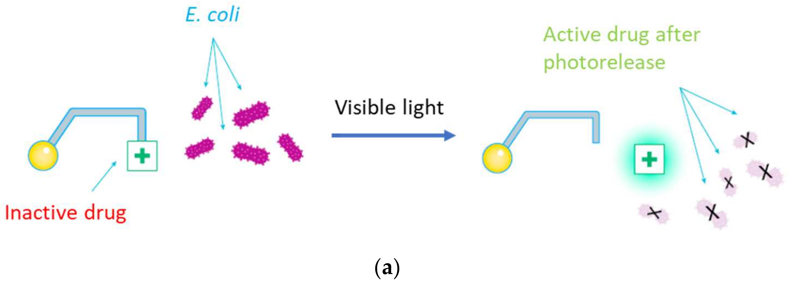
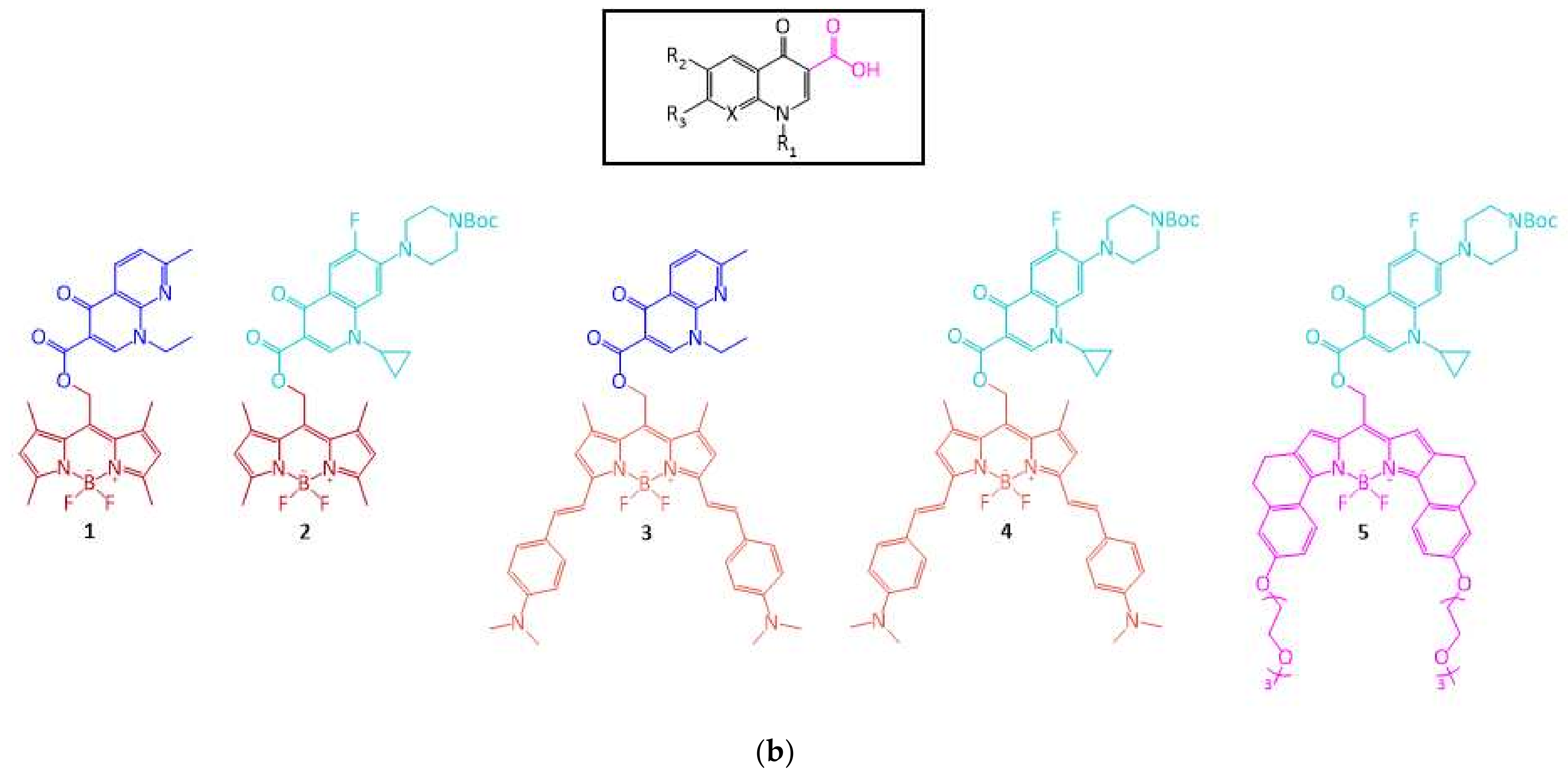
Having these excellent properties in mind, we decided to design a series of molecules combining a BODIPY moiety (acting as PPG) and a quinolone antibiotic. It is well known that the acid group in position 3 of the quinolone core for this family of compounds has a crucial role in the antimicrobial activity of the molecule [
28]. This group is required for the binding to the gyrase complex, critical for the activity of these compounds and for their antimicrobial activity. Hence, we created an irreversible system by incorporating the PPG in that position. We envisioned a strong deactivation of the antimicrobial properties once a photoactivatable moiety was attached to the quinolone structure in position 3. Doing so, the necessary free acid group would be blocked, rendering inactive the quinolone derivative. Subsequently, after irradiation of the molecule and release of the antibiotic, the activity should be recovered. Among the different alternatives for PPGs blocking position 3, we selected BODIPYs due to their optical properties which seem particularly appealing for biological applications, in strong contrast with most of the reported PPGs.
Beyond the optical properties of these photocages, water solubility is also a critical feature for biological applications. Any compound designed to be used in biological media needs to feature a reasonable water solubility or, at least, to be soluble in compatible solvents. In this respect, BODIPYs, as aromatic organic compounds present very low water solubility. Thus, the design of compounds with potential antimicrobial activity based on these PPGs should include chemical modifications which increase this value. Due to the typically low concentrations required in clinical practice for antimicrobial agents, even modest values for solubility in water or compatible solvents could be enough.
Herein, we report our efforts in the design, synthesis, photochemical characterization, computational exploration and biological use of new dyads prepared from modified BODIPYs carrying a photo-releasable quinolone derivative, activatable by visible light.
3. Results and Discussion
Our main goal was to create a system compatible with biological applications. This should imply a photorelease upon irradiation with visible light of compounds with a reasonable water solubility. With this objective in mind, we selected three BODIPY scaffolds (see
Figure 1) and combined them with two different antibiotics to prepare five different PPG-drug dyads. Two of the BODIPY moieties have been previously reported (PPG parts in dyads
1–
4) [
22]. In addition, a new PPG was specifically designed and prepared in this study with the aim of increasing the water solubility in the dyad
5. As the antibiotic parts, two different compounds have been chosen, nalidixic acid (in
1,
3) and ciprofloxacin (in
2,
4,
5). Combination of the drugs with the BODIPY-based PPGs allowed for the design of the five compounds shown in
Figure 1. Dyad
5 was designed to combine the higher antibiotic activity of ciprofloxacin with the expected improved water solubility produced by the newly designed PPG.
For the synthesis of BODIPY photocages for compounds
1,
2,
3, and
4, we based our route on a previously described procedure [
22]. However, a modification in the preparation was needed to obtain the final protected product. Specifically, we synthesized the acyl chloride of the antibiotic, either nalidixic acid or BOC-ciprofloxacin, and we made the coupling reaction with the corresponding BODIPY in the presence of a base (see Supporting Information,
Figures S1 and S2). For compound
5 and its corresponding photocage, a new synthetic route was designed (
Figure 2). First, a glycol chain was introduced in tetralone
6, through an O-alkylation to get derivative
7. Next, oxime
8 was obtained by condensation of
7 with hydroxylamine. Then, pyrrole
9 was formed via a Trofimov reaction. The synthesis of the BODIPY
10 was carried out following the standard methodology [
22]. After that, conversion to the meso-methylhydroxy BODIPY
11 was achieved upon ester hydrolysis. Finally, the coupling reaction between the acyl chloride form of BOC-ciprofloxacin and
11 gave the protected form of the antibiotic
5. More details on the synthesis of these compounds and complete characterization data are available in the Supporting Information.
Once we prepared and characterized the compounds, we studied their photochemical properties. First, we recorded their electronic absorption spectra. As expected, all of them absorb in the visible region, but more interestingly, three of them inside the optical therapeutic window (650–1350 nm, see
Figure 3 and
Table 1) in which there is a maximum depth of tissue penetration. Dyads
1 and
2 bearing the BODIPY with a simpler structure displayed absorption in the green region of the spectrum, both with the maximum absorption at 520 nm. A large bathochromic shift took place when the conjugation of the BODIPY part was elongated through the addition of two (dimethylamino)-styryl groups. Compounds
3 and
4, bearing this modified BODIPY moieties displayed absorption in the red/NIR region of the spectrum with the maximum absorption at 735 and 744 nm, respectively. Finally, compound
5 displayed absorption in the red region of the spectrum with the maximum at 671 nm. The excitation energies and electronic nature of the vertical transition have been studied computationally for compounds
2,
4 and
5 to account for the differences cause by the BODIPY moiety. Regarding the excitation energies, the same qualitative trend has been obtained: compound
2 is significantly blue-shifted compared to
4 and
5, being compound
4 the one showing the more red-shifted absorption (
Table S4). The electronic nature of the S
0 → S
1 vertical transition is characterized by a
1 (π,π*) transition localized in the BODIPY scaffolds, independently of the considered solvent. Hence, we can confidently conclude that the antibiotic moiety is not involved in the electronic transition (
Figure S10). With these results in hand, we proceeded to choose for each case the adequate source of irradiation in order to test if these compounds were indeed photoreactive.
Compounds
1 and
2 were irradiated with a 30 W RGB LED in green mode (λ
max = 520 nm), dyad
5 was irradiated in red mode (λ
max = 635 nm), and compounds
3 and
4 were irradiated with a 515 mW far-red lamp (λ
max = 730 nm, see
Figure S9 for details). Upon irradiation, the release reaction took place until completion in all cases, and no alternative products were observed by HPLC (see
Figures S3–S7, Supporting Information). Additionally, the stability of the compounds at room temperature was measured. The samples were kept in the dark for 5 days, and no release of quinolones or other decomposition products were observed by
1H-NMR or HPLC after that time.
Then, we measured the quantum yield of the release process at the different wavelengths for each compound, to evaluate the photorelease efficiency with the visible light sources previously employed. We measured the quantum yield instrumentally using a protocol already reported [
29]. The photonic flux emitted by the light source, as well as the number of photons not absorbed by the sample, were determined using an Ocean Optics USB4000-UV-Vis detector equipped with a cosine corrector (see
Figure S8). The values obtained showed a better release efficiency of compounds
1,
2, and
5. The obtained results are in line with previously reported photorelease quantum yield for BODIPYs [
22]. The low quantum yields for the photorelease is a common drawback for BODIPY PPGs [
22]. However, these molecules can fully release the antibiotic molecule with Vis/NIR light, and the quantum efficiencies (Φε) are similar to nitrobenzyl photocages because of a much larger extinction coefficient of the BODIPY chromophore compared to the nitrobenzyl chromophore.
To gather a deeper insight into the photorelease mechanism, we have studied by means of computational methods under the framework of the Time-Dependent Density Functional Theory (TD-DFT, see Computational Methods section for details) the antibiotic release pathway for compounds
2,
4 and
5. Previous reports [
30] have shown that C-O bond breaking in the triplet state may be a key step in the photocleavage mechanism for related compounds. For all the computed structures, a similar scheme has been found (
Figure 4A). In particular, after excitation to S
1 the system reaches a minimum in this state. From there, the S
1 path along the C-O bond breaking responsible of the photorelease has been scanned (
Figure 4B), finding a significantly large energy barrier compared to the excess energy obtained after excitation (red curve in
Figure 4A). In particular, the energy barriers in S
1 computed for
2,
4 and
5 when including explicit water molecules as solvent are ca. 15 kcal/mol (
Figure 4B and
Figure S11). Hence, the photorelease in S
1 is not an energetically favorable path. However, if the system is trapped in the S
1 minimum, it could also populate the first triplet state (T
1) which lies lower in energy (
Table S5), through an intersystem crossing (ISC) as shown in
Figure 4A. Once in T
1, the system evolves to a minimum from which it could complete the C-O bond breaking overcoming a barrier whose computed energy is lower than the one computed in S
1 (
Figure 4C). In particular, the energy barrier computed in T
1 is <10 kcal/mol for compounds
2,
4 and
5 (
Figure 4C and
Figure S12) when considering explicit water as solvent. The transition states corresponding to these energy barriers have been optimized, hence describing mechanistically the antibiotic release (
Figure S13). In addition, this T
1 barrier could be easily overcome by the system as the excess energy obtained after the ISC is larger than the T
1 energy barrier (blue curve in
Figure 4A). Hence, we can conclude that the photorelease of the antibiotic should take place preferentially in T
1. This conclusion is in agreement with previous studies focused on other families of BODIPY-based photocages which found the photorelease in the triplet state to be the main path [
30].
Moreover, we have evaluated the solvent effect on the mechanism. Photocleavage has been suggested to be highly affected by the solvent environment [
30]. The solvent may contribute by trapping the generated carbocation upon light irradiation or by generating a solvent-caged ion pair. In order to test the solvent effect on these compounds, we have computed the photorelease path considering three different implicit continuum solvents: dichloromethane, DMSO and water. Clearly, the ability of these solvents to stabilize carbocations or ion pairs would be different. For all compounds under study, it is observed that DMSO and water give almost identical profiles whereas the ones computed in dichloromethane are slightly different, showing larger energy barriers, both in S
1 and T
1 (
Figures S11 and S12). In addition, we have computed these paths considering two explicit water molecules, leading to two hydrogen bond interactions with the oxygen atoms of the compound (
Figure 5) while keeping the implicit description for the surroundings. A clear and significate effect of these explicit water molecules has been observed as the energy barriers computed both in S
1 and T
1 are reduced between 5 and 10 kcal/mol depending on the electronic state and the compound (
Figure 4B,C,
Figures S11 and S12).
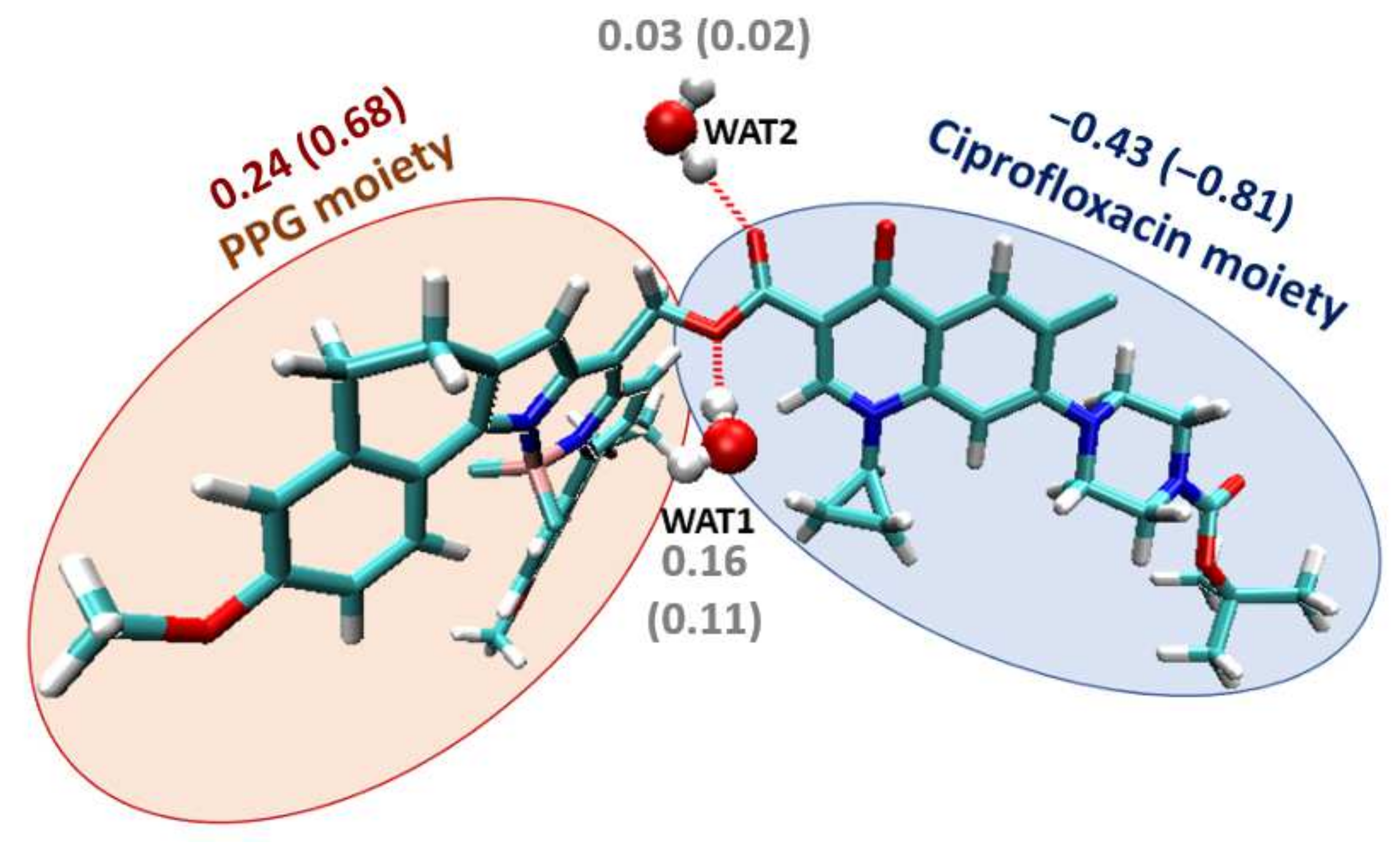
In the light of the results obtained when considering explicit water molecules, we have analyzed the charge evolution along the release path to check their role. For this aim, we have calculated the charges of ciprofloxacin and the PPG for the stationary points in T
1 (minimum, transition state and products) considering both implicit and explicit water (
Table S6). By analyzing the data, we verify that the antibiotic release generates the negatively charged ciprofloxacin moiety and the carbocation of the corresponding PPG moiety. Remarkably, it is observed that one of the explicit water molecules, in particular WAT1 (
Figure 5), has a significant positive charge along the release path. That is, this water molecule is not a passive spectator, but it acts as a nucleophile promoting the C-O breaking, and thus lowering the T
1 energy barrier. Hence, the water molecule (or probably any nucleophilic solvent) is contributing to the reaction mechanism by lowering the required activation energy. These results clearly suggest the relevance of explicit solvent molecules for the computational study of BODIPYs photorelease, in accordance with the experimental requirement for specific solvents to speed up the release process [
22].
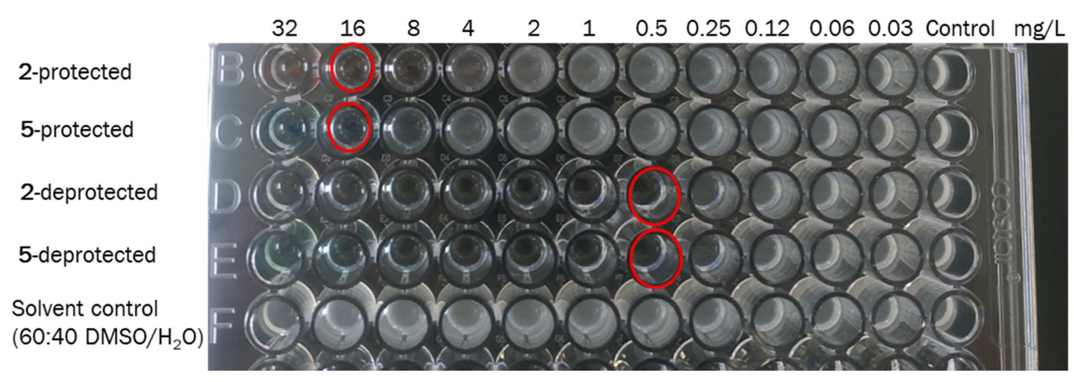
Once we proved the ability of BODIPYs as photocages of quinolone antibiotics, we tested the effect they have on the antimicrobial activity before and after irradiation (see
Table 2,
Figure 6 and
Figure 7). Firstly, the compounds were irradiated to ensure their complete photorelease. After that, an overnight culture of the quinolone susceptible
Escherichia coli ATCC 25922 strain [
31] was used to prepare an inoculum adjusted to the turbidity of a 0.5 McFarland in sterile saline (equivalent to 1 × 10
8 CFU/mL). This suspension was diluted in cation adjusted Mueller-Hinton broth to give a final organism density of 5 × 10
5 CFU/mL and was exposed to different concentrations (0.03–32 mg/L) either of the protected forms
1–
5 (non-irradiated compounds: BODIPY linked to the antibiotic) or the deprotected form (compounds previously irradiated where the antibiotic was already released) with a final volume of 0.1 mL in a 96 well microdilution tray. The microtiter plate was incubated at 37 °C for 24 h in the dark. This experiment was performed in triplicate and the same result was obtained in all experiments.
For this study, stock samples were dissolved in mixtures of water and DMSO (40:60 for
1,
2, and
5, 20:80 for
3 and
4). Subsequent serial two-fold dilutions to achieve lower concentrations were made with cation adjusted Mueller Hinton broth. A control test of solvents was made to check the survival of the bacterium in the water/DMSO mixtures used. Solvents were found to not affect the bacterium growth as both mixtures employed (80:20 and 60:40) resulted in MIC > 32 mg/L. In addition, the antimicrobial activity of both quinolones, nalidixic acid and BOC-ciprofloxacin, was tested as a reference (see
Figure 6).
The derivatives of nalidixic acid 1 and 3 showed an 8-fold change in activity when they were irradiated (MIC1-protected > 32 mg/L vs. MIC1-deprotected = 4 mg/L, MIC3-protected = 32 mg/L vs. MIC3-deprotected = 4 mg/L). These values were in agreement with the control of nalidixic acid, which reported a value of 4 mg/L, indicating that after irradiation, the antibiotic was completely released.
BOC-ciprofloxacin derivatives showed a 32-fold difference in activity in 2 and 5 (MIC2-protected = 8–16 mg/L vs. MIC2-deprotected = 0.5 mg/L, MIC5-protected = 16 mg/L vs. MIC5-deprotected = 0.5 mg/L), and 8-fold change in 4 (MIC4-protected = 16 mg/L vs. MIC4-deprotected = 2 mg/L). These results were in agreement with the controls made for BOC-ciprofloxacin which reported MIC values of 0.5 and 2 mg/L when the stock solution (256 mg/L) was dissolved in 60:40 and 80:20 DMSO:H2O respectively. The values once again demonstrated that upon irradiation the release yield of the antibiotic was 100%.
We then decided to perform the antibiotic’s release in presence of the bacteria (
Figure 7), to check the antimicrobial activity. For this experiment, we selected two of the protected compounds
2 and
5, since they showed a bigger change in activity upon irradiation (
Table 2). Like the previous experiment, 5 × 10
5 CFU/mL of
E. coli ATCC 25922 strain was exposed to different concentrations (0.03–32 mg/L) of the protected form of these compounds. Control tests of solvents and BOC-ciprofloxacin were also included. The 96 well microdilution trays were incubated at 37 °C for 24 h. During this time, the plates were irradiated for a period of 1 h with green light in the case of
2, and red light for
5.
The difference found in the MIC values shown in
Table 2 and
Table 3 are due to the different experimental conditions. However, it was shown that the release process could be successfully induced also in a biological environment, although a smaller antimicrobial activity was observed in this case. The experimental irradiation conditions (see
Figure S9) implied the irradiation of the well plates from the bottom with the corresponding LED, green for
2, and red for
5. Therefore, the light was not directly applied to the sample, but it went through the well plate material, which allows for a ca. 60% of light transmittance. Thus, light intensity effectively reaching the sample is lower. Besides, the culture medium is non-transparent, and as the bacteria grows, turbidity appears, which also implies a decrease in the amount of light that reaches the photoactive compound. Moreover, while the release process takes place, the bacteria are being incubated and therefore keep growing. This implies a different time scale between results shown in
Table 2 and
Table 3. These practical constraints are due to the set-up used and we plan to modify it in the following studies. However, these experiments clearly manifest that we could use the prepared compounds in a biological environment.


 ,
, 


