Communication

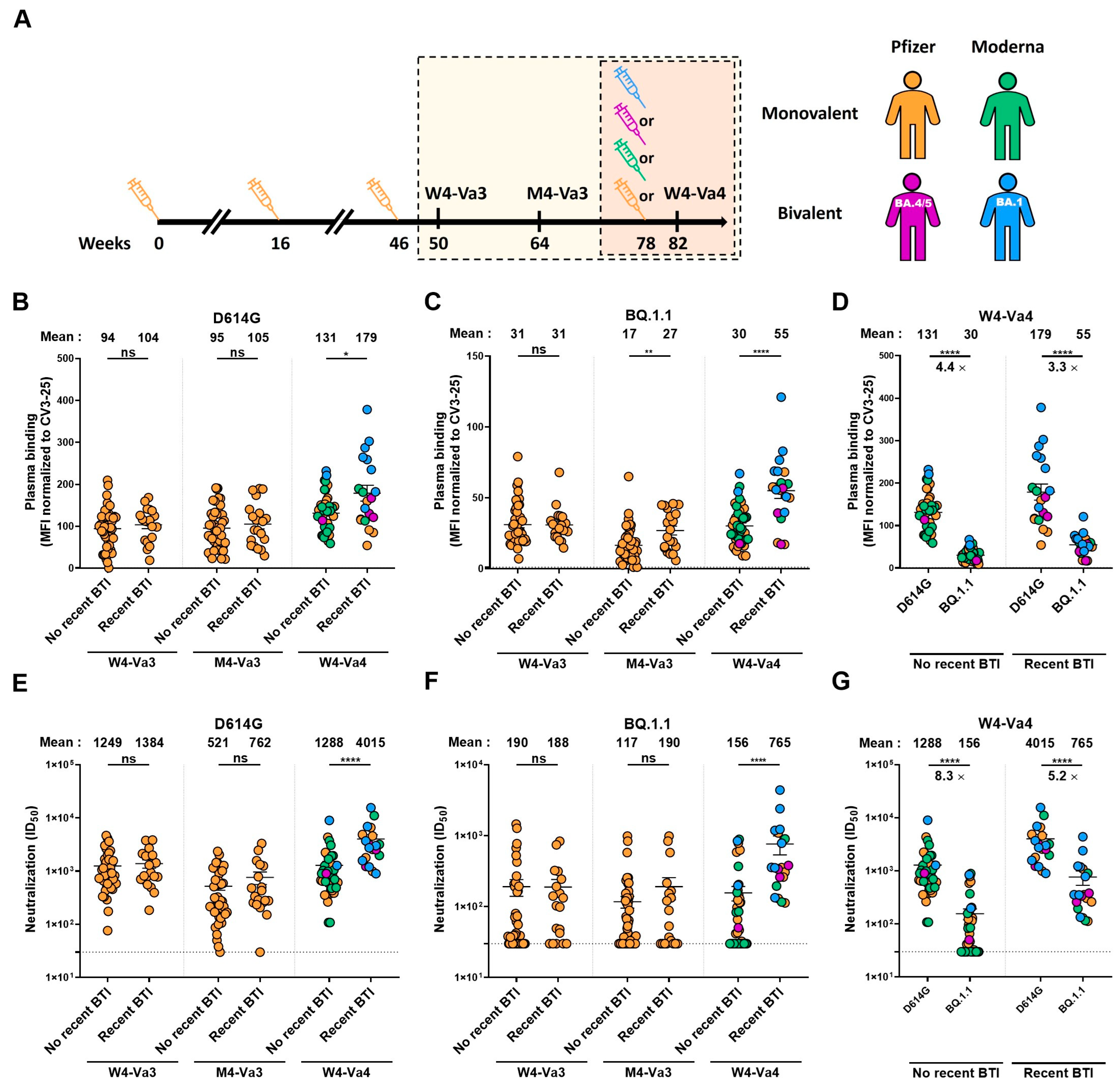
Humoral Responses against BQ.1.1 Elicited after Breakthrough Infection and SARS-CoV-2 mRNA Vaccination

1
Centre de Recherche du CHUM, Montreal, QC H2X 0A9, Canada
2
Département de Microbiologie, Infectiologie et Immunologie, Université de Montréal, Montreal, QC H2X 0A9, Canada
3
Héma-Québec, Affaires Médicales et Innovation, Quebec, QC G1V 5C3, Canada
4
Département de Médecine, Université de Montréal, Montreal, QC H3T 1J4, Canada
5
Division of Infectious Diseases, Department of Medicine, University Hospital of Lausanne and University of Lausanne, 1011 Lausanne, Switzerland
6
Laboratoire de Santé Publique du Québec, Institut National de Santé Publique du Québec, Sainte-Anne-de-Bellevue, QC H9X 3R5, Canada
7
Department of Biochemistry, Microbiology and Immunology, and Centre for Infection, Immunity, and Inflammation, University of Ottawa, Ottawa, ON K1H 8M5, Canada
8
Institut National de Santé Publique du Québec, Quebec, QC H2P 1E2, Canada
*
Author to whom correspondence should be addressed.
Vaccines 2023, 11(2), 242; https://doi.org/10.3390/vaccines11020242
Submission received: 20 December 2022 / Revised: 18 January 2023 / Accepted: 20 January 2023 / Published: 21 January 2023
(This article belongs to the Special Issue The Immune Response to Viral Infection)

Abstract

:
The Omicron BQ.1.1 variant is now the major SARS-CoV-2 circulating strain in many countries. Because of the many mutations present in its Spike glycoprotein, this variant is resistant to humoral responses elicited by monovalent mRNA vaccines. With the goal to improve immune responses against Omicron subvariants, bivalent mRNA vaccines have recently been approved in several countries. In this study, we measure the capacity of plasma from vaccinated individuals, before and after a fourth dose of mono- or bivalent mRNA vaccine, to recognize and neutralize the ancestral (D614G) and the BQ.1.1 Spikes. Before and after the fourth dose, we observe a significantly better recognition and neutralization of the ancestral Spike. We also observe that fourth-dose vaccinated individuals who have been recently infected better recognize and neutralize the BQ.1.1 Spike, independently of the mRNA vaccine used, than donors who have never been infected or have an older infection. Our study supports that hybrid immunity, generated by vaccination and a recent infection, induces higher humoral responses than vaccination alone, independently of the mRNA vaccine used.

1. Introduction

The Omicron BQ.1.1 variant is a sublineage of the BA.5 variant that spreads very rapidly and is now the major circulating lineage in several countries [1,2]. Recent studies have shown that original SARS-CoV-2 mRNA vaccines, based on the ancestral Wuhan strain Spike (S), lead to poor humoral responses against several Omicron subvariants, including the BQ.1.1 variant [3,4,5]. With the goal to improve immune responses against these subvariants, Moderna and Pfizer bivalent vaccines have recently been approved by health authorities in many countries [6,7,8]. These updated versions of the vaccines are composed of mRNA coding for the expression of both the ancestral and an Omicron subvariant S [9,10]. However, the continued evolution of SARS-CoV-2 has resulted in the emergence of multiple Omicron sub-lineages showing signs of convergent evolution by the acquisition of the same immune escape mutation in the RBD region of the Spike protein. Notably, all five recent convergent mutations are present in BQ. 1.1: R346T, K444T, L452R, N460K, or F486V [3]. Because of these newly acquired mutations, the benefits of bivalent compared to monovalent vaccines against this lineage remain to be established.
It is well accepted now that hybrid immunity, conferred by both infection and vaccination, leads to better immune responses and protection from severe outcomes than vaccination alone [11,12,13,14,15,16,17]. Because the original mRNA vaccines poorly prevent viral transmission of the recent emerging variants compared to the original strain [12,18,19,20,21,22], an important part of the vaccinated population have been recently infected by Omicron subvariants, leading to improved immune responses in these individuals compared to SARS-CoV-2 naïve individuals who have just been vaccinated [23].
In this study, we evaluated the capacity of plasma antibodies to recognize and neutralize the original D614G and the Omicron BQ.1.1 subvariant S four weeks (W4-Va3) and four months (M4-Va3) after the third dose and four weeks after the fourth dose (W4-Va4) of mRNA vaccines (Figure 1A). These participants mainly received as their first three doses of vaccine the Pfizer monovalent vaccine, and as the fourth dose either the Pfizer or Moderna monovalent or Pfizer (BA.4/5) or Moderna (BA.1) bivalent vaccines. Because breakthrough infection (BTI) strongly improved humoral responses, and may impact the responses measured after vaccination, we also measured the anti-nucleocapsid (N) level at these three time points to determine if the donors have been infected between their third and fourth doses of vaccine (by an Omicron sublineage). Based on anti-N results, the donors were separated in two groups, donors without recent BTI for whom we did not observe an increase in anti-N over time, and donors with recent BTI for whom we measured a significant increase in anti-N between the third and fourth doses of vaccine (Figure 2A), according to a recent study [24]. Basic demographic characteristics of the cohort are summarized in Table 1.

2. Materials and Methods

2.1. Ethics Statement

The study was conducted in accordance with the Declaration of Helsinki in terms of informed consent and approval by an appropriate institutional board. The protocol was approved by the Ethics Committee of CHUM (19.381, approved on 28 February 2022) and Héma-Québec (2022-016, approved on 7 October 2022).

2.2. Human Subjects

The study was conducted in 63 individuals (25 males and 38 females; age range: 24–84 years). In total, 20 of these individuals had recent breakthrough infection with an Omicron sublineage (9 males and 11 females; age range: 24–67 years), i.e., as determined by the increase in anti-N levels between W4-Va3 and M4-Va3 or between M4-Va3 and W4-Va4 (ratio M4-Va3/W4-Va3 and/or W4-Va4/M4-Va3 higher than 1.5) using a recently described analytical approach [24] (Figure 2). For the other donors (16 males and 27 females; age range: 31–84 years), we did not observe a significant increase in the anti-N levels, although some of them have a history of infection (to our knowledge, 12 donors had a history of infection: 3 were infected during the first wave of COVID-19 in winter/spring 2020, and 9 of them were tested anti-N positive at W4-Va3). No other specific criteria, such as number of patients (sample size), sex, clinical or demographic were used for inclusion.

2.3. Plasma Samples and Antibodies

Plasma samples were either recovered from whole blood or directly obtained from the PlasCov biobank [25], heat-inactivated for 1 h at 56 °C and stored at −80 °C until use in subsequent experiments. Pre-pandemic plasma samples were used as negative controls in cytometry assays (data not shown). The conformationally independent S2-specific monoclonal antibody CV3-25 was used as a positive control and to normalize Spike expression in flow cytometry assays, as described [4,26,27,28,29]. Alexa Fluor-647-conjugated goat anti-human antibodies (Abs) able to detect all Ig isotypes (anti-human IgM, IgG, IgA; Jackson ImmunoResearch Laboratories, Cat # 109-605-064) were used as secondary Abs to detect plasma binding in flow cytometry experiments.

2.4. Plasmids

The plasmids encoding the SARS-CoV-2 D614G and BQ.1.1 Spike variants were previously described [4]. The pNL4.3 R-E-Luc plasmid was obtained from the NIH AIDS Reagent Program (Cat# 3418). The pIRES2-EGFP expressing plasmid was purchased from Clontech, Mountain View, CA, USA (Cat# 6029-1).

2.5. Cell Lines

For this, 293T human embryonic kidney cells (obtained from ATCC, Cat# CRL-3216) were maintained at 37 °C under 5% CO2 in Dulbecco’s modified Eagle’s medium (DMEM) (Wisent) containing 5% fetal bovine serum (FBS) (VWR) and 100 μg/Ml of penicillin–streptomycin (Wisent). The 293T-ACE2 cell line was previously reported [30].

2.6. Enzyme-Linked Immunosorbent Assay (ELISA)

All samples were tested for anti-N total immunoglobulin levels using an in-house anti-N ELISA. The assay protocol is similar to the anti-SARS-CoV-2 RBD ELISA previously developed by our group [31], except that recombinant N (Centre National en Électrochimie et en Technologies Environnementales Inc., Shawinigan, QC, Canada) was used (0.25 µg/mL) in lieu of the RBD antigen (2.5 µg/mL).

2.7. Cell Surface Staining and Flow Cytometry Analysis

For this, 293T were transfected with full-length SARS-CoV-2 Spikes and a green fluorescent protein (GFP) expressor (pIRES2-eGFP) using the calcium–phosphate method. Two days post-transfection, Spike-expressing 293T cells were stained with the CV3-25 Ab (5 μg/mL) as control or plasma (1:250 dilution) for 45 min at 37 °C. AlexaFluor-647-conjugated goat anti-human IgM, IgG, IgA (1/800 dilution) were used as secondary Abs. The percentage of Spike-expressing cells (GFP + cells) was determined by gating the living cell population based on viability dye staining (Aqua Vivid, Invitrogen, Waltham, MA, USA). Samples were acquired on a LSRFortessa cytometer (BD Biosciences, Franklin Lakes, NJ, USA), and data analysis was performed using FlowJo v10.7.1 (Tree Star). The conformationally independent anti-S2 antibody CV3-25, effective against all Spike variants, was used to normalize Spike expression, as reported [4,26,28,29]. The Median Fluorescence intensities (MFI) obtained with plasma were normalized to the MFI obtained with CV3-25 and presented as percentage of CV3-25 binding.

2.8. Virus Neutralization Assay

For this, 293T cells were transfected with the lentiviral vector pNL4.3 R-E− Luc and a plasmid encoding the D614G or the BQ.1.1 S glycoprotein at a ratio of 10:1 to produce SARS-CoV-2 pseudoviruses. Two days post-transfection, cell supernatants were harvested and stored at −80 °C until use. For the neutralization assay, 293T-ACE2 target cells were seeded at a density of 1 × 10⁴ cells/well in 96-well luminometer-compatible tissue culture plates (PerkinElmer, Waltham, MA, USA) 24 h before infection. Pseudoviral particles were incubated with several plasma dilutions (1/50; 1/250; 1/1250; 1/6250; 1/31250) for 1 h at 37 °C and were then added to the target cells followed by incubation for 48 h at 37 °C. Cells were lysed by the addition of 30 μL of passive lysis buffer (Promega, Madison, WI, USA) followed by one freeze–thaw cycle. An LB942 TriStar luminometer (Berthold Technologies, Bad Wildbad, Germany) was used to measure the luciferase activity of each well after the addition of 100 μL of luciferin buffer (15 mM MgSO4, 15 mM KH2PO4 [pH 7.8], 1 mM ATP, and 1 mM dithiothreitol) and 50 μL of 1 mM d-luciferin potassium salt (Prolume). The neutralization half-maximal inhibitory dilution (ID50) represents the plasma dilution to inhibit 50% of the infection of 293T-ACE2 cells by pseudoviruses.

2.9. Statistical Analysis
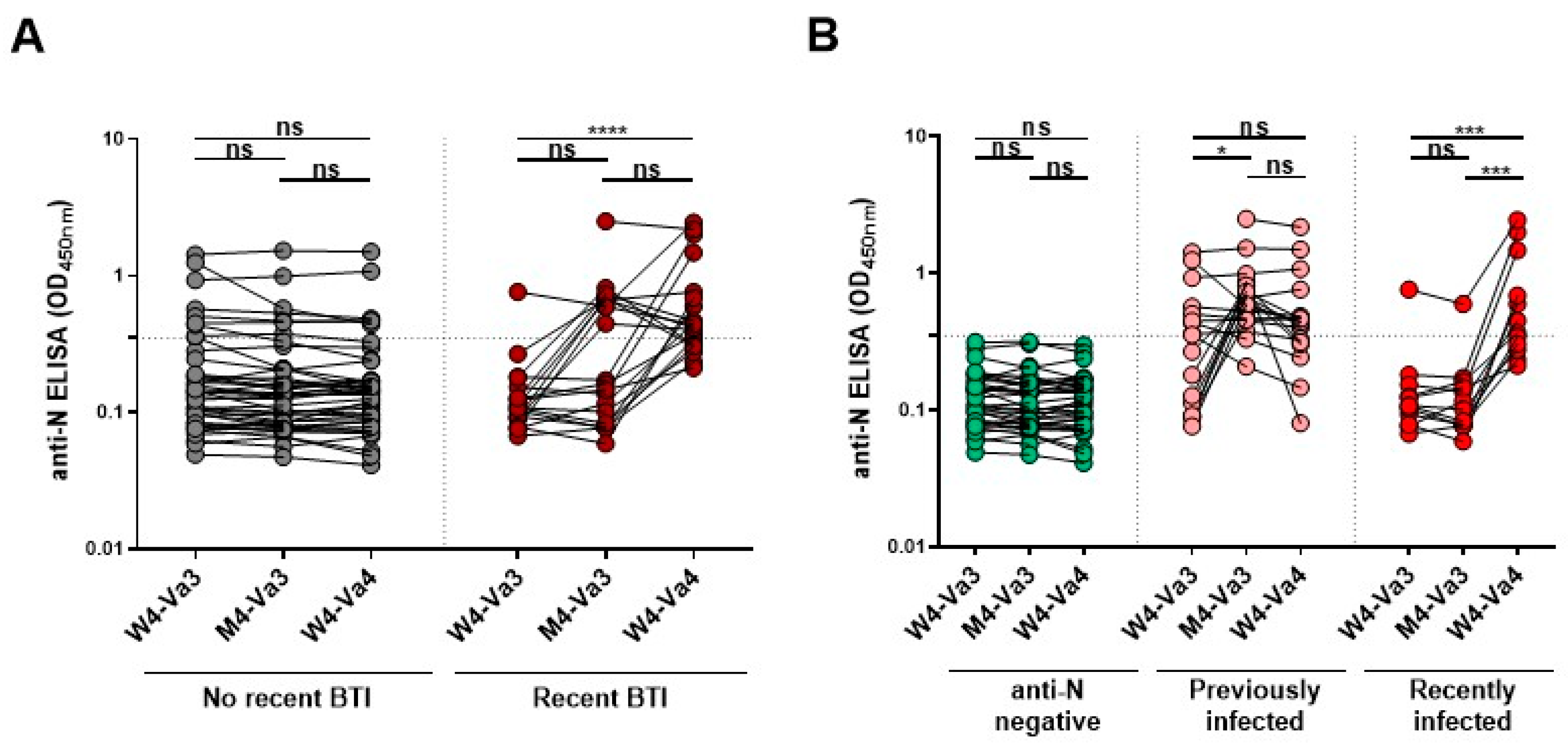
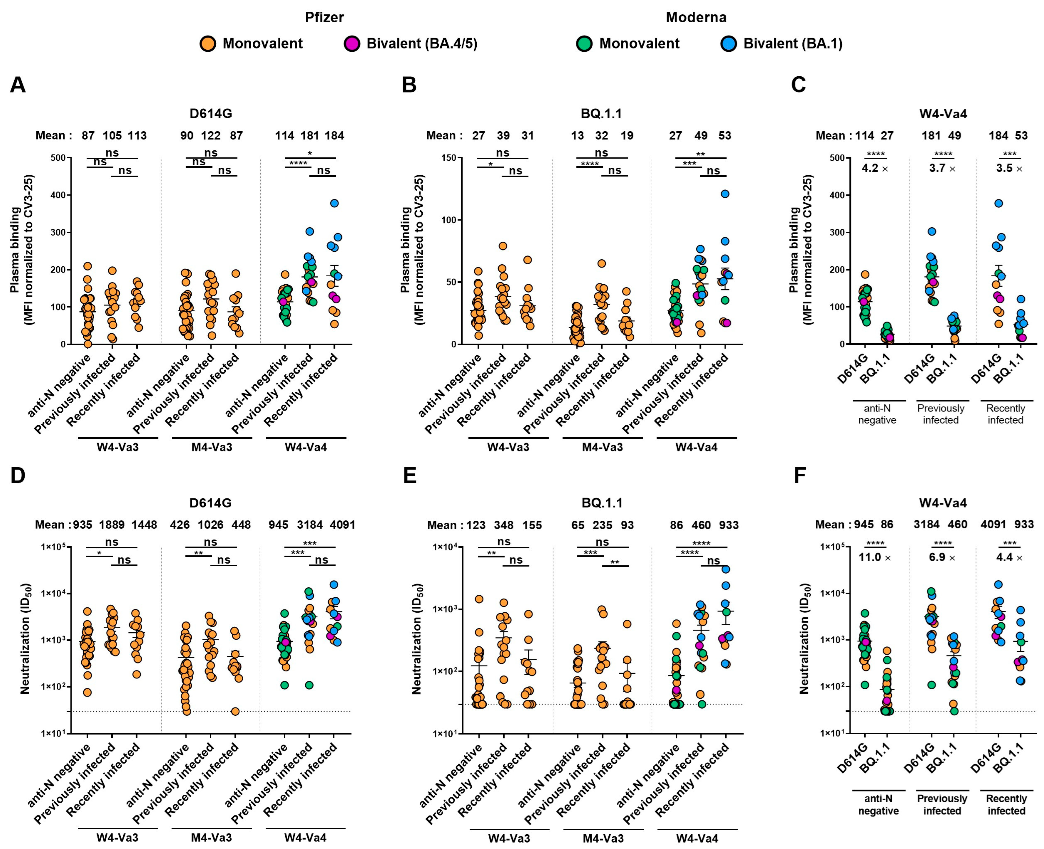
Symbols represent biologically independent samples from individuals. Statistics were analyzed using GraphPad Prism version 8.0.1 (GraphPad, San Diego, CA, USA). Each dataset was tested for statistical normality and this information was used to apply the appropriate (parametric or nonparametric) statistical test. Differences in responses at every time point between no recent BTI and recent BTI groups were performed by Mann–Whitney unpaired tests. Differences in responses against the D614G and BQ.1.1 Spikes for the same patient were measured by Wilcoxon paired tests. Differences in responses at every time points between anti-N negative, previously infected and recently infected groups, were performed by Kruskal–Wallis tests. Differences in responses for the same patient between the three timepoints were performed using Friedman tests. Differences in responses between the different vaccine platforms were performed by Kruskal–Wallis tests. p values < 0.05 were considered significant; significance values are indicated as * p < 0.05, ** p < 0.01, *** p < 0.001, *** p < 0.0001, ns, non-significant.

3. Results

We first monitored the capacity of plasma to recognize the D614G and BQ.1.1 Spikes after the third and fourth doses of mRNA vaccine by flow cytometry (Figure 1B–D). For the D614G S, no significant differences were observed four weeks and four months after the third dose of vaccine between individuals with or without recent BTI. In contrast, four weeks after the fourth dose of mRNA vaccine, individuals with recent BTI recognized better the D614G S than donors with no recent BTI, although this difference was mainly observed with the Moderna bivalent vaccine (Figure 1B and Figure S1A). For the BQ.1.1 S, at the M4-Va3 timepoint donors with recent BTI better recognized the S than individuals with no recent infection, and this difference in recognition was more significant four weeks after the fourth dose (Figure 1C). The level of BQ.1.1 S recognition was significantly lower compared to D614G S in donors without recent BTI (Figure 1D), in agreement with recent reports [4,5]. In donors who had recently been infected, there was a significant but smaller difference in the level of recognition between the two Spikes compared to the other group.
We also measured the neutralizing activity of plasma against the D614G and BQ.1.1 S (Figure 1E–G). We observed patterns similar to that measured for Spike recognition. No significant differences were observed between the two groups at W4-Va3 and M4-Va3 timepoints (Figure 1E,F). In contrast, four weeks after the fourth dose, donors with recent BTI had a significantly higher level of neutralizing activity against D614G and BQ.1.1 S. All donors with recent BTI who received a fourth dose developed neutralizing antibodies against BQ.1.1 S, while some donors who just received four doses of vaccine were still not able to neutralize this Spike. As observed for S recognition (Figure 1D), BQ.1.1 Spike was significantly less neutralized than D614G S, even after four doses of mRNA vaccine (Figure 1G). However, the difference in neutralization between the two S was smaller in the group with recent BTI.
The Moderna BA.1 bivalent vaccine (blue points) tended to induce better recognition and neutralization than the other vaccine platforms, including the Pfizer BA.4/5 bivalent vaccine with lesser decrease in recognition and neutralization of BQ.1.1 S (Figure 1B–G and Figure S1). These differences did not reach statistical significance; whether this is due to the relatively low number of samples tested remains to be determined.

4. Discussion

Since its emergence in late 2021, the Omicron variant continues to evolve into new subvariants that are increasingly resistant to monoclonal antibodies and vaccination [5,32,33,34,35,36]. To address vaccine resistance, bivalent mRNA vaccines, expressing both the original Spike and one of the parental lineages of Omicron (BA.1 or BA.4/5) Spike, have been developed and are now being administered in several jurisdictions worldwide. However, although the bivalent mRNA vaccine has been shown to increase the level of protection against BA.5 variant in mice [37], evidence of its superior effectiveness in the human population remains to be demonstrated, especially against sub-lineages with newly acquired immune escape mutations. Recent studies showed that both monovalent and bivalent vaccines induced low humoral responses against BQ.1.1, but recent breakthrough infection before vaccination strongly improved these responses [38,39]. The results presented herein support these observations.
As previously reported in numerous studies, including ours, hybrid immunity led to better humoral responses against the BQ.1.1 and other recent variants than just vaccination [4,5,38]. Moreover, we observed that after four doses of mRNA vaccine and no recent BTI, some donors did not have neutralizing activity against pseudoviral particles bearing the BQ.1.1 Spike. Whether these changes of recognition and neutralization translate into greater risk of severe disease is currently unknown. In contrast, BTI likely increased the breadth of neutralizing antibodies since all donors had detectable levels of neutralization against BQ.1.1.
In our study, some donors were infected before or just after their third dose of vaccine before May 2022, when BA.1 and BA.2 variants were the major circulating strains in Quebec. The other donors were infected later, before their fourth dose and after May 2022 when BA.4/5 and then BQ.1.1 replaced the BA.2 variant as major circulating strains [2,40]. To test whether infection with a specific Omicron subvariant as well as the interval between infection and vaccination impacted the humoral response, we divided our cohort into three groups: individuals with anti-N negative at W4-Va3 and who have not been infected between the third and fourth doses of vaccine; individuals seropositive at W4-Va3 or who have been infected between W4-Va3 and M4-Va3; and individuals who have been infected between M4-Va3 and W4-Va4 (Figure 2B). We did not observe significant differences for Spike recognition and neutralization capacity between donors who were infected long ago or more recently (Figure 3, Figure S1). Both groups had better humoral responses than anti-N negative donors. We also noted that recently infected donors had higher levels of neutralizing antibodies than previously infected donors, although we did not measure significant differences.

5. Conclusions

These results indicate that further efforts have to be devoted to improving vaccines against new SARS-CoV-2 variants of concern. Whether immune responses comparable to those observed with breakthrough infections could be obtained with new vaccine formulations remains to be determined.

Supplementary Materials

The following supporting information can be downloaded at: https://www.mdpi.com/article/10.3390/vaccines11020242/s1, Figure S1: Recognition and neutralization of the D614G and BQ.1.1 Spikes after the fourth doses of SARS-CoV-2 vaccine in individuals with or without a recent breakthrough infection.

Author Contributions

A.T., R.B. and A.F. conceived the study. A.T., M.B., H.M., J.P., G.G.-L., R.B. and A.F. performed, analyzed, and interpreted the experiments. A.T. performed statistical analysis. H.M., G.G.-L., M.C. and A.F. contributed unique reagents. L.G., P.A., C.M., C.T., D.E.K., Y.G. and V.M.-L. collected and provided clinical samples. R.B., G.D.S. and I.L. provided scientific input related to VOCs and vaccine efficacy. A.T. and A.F. wrote the manuscript with input from others. Every author has read, edited, and approved the final manuscript. All authors have read and agreed to the published version of the manuscript.

Funding

This work was supported by le Ministère de l’Economie et de l’Innovation du Québec, Programme de soutien aux organismes de recherche et d’innovation to A.F. and by the Fondation du CHUM. This work was also supported by a CIHR foundation grant #352417, by a CIHR operating Pandemic and Health Emergencies Research grant #177958, by an Exceptional Fund COVID-19 from the Canada Foundation for Innovation (CFI) #41027 to A.F. The PlasCov biobank was supported by funding from the COVID-19 Immunity Task Force (CITF) which is supported by the Public Health Agency of Canada (PHAC). Work on variants presented was also supported by the Sentinelle COVID Quebec network led by the LSPQ in collaboration with Fonds de Recherche du Québec Santé (FRQS) to A.F. A.F. is the recipient of Canada Research Chair on Retroviral Entry no. RCHS0235 950-232424. C.T. is the Pfizer/Université de Montréal Chair on HIV translational research. V.M.-L is supported by a FRQS Junior 2 salary award. A.T. was supported by MITACS Accélération postdoctoral fellowship. M.B. was the recipient of a CIHR master’s scholarship award. The funders had no role in study design, data collection and analysis, decision to publish, or preparation of the manuscript. We declare no competing interests.

Institutional Review Board Statement

The study was conducted in accordance with the Declaration of Helsinki in terms of informed consent and approval by an appropriate institutional board. The protocol was approved by the Ethics Committee of CHUM (19.381, approved on 28 February 2022) and Héma-Québec (2022-016, approved on 7 October 2022).

Informed Consent Statement

Informed consent was obtained from all subjects involved in the study.

Data Availability Statement

Further information, data reported in this paper, and requests for resources and reagents should be directed to and will be fulfilled by the lead contact, Andrés Finzi ([email protected]) upon request.

Acknowledgments

The authors are grateful to the donors who participated in this study. The authors thank the CRCHUM BSL3 and Flow Cytometry Platforms for technical assistance.

Conflicts of Interest

The authors declare no conflict of interest.

References

  1. Données sur les Variants du SRAS-CoV-2 au Québec. Available online: https://www.inspq.qc.ca/covid-19/donnees/variants (accessed on 6 December 2022).
  2. CDC. COVID Data Tracker Weekly Review. Available online: https://www.cdc.gov/coronavirus/2019-ncov/covid-data/covidview/index.html (accessed on 6 December 2022).
  3. Qu, P.; Evans, J.P.; Faraone, J.; Zheng, Y.-M.; Carlin, C.; Anghelina, M.; Stevens, P.; Fernandez, S.; Jones, D.; Lozanski, G.; et al. Enhanced Neutralization Resistance of SARS-CoV-2 Omicron Subvariants BQ.1, BQ.1.1, BA.4.6, BF.7 and BA.2.75.2. Cell Host Microbe 2022, 31, 9–17.e3. [Google Scholar] [CrossRef] [PubMed]
  4. Tauzin, A.; Nicolas, A.; Ding, S.; Benlarbi, M.; Medjahed, H.; Chatterjee, D.; Dionne, K.; Gong, S.Y.; Gendron-Lepage, G.; Bo, Y.; et al. SARS-CoV-2 Omicron Subvariants Spike Recognition and Neutralization Elicited after the Third Dose of MRNA Vaccine. Cell Rep. 2023, 42, 111998. [Google Scholar] [CrossRef] [PubMed]
  5. Kurhade, C.; Zou, J.; Xia, H.; Liu, M.; Chang, H.C.; Ren, P.; Xie, X.; Shi, P.-Y. Low Neutralization of SARS-CoV-2 Omicron BA.2.75.2, BQ.1.1, and XBB.1 by Parental MRNA Vaccine or a BA.5-Bivalent Booster. Nat. Med. 2022. [Google Scholar] [CrossRef] [PubMed]
  6. FDA. Coronavirus (COVID-19) Update: FDA Authorizes Moderna, Pfizer-BioNTech Bivalent COVID-19 Vaccines for Use as a Booster Dose. Available online: https://www.fda.gov/news-events/press-announcements/coronavirus-covid-19-update-fda-authorizes-moderna-pfizer-biontech-bivalent-covid-19-vaccines-use (accessed on 6 December 2022).
  7. EMA. First Adapted COVID-19 Booster Vaccines Recommended for Approval in the EU. Available online: https://www.ema.europa.eu/en/news/first-adapted-covid-19-booster-vaccines-recommended-approval-eu (accessed on 6 December 2022).
  8. Health Canada. Health Canada Authorizes COVID-19 Vaccine Booster Targeting the Omicron BA.4/BA.5 Subvariants. Available online: https://www.canada.ca/en/health-canada/news/2022/10/health-canada-authorizes-covid-19-vaccine-booster-targeting-the-omicron-ba4ba5-subvariants.html (accessed on 6 December 2022).
  9. Pfizer and BioNTech Complete Submission to European Medicines Agency for Omicron BA.4/BA.5 Adapted Bivalent Vaccine|Pfizer. Available online: https://www.pfizer.com/news/press-release/press-release-detail/pfizer-and-biontech-complete-submission-european-medicines (accessed on 6 December 2022).
  10. Canada|SPIKEVAXTM Information. Available online: https://www.modernacovid19global.com/ca/ (accessed on 6 December 2022).
  11. Ntziora, F.; Kostaki, E.G.; Karapanou, A.; Mylona, M.; Tseti, I.; Sipsas, N.V.; Paraskevis, D.; Sfikakis, P.P. Protection of Vaccination versus Hybrid Immunity against Infection with COVID-19 Omicron Variants among Health-Care Workers. Vaccine 2022, 40, 7195–7200. [Google Scholar] [CrossRef] [PubMed]
  12. Altarawneh, H.N.; Chemaitelly, H.; Ayoub, H.H.; Tang, P.; Hasan, M.R.; Yassine, H.M.; Al-Khatib, H.A.; Smatti, M.K.; Coyle, P.; Al-Kanaani, Z.; et al. Effects of Previous Infection and Vaccination on Symptomatic Omicron Infections. N. Engl. J. Med. 2022, 387, 21–34. [Google Scholar] [CrossRef]
  13. Carazo, S.; Skowronski, D.M.; Brisson, M.; Barkati, S.; Sauvageau, C.; Brousseau, N.; Gilca, R.; Fafard, J.; Talbot, D.; Ouakki, M.; et al. Protection against Omicron (B.1.1.529) BA.2 Reinfection Conferred by Primary Omicron BA.1 or Pre-Omicron SARS-CoV-2 Infection among Health-Care Workers with and without MRNA Vaccination: A Test-Negative Case-Control Study. Lancet Infect. Dis. 2022, 23, 45–55. [Google Scholar] [CrossRef]
  14. Tauzin, A.; Nayrac, M.; Benlarbi, M.; Gong, S.Y.; Gasser, R.; Beaudoin-Bussières, G.; Brassard, N.; Laumaea, A.; Vézina, D.; Prévost, J.; et al. A Single Dose of the SARS-CoV-2 Vaccine BNT162b2 Elicits Fc-Mediated Antibody Effector Functions and T Cell Responses. Cell Host Microbe 2021, 29, 1137–1150.e6. [Google Scholar] [CrossRef] [PubMed]
  15. Tauzin, A.; Gong, S.Y.; Beaudoin-Bussières, G.; Vézina, D.; Gasser, R.; Nault, L.; Marchitto, L.; Benlarbi, M.; Chatterjee, D.; Nayrac, M.; et al. Strong Humoral Immune Responses against SARS-CoV-2 Spike after BNT162b2 MRNA Vaccination with a 16-Week Interval between Doses. Cell Host Microbe 2022, 30, 97–109.e5. [Google Scholar] [CrossRef]
  16. Stamatatos, L.; Czartoski, J.; Wan, Y.-H.; Homad, L.J.; Rubin, V.; Glantz, H.; Neradilek, M.; Seydoux, E.; Jennewein, M.F.; MacCamy, A.J.; et al. MRNA Vaccination Boosts Cross-Variant Neutralizing Antibodies Elicited by SARS-CoV-2 Infection. Science 2021, 372, 1413–1418. [Google Scholar] [CrossRef] [PubMed]
  17. Tauzin, A.; Gong, S.Y.; Chatterjee, D.; Ding, S.; Painter, M.M.; Goel, R.R.; Beaudoin-Bussières, G.; Marchitto, L.; Boutin, M.; Laumaea, A.; et al. A Boost with SARS-CoV-2 BNT162b2 MRNA Vaccine Elicits Strong Humoral Responses Independently of the Interval between the First Two Doses. Cell Rep. 2022, 41, 111554. [Google Scholar] [CrossRef] [PubMed]
  18. Tan, S.T.; Kwan, A.T.; Rodríguez-Barraquer, I.; Singer, B.J.; Park, H.J.; Lewnard, J.A.; Sears, D.; Lo, N.C. Infectiousness of SARS-CoV-2 Breakthrough Infections and Reinfections during the Omicron Wave. Nat. Med. 2023, 1–8. [Google Scholar] [CrossRef] [PubMed]
  19. Woodbridge, Y.; Amit, S.; Huppert, A.; Kopelman, N.M. Viral Load Dynamics of SARS-CoV-2 Delta and Omicron Variants Following Multiple Vaccine Doses and Previous Infection. Nat. Commun. 2022, 13, 6706. [Google Scholar] [CrossRef] [PubMed]
  20. Sun, K.; Tempia, S.; Kleynhans, J.; von Gottberg, A.; McMorrow, M.L.; Wolter, N.; Bhiman, J.N.; Moyes, J.; du Plessis, M.; Carrim, M.; et al. SARS-CoV-2 Transmission, Persistence of Immunity, and Estimates of Omicron’s Impact in South African Population Cohorts. Sci. Transl. Med. 2022, 14, eabo7081. [Google Scholar] [CrossRef] [PubMed]
  21. Shah, A.S.V.; Gribben, C.; Bishop, J.; Hanlon, P.; Caldwell, D.; Wood, R.; Reid, M.; McMenamin, J.; Goldberg, D.; Stockton, D.; et al. Effect of Vaccination on Transmission of SARS-CoV-2. N. Engl. J. Med. 2021, 385, 1718–1720. [Google Scholar] [CrossRef] [PubMed]
  22. Harris, R.J.; Hall, J.A.; Zaidi, A.; Andrews, N.J.; Dunbar, J.K.; Dabrera, G. Effect of Vaccination on Household Transmission of SARS-CoV-2 in England. N. Engl. J. Med. 2021, 385, 759–760. [Google Scholar] [CrossRef]
  23. COVID-19 Immunity Task Force. Seroprevalence in Canada. Available online: https://www.Covid19immunitytaskforce.ca/Seroprevalence-in-Canada/.COVID-19 Immunity Task Force (accessed on 30 November 2022).
  24. Bazin, R.; Rochette, S.; Perreault, J.; Fournier, M.-J.; Grégoire, Y.; Boivin, A.; Lewin, A.; Germain, M.; Renaud, C. Development and Use of a Method Based on the Anti-N Reactivity of Longitudinal Samples to Better Estimate SARS-CoV-2 Seroprevalence in a Vaccinated Population. medRxiv 2022. [Google Scholar] [CrossRef]
  25. Germain, M.; Lewin, A.; Bazin, R.; Dieudé, M.; Perreault, J.; Boivin, A.; Grégoire, Y.; Renaud, C. Cohort Profile: A Québec-Based Plasma Donor Biobank to Study COVID-19 Immunity (PlasCoV). medRxiv 2022. [Google Scholar] [CrossRef]
  26. Prévost, J.; Richard, J.; Gasser, R.; Ding, S.; Fage, C.; Anand, S.P.; Adam, D.; Vergara, N.G.; Tauzin, A.; Benlarbi, M.; et al. Impact of Temperature on the Affinity of SARS-CoV-2 Spike Glycoprotein for Host ACE2. J. Biol. Chem. 2021, 297, 101151. [Google Scholar] [CrossRef]
  27. Jennewein, M.F.; MacCamy, A.J.; Akins, N.R.; Feng, J.; Homad, L.J.; Hurlburt, N.K.; Seydoux, E.; Wan, Y.-H.; Stuart, A.B.; Edara, V.V.; et al. Isolation and Characterization of Cross-Neutralizing Coronavirus Antibodies from COVID-19+ Subjects. Cell Rep. 2021, 36, 109353. [Google Scholar] [CrossRef]
  28. Gong, S.Y.; Chatterjee, D.; Richard, J.; Prévost, J.; Tauzin, A.; Gasser, R.; Bo, Y.; Vézina, D.; Goyette, G.; Gendron-Lepage, G.; et al. Contribution of Single Mutations to Selected SARS-CoV-2 Emerging Variants Spike Antigenicity. Virology 2021, 563, 134–145. [Google Scholar] [CrossRef]
  29. Chatterjee, D.; Tauzin, A.; Marchitto, L.; Gong, S.Y.; Boutin, M.; Bourassa, C.; Beaudoin-Bussières, G.; Bo, Y.; Ding, S.; Laumaea, A.; et al. SARS-CoV-2 Omicron Spike Recognition by Plasma from Individuals Receiving BNT162b2 MRNA Vaccination with a 16-Week Interval between Doses. Cell Rep. 2022, 38, 110429. [Google Scholar] [CrossRef] [PubMed]
  30. Prévost, J.; Gasser, R.; Beaudoin-Bussières, G.; Richard, J.; Duerr, R.; Laumaea, A.; Anand, S.P.; Goyette, G.; Benlarbi, M.; Ding, S.; et al. Cross-Sectional Evaluation of Humoral Responses against SARS-CoV-2 Spike. Cell Rep. Med. 2020, 1, 100126. [Google Scholar] [CrossRef]
  31. Perreault, J.; Tremblay, T.; Fournier, M.-J.; Drouin, M.; Beaudoin-Bussières, G.; Prévost, J.; Lewin, A.; Bégin, P.; Finzi, A.; Bazin, R. Waning of SARS-CoV-2 RBD Antibodies in Longitudinal Convalescent Plasma Samples within 4 Months after Symptom Onset. Blood 2020, 136, 2588–2591. [Google Scholar] [CrossRef] [PubMed]
  32. Qu, P.; Faraone, J.; Evans, J.P.; Zou, X.; Zheng, Y.-M.; Carlin, C.; Bednash, J.S.; Lozanski, G.; Mallampalli, R.K.; Saif, L.J.; et al. Neutralization of the SARS-CoV-2 Omicron BA.4/5 and BA.2.12.1 Subvariants. N. Engl. J. Med. 2022, 386, 2526–2528. [Google Scholar] [CrossRef] [PubMed]
  33. Tuekprakhon, A.; Nutalai, R.; Dijokaite-Guraliuc, A.; Zhou, D.; Ginn, H.M.; Selvaraj, M.; Liu, C.; Mentzer, A.J.; Supasa, P.; Duyvesteyn, H.M.E.; et al. Antibody Escape of SARS-CoV-2 Omicron BA.4 and BA.5 from Vaccine and BA.1 Serum. Cell 2022, 185, 2422–2433.e13. [Google Scholar] [CrossRef] [PubMed]
  34. Wang, Q.; Guo, Y.; Iketani, S.; Nair, M.S.; Li, Z.; Mohri, H.; Wang, M.; Yu, J.; Bowen, A.D.; Chang, J.Y.; et al. Antibody Evasion by SARS-CoV-2 Omicron Subvariants BA.2.12.1, BA.4, & BA.5. Nature 2022, 4, 2022–2025. [Google Scholar] [CrossRef]
  35. Sheward, D.J.; Kim, C.; Fischbach, J.; Sato, K.; Muschiol, S.; Ehling, R.A.; Björkström, N.K.; Hedestam, G.B.K.; Reddy, S.T.; Albert, J.; et al. Omicron Sublineage BA.2.75.2 Exhibits Extensive Escape from Neutralising Antibodies. Lancet Infect. Dis. 2022, 22, 1538–1540. [Google Scholar] [CrossRef]
  36. Arora, P.; Kempf, A.; Nehlmeier, I.; Schulz, S.R.; Jäck, H.-M.; Pöhlmann, S.; Hoffmann, M. Omicron Sublineage BQ.1.1 Resistance to Monoclonal Antibodies. Lancet Infect. Dis. 2022, 23, 22–23. [Google Scholar] [CrossRef]
  37. Scheaffer, S.M.; Lee, D.; Whitener, B.; Ying, B.; Wu, K.; Liang, C.-Y.; Jani, H.; Martin, P.; Amato, N.J.; Avena, L.E.; et al. Bivalent SARS-CoV-2 MRNA Vaccines Increase Breadth of Neutralization and Protect against the BA.5 Omicron Variant in Mice. Nat. Med. 2022, 1–11. [Google Scholar] [CrossRef]
  38. Hoffmann, M.; Behrens, G.M.N.; Arora, P.; Kempf, A.; Nehlmeier, I.; Cossmann, A.; Manthey, L.; Dopfer-Jablonka, A.; Pöhlmann, S. Effect of Hybrid Immunity and Bivalent Booster Vaccination on Omicron Sublineage Neutralisation. Lancet Infect. Dis. 2022, 23, 25–28. [Google Scholar] [CrossRef]
  39. Davis-Gardner, M.E.; Lai, L.; Wali, B.; Samaha, H.; Solis, D.; Lee, M.; Porter-Morrison, A.; Hentenaar, I.T.; Yamamoto, F.; Godbole, S.; et al. MRNA Bivalent Booster Enhances Neutralization against BA.2.75.2 and BQ.1.1. Biorxiv 2022. [Google Scholar] [CrossRef]
  40. WHO. COVID-19 Weekly Epidemiological Update; WHO: Geneva, Switzeland, 2022; Available online: https://www.who.int/publications/m/item/covid-19-weekly-epidemiological-update---21-december-2022 (accessed on 21 December 2022).
Figure 1. Recognition and neutralization of the D614G and BQ.1.1 Spikes after the third and fourth doses of SARS-CoV-2 vaccine in individuals with or without a recent breakthrough infection. (A) SARS-CoV-2 vaccine cohort design. The yellow box identifies the three timepoints under study shown in panels B, C, E and F and the red box the period presented in panels D and G. (BD) 293T cells were transfected with the full-length D614G or BQ.1.1 S, stained with the CV3-25 mAb or with plasma from vaccinated individuals and analyzed by flow cytometry. The values represent the MFI normalized by CV3-25 mAb binding. (EG) Neutralization activity was measured by incubating pseudoviruses bearing SARS-CoV-2 S glycoproteins, with serial dilutions of plasma for 1 h at 37 °C before infecting 293T-ACE2 cells. Neutralization half maximal inhibitory serum dilution (ID50) values were determined using a normalized non-linear regression using GraphPad Prism software. Individuals vaccinated with Pfizer monovalent, Moderna monovalent, Pfizer bivalent (BA.4/5) or Moderna bivalent (BA.1) fourth dose are represented by orange, green, purple, and blue points, respectively. Limits of detection are plotted. Error bars indicate means ± SEM (* p < 0.05; ** p < 0.01; **** p < 0.0001; ns, non-significant).
Vaccines 11 00242 g001
Figure 2. Anti-N level measured after the third and fourth doses of SARS-CoV-2 vaccine. Anti-N level was measured in plasma from vaccinated donors by ELISA. (A) Donors are considered to have a recent BTI when a significant increase in anti-N Abs level between W4-Va3 and M4-Va3 or between M4-Va3 and W4-Va4 is observed (ratio M4-Va3/W4-Va3 and/or ratio W4-Va4/M4-Va3 higher than 1.5), according to a recently described analytical approach based on the ratio of anti-N absorbance [18]. (B) Infections were defined using either the absorbance result of anti-N ELISA (cut-off for seropositivity of 0.350) for the W4-Va3 samples or the ratio-based approach [18] when two consecutive samples were available (W4-Va3 and M4-Va3 or M4-Va3 and W4-Va4). Samples with absorbance below the cut-off at the W4-Va3 timepoint and no significant increase in anti-N absorbance ratio were included in the anti-N negative group (n = 33). Individuals seropositive at W4-Va3 (absorbance > 0.350) or with a significant increase in anti-N absorbance between W4-Va3 and M4-Va3 (ratio M4-Va3/W4-Va3 higher than 1.5) were included in the previously infected group (n = 18). Finally, individuals with a significant increase in anti-N absorbance between the M4-Va3 and W4-Va4 (ratio > 1.5) were included in the recently infected group (n = 12). Seropositivity thresholds are plotted (* p < 0.05; *** p < 0.001; **** p < 0.0001; ns, non-significant).
Vaccines 11 00242 g002
Figure 3. Recognition and neutralization of the D614G and BQ.1.1 Spikes in anti-N negative, previously infected or recently infected individuals. (AC) 293T cells were transfected with the full-length D614G or BQ.1.1 S, stained with the CV3-25 mAb or with plasma from vaccinated individuals and analyzed by flow cytometry. The values represent the MFI normalized by CV3-25 mAb binding. (DF) Neutralization activity was measured by incubating pseudoviruses bearing SARS-CoV-2 S glycoproteins, with serial dilutions of plasma for 1 h at 37 °C before infecting 293T-ACE2 cells. Neutralization half maximal inhibitory serum dilution (ID50) values were determined using a normalized non-linear regression using GraphPad Prism software. Donors were separated in three different groups, anti-N negative group, previously infected group, or recently infected group (see Figure 2). Individuals vaccinated with Pfizer monovalent, Moderna monovalent, Pfizer bivalent (BA.4/5) or Moderna bivalent (BA.1) fourth dose are represented by orange, green, purple, and blue points, respectively. Limits of detection are plotted. Error bars indicate means ± SEM (* p < 0.05; ** p < 0.01; *** p < 0.001; **** p < 0.0001; ns, non-significant).
Vaccines 11 00242 g003
Table 1. Characteristics of the SARS-CoV-2 vaccinated cohort.
Entire CohortNo Recent BTIRecent BTI
Number (n) a634320
Age ** b59 (47–65)63 (49–67)54 (41–60)
Sex aFemale (n)382711
Male (n)25169
Days between the third and fourth doses **** b186 (134–229)155 (121–215)268 (209–288)
Fourth dose (n) aPfizer monovalent28226
Moderna monovalent21183
Pfizer BA.4/5413
Moderna BA.11028
Days between the third dose and W4-Va3 b26 (22–33)25 (21–31)28 (22–34)
Days between the third dose and M4-Va3 b120 (113–126)120 (111–126)122 (117–129)
Days between the fourth dose and W4-Va4 b28 (22–35)28 (23–36)25 (21–30)
a Values displayed are numbers. b Values displayed are medians, with interquartile ranges in parentheses. Continuous variables between individuals with no recent and recent BTI were compared by using Mann–Whitney tests. p < 0.05 was considered statistically significant for all analyses. Statistical differences between the two groups were found for the age of the donors and the interval between the third and fourth doses of vaccine (** p < 0.01; **** p < 0.0001).
Disclaimer/Publisher’s Note: The statements, opinions and data contained in all publications are solely those of the individual author(s) and contributor(s) and not of MDPI and/or the editor(s). MDPI and/or the editor(s) disclaim responsibility for any injury to people or property resulting from any ideas, methods, instructions or products referred to in the content.

