
Evaluation of the alveolar bone and lingual concavity in the posterior mandibular region based on cone-beam computed tomography data

Article Sidebar
-
Alveolar bone, alveolar crest, lingual concavity, submandibular fossa, implant, cone-beam computed tomography
Abstract
Aim: The objective of this study is to evaluate the morphology of the alveolar bone in the posterior mandibular region and its relationship with age and sex.
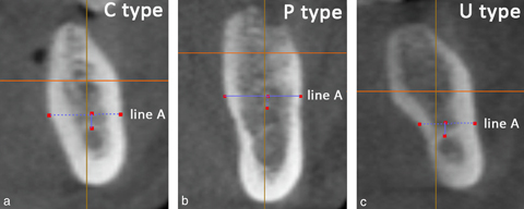
Methodology: In the present study, the reports of 500 patients over 18 years of age who were admitted to our faculty with an existing second premolar and missing first molar and who underwent cone-beam computed tomography (CBCT) imaging were randomly selected and retrospectively evaluated. In the study, alveolar crest types, the buccolingual width of the alveolar crest, crest height, lingual concavity depth, and lingual concavity angle were measured.
Results: U-type crest was detected in 47.8% of 500 individuals evaluated using CBCT. The mean depth of the lingual concavities was 2.36 ± 1.11 mm, and the mean angle of the lingual concavities was 61.09 ± 11.33°. No statistically significant relationship was found between age and alveolar crest width, alveolar crest height, lingual concavity depth, and lingual concavity angle. No significant difference was found between genders in terms of lingual concavity depth, whereas alveolar crest width, alveolar crest height, and lingual concavity angle were significantly higher in males.
Conclusion: The alveolar crest height, alveolar crest width, and lingual concavity angle of edentulous crests in the mandibular first molar region were statistically significantly higher in males compared with females. It can be beneficial to evaluate gender-related differences using CBCT to prevent complications before performing implantation and other oral surgical procedures in the related region.
How to cite this article:
Dala A, İpek F. Evaluation of the alveolar bone and lingual concavity in the posterior mandibular region based on cone-beam computed tomography data. Int Dent Res 2023;13(S1):11-19. https://doi.org/10.5577/idr.2023.vol13.s1.2
Linguistic Revision: The English in this manuscript has been checked by at least two professional editors, both native speakers of English.
Full text article
Authors
Copyright © 2023 International Dental Research

This work is licensed under a Creative Commons Attribution-NonCommercial-NoDerivatives 4.0 International License.
![]()
This is an Open Access article distributed under the terms of the Creative Commons Attribution 4.0 International License (CC BY 4.0), which permits unrestricted use, distribution, and reproduction in any medium, provided the original work is properly cited.